http://doi.org/10.1590/0004-282X20160200
ARTICLE
Reliability and safety of a new upper cervical
spine injury treatment algorithm
Avaliação de reprodutibilidade e segurança de um novo algoritmo de tratamento das
lesões cervicais altas
Andrei Fernandes Joaquim1,2, Roger Schmidt Brock1, Vinicius Monteiro de Paula Guirado1, Luis Henrique Sandon1,
Otávio Turolo da Silva2, Mário Augusto Taricco1, Manoel Jacobsen Teixeira1, Eberval Gadelha Figueiredo1
Upper cervical spine injuries are the most severe traumatic lesions that afect the spine, and are potentially associated with tetraplegia, respiratory dysfunction and even sudden death1,2,3,4.
hese include injuries that may afect the occipital condyles, the atlas and the axis, as well as their adjacent ligamentous and facet joints. he stability of most of this region relies on power-ful and complex ligamentous support, which allows the major -ity of cervical rotation (especially in the atlanto-axial joints) and lexion-extension (especially between the occipital con -dyles and the lateral masses of the atlas)5,6.
Treatment goals are relatively well established and include: 1) maintenance or restoration of spinal stability, 2) protection
and/or decompression of the spinal cord, 3) correction or avoid -ance of progressive spinal deformities. In the last few years, many new surgical techniques and spinal instrumentation sys -tems have been developed, providing immediate stability with selective fusion of the involved levels3,7,8.
However, due to the complexity of its anatomy and a mul-titude of possible injury patterns that afects this region, many classiication schemes have been proposed for upper cervi-cal spine injuries in the past decades, precluding an objec -tive and standardized treatment. his context may result in heterogeneous treatment and complex classiications, some -times not easily applied in the decision-making process of
1Universidade de São Paulo, Divisão de Neurocirurgia, São Paulo SP, Brasil; 2Universidade Estadual de Campinas, Divisão de Neurocirurgia, Campinas SP, Brasil.
Correspondence: Andrei F. Joaquim; Divisão de Neurocirurgia, UNICAMP; Rua Tessália Vieira de Camargo, 126; 13083-872 Campinas SP, Brasil; E-mail: [email protected]
Conflict of interest: There is no conlict of interest to declare.
Received 22 September 2016; Received in inal form 09 November 2016; Accepted 09 November 2016.
ABSTRACT
In the present study, we evaluated the reliability and safety of a new upper cervical spine injury treatment algorithm to help in the selection of the best treatment modality for these injuries. Methods: Thirty cases, previously treated according to the new algorithm, were presented to four spine surgeons who were questioned about their personal suggestion for treatment, and the treatment suggested according to the application of the algorithm. After four weeks, the same questions were asked again to evaluate reliability (intra- and inter-observer) using the Kappa index.
Results: The reliability of the treatment suggested by applying the algorithm was superior to the reliability of the surgeons’ personal suggestion for treatment. When applying the upper cervical spine injury treatment algorithm, an agreement with the treatment actually performed was obtained in more than 89% of the cases. Conclusion: The system is safe and reliable for treating traumatic upper cervical spine injuries. The algorithm can be used to help surgeons in the decision between conservative versus surgical treatment of these injuries.
Keywords: spinal injuries; spinal cord injuries; therapeutics; classiication. RESUMO
Avaliamos a reprodutibilidade e segurança do algoritmo Upper Cervical Spine Injuries Treatment Algorithm (UCITA) recém proposto para a escolha do tratamento das lesões traumáticas da junção crânio-cervical. Métodos: Trinta casos previamente tratados de acordo com o algoritmo foram apresentados a quatro cirurgiões de coluna, sendo questionada a conduta pessoal dos mesmos e a conduta segundo a aplicação do algoritmo. Após 4 semanas, foram refeitas as mesmas perguntas para avaliar a reprodutibilidade (intra e interobservador) do algoritmo, através do índice estatístico “Kappa”. Resultados: A reprodutibilidade da conduta com o uso do algoritmo foi superior a reprodutibilidade da conduta pessoal dos cirurgiões. Com o uso do UCITA, a concordância do tratamento realmente efetivado foi encontrada em mais de 89% dos casos. Conclusão: O uso do UCITA foi seguro e reprodutível, podendo ser usado como ferramenta auxiliar na tomada de decisão entre tratamento cirúrgico versus conservador dos traumatismos da junção crâniocervical.
conservative versus surgical treatment9. Among numerous
schemes some deserve attention, such as the Anderson and D’Alonzo classiication, published in 1974, for odontoid frac-tures, the Efendi et al.2 and the Levine and Edwards classi
-ication for injuries of the posterior elements of the axis, the Anderson and Montesano classiication for occipital condyle fractures, among many others1,4,10. Most of them are complex,
which may result in diferent classiications for the same spe -ciic injury pattern, as well as diferent treatment modalities. Also important is that the majority of these systems were proposed in the era of plain radiographs, without the details of recent 3D CT reconstructions that may display these inju -ries with higher sensitivity and speciicity. In some cases, where soft tissue injuries cannot clearly be identiied using CT imaging, an MRI provides additional information about the spinal cord and nerve roots, even though this informa -tion is not included in the vast majority of the classical clas-siication systems3,9.
In this scenario, a uniied and simpliied classiication system for upper cervical spine injuries became necessary. In 2014, Joaquim et al.9 proposed a new upper cervical spine
injury treatment algorithm for choosing between conserva -tive and surgical treatment for upper cervical spine injuries, based on a literature review of the accepted surgical indica -tions for traumatic injuries of the upper cervical spine and
craniovertebral junction. he idea of this new system is to classify injuries according to: 1) integrity of their ligamentous injuries – disrupted ligaments (with or without fractures) may preferentially be treated with surgical ixation due to their high risk of instability and neurological deterioration; and 2) iso-lated fractures, which should be managed conservatively, with surgery reserved for those who have had a high rate of non-healing or failure of conservative treatment (with deformity, misalignment or neurological risk). An adapted version of the algorithm is presented in the Figure.
However, although promising, this proposed algorithm requires further validation. he main goals of this study were to evaluate the reproducibility and the safety of this new algo-rithm in supporting surgeons to choose between conserva -tive versus surgical treatment of upper cervical spine injuries.
METHODS
hirty cases, previously treated according to the new algorithm, were presented to four spine surgeons. Of the 30 cases included in our study, 19 were treated conservatively, achieving good bone healing and also maintaining normal cervical alignment, whereas 11 were referred for surgical ixa -tion according to the upper cervical spine injury treatment
Traumatic Upper Cervical Injuries
Evaluate if there is a ligamentous Injury:
1) abnormal misalignment, outside the normative ranges of alignment, 2) perched or locked facet joints, 3) increase atlanto-dens interval > 3.5 mm
Yes No
Surgery according to the injury type:
-Occipto-cervical dislocation: OC fusion - Atlantoaxial instability: C1-2 fusion
- Ligament Transverse injury in its substance: C1-2 fusion - C2-3 ligament injury: C2-3 fusion
Evaluate fracture morphology
Occipital Condyle and Atlas Fractures: Initial conservative treatment with a cervical orthosis
Axis fractures: Initial Conservative treatment. Considering surgical treatment in:
1) fractures in the base of the odontoid process, especially in patients older than 50 years, fracture with dens displacement greater than 5 mm or severe comminution in the dens base.
2) fractures that do not heal after conservative management or do not achieve a good cervical alignment.
OCD: Occipto-cervical dislocation; OC: Occipto-cervical; AA: Atlanto-axial Instabilit; LT: Transverse Ligament.
algorithm applied by one of the authors. here were 23 men (76.7%) and seven women (23.3%) in this series. Ages ranged from 16 to 77 years (mean 38.4, median 37, SD ± 14.41 years). he mean follow up was 13.5 months (ranging from three to 36 months, with a median of 10.5, SD ± 11.2 months). he mean follow up was 21 months in the surgical group com-pared with 9.1 months in the group managed conservatively. After institutional review board approval (CAAE: 53542416.2.0000.0065), the algorithm description was presented by one of the authors to four spine surgeons with expertise in the management of spinal cord injuries. All four evaluators were board-certiied neurosurgeons. After that, 30 consecutive cases (> 16 years old) of upper cervical spine injuries, treated by one of the authors, were presented digi-tally with high resolution images, with age and neurologi -cal status (assessed on the American Spine Injury Association Impairment Scale – AIS), to the four surgeons (RSB, VMPG, LHS, MAT). hese patients had been treated according to the algorithm and the treatment was blinded to the four evalu-ators. All the patients were followed up after hospital dis-charge by the same surgeon (AFJ) with routine radiological and clinical follow up (two weeks, one month, three months and then every six months after hospital discharge, with dynamic plain radiographs and CT scans when necessary). A successful conservative treatment was considered when there was evident bone healing and a good cervical align -ment on post-injury images at least three months after the trauma, without incapacitating local pain.
he evaluators were questioned about: 1) the speciic diagnosis of the upper cervical spine injury (injury classii-cation according to the evaluator’s preference), 2) their per -sonal treatment proposal (conservative versus surgical), based on their own clinical experience, and 3) management option according to the application of the algorithm, with a total of three answers for each patient. After four weeks, the same questions were presented again. Both intra- and inter-observer agreements were assessed using the kappa coeicient (Table 1), calculated with the STATA software for Windows®, version 13.
In order to evaluate safety, we compared the actual treat -ment for the 30 patients with the treat-ment proposed by the each of the evaluators.
RESULTS
General results
he mechanisms of the injuries were as a result of 24 patients (80%) being involved in motor vehicle accidents, ive (16.7%) falling from a height and one diving into shallow water (3.3%). One patient, with an odontoid fracture in the dens base, without risk factors for nonunion, was initially con -servatively managed, but then required late surgery for non-healing (three months) and mild persistent cervical pain and
was, therefore, included in the surgical group. In 19 patients treated conservatively, all were AIS E at the index level (except for one patient with a concomitant thoracic fracture and AIS A at T3). Of the 11 patients who underwent surgical treatment, seven were AIS E and four were AIS C by the time surgical treatment was indicated. Posterior atlanto-axial fusion was performed in seven patients, two patients had an occipito-C2-3 fusion, one had an anterior C2-3 ixation and fusion and one had a C1-2-3 instrumented fusion. he clinical data of all patients included in this study are summarized in Table 2 (conservative treatment) and Table 3 (surgical treat-ment). No patient had additional surgery for local pain or severe disability during the follow up.
Classification systems used for the evaluators to guide treatment of upper cervical spine injuries
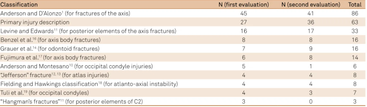
To describe injury characteristics in the irst and in the second evaluation, the evaluators used a total of ten classi -ications systems/eponyms for upper cervical spine injuries, as shown in Table 4.
Evaluation of reliability
Intra-observer analysis
Table 5 shows the results of intra-observer reliability assessed for treatment proposal in each round, and sug -gested treatment according to the algorithm.
Table 6 shows the results of intra-observer reliability assessed for treatment according to the application of the algorithm, and the treatment actually performed.
Inter-observer analysis
Table 7 shows the inter-observer reliability, assessed for personal treatment option and for the treatment proposed by the application of the algorithm.
Validity
Table 8 shows the agreement rates according to three variables: 1) treatment proposal by the evaluator and the application of the algorithm, 2) application of the algorithm and treatment actually performed and 3) treatment proposal by the evaluator and the treatment actually performed.
Of the 34 answers where there was disagreement between the treatment suggestion by the evaluator and the application of the algorithm, 20 (58.8%) occurred in odontoid fractures, attest-ing to the controversies in the management of these injuries.
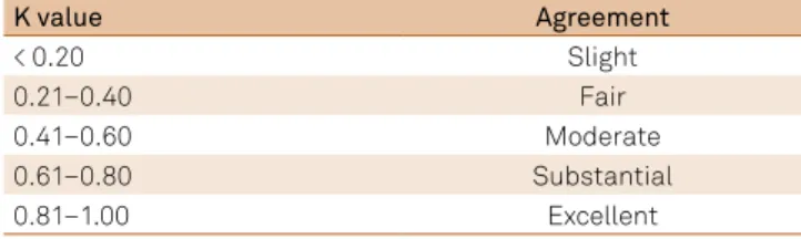
Table 1. Kappa values according the Landis and Koch grading system15.
K value Agreement
< 0.20 Slight
0.21–0.40 Fair
0.41–0.60 Moderate
0.61–0.80 Substantial
Table 2. Summary of 19 patients treated conservatively.
Case Age Injury description Etiology AIS - Observations
1 21 Linear fracture of C2 body Dive in shallow water AIS E
2 58 Fracture of the anterior arch of C1 and also a linear fracture of C2 body MVA AIS E 3 28 Linear fracture of the posterior elements of C2 without displacement MVA AIS E
4 33 Linear fracture of the posterior elements of C2 MVA
AIS E AIS A - thoracic level
(T3 fracture)
5 38 Fracture of the dens base without displacement MVA AIS E
6 60 Fracture of the body of C2 Fall from a height AIS E
7 38 Fracture of the body of C2 MVA AIS E
8 38 Fracture of the dens base MVA AIS E
9 26 Linear right side condyle fracture MVA AIS E
10 37 Fracture of both anterior and posterior arches of C1 MVA AIS E
11 44 Linear fracture of the posterior elements of C2 MVA AIS E
12 17 Fracture of the body of C2 MVA AIS E
13 35 Fracture of the anterior arch of C1 Fall from the height AIS E
14 43 Linear fracture of the body of C2 MVA AIS E
15 36 Fracture of the posterior elements of C2 and mild increase
of the angulation of C2 over C3 MVA AIS E
16 43 Fracture of the body of C2 involving the left C12 joint but without any
displacement MVA AIS E
17 48 Linear left side occipital condyle fracture without displacement of the
facet joints MVA AIS E
18 59 Linear fracture of the posterior arch of C1 without displacement MVA AIS E 19 45 Linear fracture of the posterior elements of C2 without displacement MVA AIS E
AIS: American Spine Injury Association Impairment Scale, MVA: motor vehicle accidents.
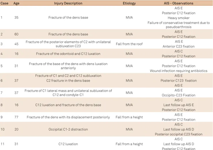
Table 3. Summary of 11 patients operated on.
Case Age Injury Description Etiology AIS - Observations
1 35 Fracture of the dens base MVA
AIS E Posterior C12 ixation
Heavy smoker
Failure of conservative treatment due to pseudoarthrosis
2 60 Fracture of the dens base MVA AIS E
Posterior C12 ixation 3 45 Fracture of the posterior elements of C2 with unilateral
subluxation C23 Fall from the roof
AIS E Anterior C23 ixation
4 16 Fracture of the odontoid and C12 luxation MVA AIS C
Posterior C12 ixation 5 31 Fracture of the base of the dens with dens luxation
anteriorly MVA
AIS E Posterior C12 ixation Wound infection requiring antibiotics
6 37
Fracture of C1 and C2 and C12 subluxation
MVA
AIS E
C2 fracture in the dens base Posterior C123 ixation
AIS E
7 37 Fracture of C1 lateral mass and unilateral subluxation of
C12 and condyle-C1 MVA
AIS E Occipito-C23 Fixation
8 16 C12 luxation and fracture of the dens base MVA
AIS C Last follow up AIS E Posterior C12 ixation 9 77 Fracture of the dens with its displacement posteriorly Fall from a height AIS E
Posterior C12 ixation
10 20 Occipital C1-2 distraction MVA
AIS C Last follow up AIS D Posterior occipital C23 ixation
11 31 C12 luxation Fall from a height
AIS C Last follow up AIS D Posterior C12 ixation
Table 4. Most-used systems for treatment of upper cervical spine injuries (note: some injuries were classiied more than once).
Classification N (first evaluation) N (second evaluation) Total
Anderson and D’Alonzo1 (for fractures of the axis) 45 41 86
Primary injury description 27 36 63
Levine and Edwards11 (for posterior elements of the axis fractures) 16 17 33
Benzel et al.16 (for axis body fractures) 8 8 16
Grauer et al.14 (for odontoid fractures) 7 9 16
Fujimura et al.17 (for axis body fractures) 6 8 14
Anderson and Montesano10 (for occipital condyle injuries) 5 1 6
“Jefferson” fracture12, 13 (for atlas injuries) 4 4 8
Fielding and Hawkings classiication18 (for atlanto-axial instability) 4 4 8
Tuli et al.19 (for occipital condyles) 4 3 7
“Hangman’s fractures”11 (for posterior elements of C2) 3 0 3
Table 5. Intra-observer reliability assessment of each evaluator according to personal treatment proposal and the treatment suggested by the algorithm.
Evaluator Kappa – first evaluation Kappa – second evaluation
1st 0.4828 (Moderate) 0.6667 (Substantial)
2nd 0.7964 (Substantial) 0.6637 (Substantial)
3rd 0.9333 (Excellent) 0.7183 (Substantial)
4th 0.7945 (Substantial) 0.6666 (Substantial)
Table 6. Intra-observer reliability assessment of each evaluator according to treatment proposed by the application of the algorithm and the treatment actually performed
Evaluator Kappa – first evaluation Kappa – second evaluation
1st 0.6193 (Substantial) 0.6479 (Substantial)
2nd 0.8565 (Excellent) 0.7235 (Substantial)
3rd 0.7964 (Substantial) 0.8507 (Excellent)
4th 0.9296 (Excellent) 0.7333 (Substantial)
Table 7. Reliability assessment of treatment proposal by the evaluator and the treatment proposed by the application of the algorithm.
Evaluation Kappa – personal treatment proposal Kappa – treatment proposed by the algorithm
1st 0.5996 (Moderate) 0.6326 (Substantial)
2nd 0.4661 (Moderate) 0.5378 (Moderate)
1st and 2nd rounds together 0.5662 (Moderate) 0.6292 (Substantial)
Table 8. Evaluation of the agreement rates according to three variables: 1) treatment proposal by the evaluator and the application of the algorithm, 2) application of the algorithm and treatment actually performed and 3) treatment proposal by the evaluator and the treatment actually performed.
Evaluator
Agreement of treatment proposal by the evaluator and the application of the algorithm
Application of the algorithm and treatment performed
Treatment proposal by the evaluator and treatment performed
1st Round
1st 22/30 (73.33%) 26/30 (86.67%) 23/30 (76.67%)
2nd 27/30 (90%) 27/30 (90%) 27/30 (90%)
3rd 27/30 (90%) 29/30 (96.67%) 28/30 (93.33%)
4th 29/30 (96.67%) 27/30 (90%) 26/30 (86.67%)
2nd Round
1st 25/30 (83.33%) 25/30 (83.33%) 24/30 (80%)
2nd 25/30 (83.33%) 26/30 (86.67%) 25/30 (83.33%)
3rd 25/30 (83.33%) 26/30 (86.67%) 26/30 (86.67%)
4th 26/30 (86.67%) 28/30 (93.33%) 24/30 (80%)
DISCUSSION
In 2014, the upper cervical spine injury treatment algorithm used in our study based on a literature review and expert opinion was published. he algorithm, divided upper cervical spine inju-ries into ligamentous injuinju-ries (with or without concomitant frac-tures) and isolated fractures, in an attempt to guide toward the best treatment option9. In 2015, preliminary results of a cohort
of patients with upper cervical spine injuries, treated according to this rational treatment guide, was published, with 23 patients treated conservatively and 15 surgically managed. During the follow up, the authors reported that there was no neurologi-cal worsening and patients with incomplete deicits had some improvement3. However, evaluation of the reliability and validity of this system has not been performed since its publication.
In the present series, the majority of the patients were men (76.7%), mostly with injuries secondary to motor vehicle accidents (80%). All patients treated conservatively were neu-rologically intact (19/19 – 100%). However, four in the surgi-cal group (4/11 – 36.36%) had incomplete deicits. Although neurological deicits are not criteria for instability, they may be associated with more severe injuries that potentially would require surgical treatment.
As noted, a wide range of diferent classiication systems were used by the four spine surgeons, even for similar injury patterns, such as axis fractures. Additionally, we observed that the injury description by itself, or classic eponyms (such as “Jeferson’s” or “Hangman’s” fractures), were used to describe upper cervical spine injuries, suggesting a heteroge-neous classiication and potentially diicult comparison of treatment modalities2,11,12,13.
he evaluators’ treatment options had substantial agree-ment with the treatagree-ment suggested by the application of the algorithm in the majority of the cases (with a substantial kappa value obtained in six of eight comparisons, and moder-ate and excellent in one comparison each, as shown in Table 5). However, when intra-observer reliability was assessed for the treatment suggested by the application of the algorithm and the actual treatment performed, we obtained an even higher kappa value (a substantial kappa value was obtained in ive of eight comparisons and an excellent kappa value in three of eight com-parisons, as shown in Table 6). herefore, the use of a global and more uniform system improves classiication reproducibility.
Finally, the inter-observer reliability for the application of the algorithm was substantial (0.63) compared with moderate (0.57) reliability for the evaluators’ personal treatment option. Based on this, we infer that the system is more reliable than the surgeon’s own opinion about the treatment proposed.
When evaluating safety, we obtained a higher rate of agree -ment between the application of the algorithm and the treat-ment actually performed, ranging from 83.3% to 93.3%, as shown in Table 8. his suggested that the use of this new system was reliable and safe. Of note, in the 240 evaluations, 20 (58.8%) of the 34 answers where there was disagreement between the personal treatment option and the algorithm refer to odontoid fracture management. Management of odontoid fractures is controversial, especially for fractures in the dens base, classiied according to Anderson and D’Alonzo as type 27,14. hese injuries had a higher rate of
pseudo-arthrosis, especially when risk factors for nonunion are present7.
However, even in the absence of these risk factors, surgical treat-ment is acceptable. As a consequence, we proposed in our inal version of the algorithm that odontoid fractures in the dens base may be treated surgically or conservatively, despite the risk factors for nonunion, based on the surgeon’s preference and patient’s char-acteristics (preference, comorbidities, age, etc.), until further evi -dence for the best treatment option of these injuries is available.
Limitations of the study
he retrospective application of the algorithm, with limited information, may result in potential bias for a treatment deci-sion. Additionally, it is a guide to treatment, not a descriptive injury system, which may still result in an imprecise description of upper cervical spine injuries. Finally, disability and pain were not speciically addressed, which may alter potential surgical indications. Nonetheless, the patients required no further sur -gery or intervention for pain management. Due to its practical nature, the algorithm may guide surgical indication, helping to identify the most important factors that lead to conservative or surgical management of these complex injuries.
In conclusion, an acceptable intra- and inter-reliability appli-cation of the upper cervical spine injury treatment algorithm is reported in the current study. Additionally, the algorithm was safe to guide treatment of upper cervical spine injuries with respect to neurological morbidity. he management of odontoid fractures in the dens base is still controversial. Further studies evaluating the results of treatment of upper cervical spine injuries are necessary.
References
1. Anderson LD, D’Alonzo RT. Fractures of the odontoid process of the axis. J Bone Joint Surg Am. 1974;56(8):1663-74. http://doi.org/10.2106/00004623-197456080-00017
2. Effendi B, Roy D, Cornish B, Dussault RG, Laurin CA. Fractures of the ring of the axis. A classiication based on the analysis of 131 cases. J Bone Joint Surg Br. 1981;63-B(3):319-27.
3. Joaquim AF, Ghizoni E, Tedeschi H, Yacoub AR, Brodke DS, Vaccaro AR et al. Upper cervical injuries: clinical results using
a new treatment algorithm. J Craniovertebr Junction Spine. 2015;6(1):16-20. http://doi.org/10.4103/0974-8237.151585 4. Traynelis VC, Marano GD, Dunker RO, Kaufman HH. Traumatic
atlanto-occipital dislocation: case report. J Neurosurg. 1986;65(6):863-70. http://doi.org/10.3171/jns.1986.65.6.0863 5. Menezes AH, Traynelis VC. Anatomy and biomechanics of normal
6. Sances A, Jr., Myklebust JB, Maiman DJ, Larson SJ, Cusick JF, Jodat RW. The biomechanics of spinal injuries. Crit Rev Biomed Eng. 1984;11(1):1-76. 7. Lewis E, Liew S, Dowrick A. Risk factors for non-union in the
non-operative management of type II dens fractures. ANZ J Surg. 2011;81(9):604-7. http://doi.org/10.1111/j.1445-2197.2010.05586.x 8. Middendorp JJ, Slooff WB, Nellestein WR, Oner FC. Incidence of and risk
factors for complications associated with halo-vest immobilization: a prospective, descriptive cohort study of 239 patients. J Bone Joint Surg Am. 2009;91(1):71-9. http://doi.org/10.2106/JBJS.G.01347
9. Joaquim AF, Ghizoni E, Tedeschi H, Lawrence B, Brodke DS, Vaccaro AR et al. Upper cervical injuries - a rational approach to guide surgical management. J Spinal Cord Med. 2014;37(2):139-51. http://doi.org/10.1179/2045772313Y.0000000158
10. Anderson PA, Montesano PX. Morphology and treatment of occipital condyle fractures. Spine. 1988;13(7):731-6. http://doi.org/10.1097/00007632-198807000-00004 11. Levine AM, Edwards CC. The management of traumatic
spondylolisthesis of the axis. J Bone Joint Surg Am. 1985;67(2):217-26. http://doi.org/10.2106/00004623-198567020-00007
12. Hays MB, Alker GJ, Jr. Fractures of the atlas vertebra. The two-part burst fracture of Jefferson. Spine. 1988;13(6):601-3. http://doi.org/10.1097/00007632-198813060-00001
13. Levine AM, Edwards CC. Fractures of the atlas. J Bone Joint Surg Am. 1991;73(5):680-91.
14. Grauer JN, Shafi B, Hilibrand AS, Harrop JS, Kwon BK, Beiner JM et al. Proposal of a modified, treatment-oriented classification of odontoid fractures. Spine J. 2005;5(2):123-9. http://doi.org/ 0.1016/j.spinee.2004.09.014
15. Landis JR, Koch GG. An application of hierarchical kappa-type statistics in the assessment of majority agreement among multiple observers. Biometrics. 1977;33(2):363-74. http://doi.org/10.2307/2529786
16. Benzel EC, Hart BL, Ball PA, Baldwin NG, Orrison WW, Espinosa M. Fractures of the C-2 vertebral body. J Neurosurg. 1994;81(2):206-12. http://doi.org/10.3171/jns.1994.81.2.0206
17. Fujimura Y, Nishi Y, Kobayashi K. Classiication and treatment of axis body fractures. J Orthop Trauma. 1996;10(8):536-40. http://doi.org/10.1097/00005131-199611000-00005 18. Fielding JW, Hawkins RJ, Ratzan SA. Spine fusion for
atlanto-axial instability. The J Bone Joint Surg Am. 1976;58(3):400-7. http://doi.org/10.2106/00004623-197658030-00020 19. Tuli S, Tator CH, Fehlings MG, Mackay M. Occipital