Long-term Clinical and Angiographic Follow-up
of the New Non-Polymeric Paclitaxel-Eluting Stent
for the Treatment of
De Novo
Coronary Lesions:
Outcomes of the PAX-B Study
Erlon O. de Abreu-Silva
1, Ricardo A. Costa
2, Andrea Abizaid
3, Angelo Ramondo
4, Philippe Brenot
5,
Hakim Benamer
6, Alessandro Desideri
7, Jacques Berland
8, Breno O. Almeida
9, Franck Digne
10,
Marco A. Perin
11, Juliana P. de Castro
12, J. Ribamar Costa Jr.
13, Rodolfo Staico
13, Luiz F. Tanajura
14,
Alexandre Abizaid
15, on behalf of the PAX-B study Investigators
ABSTRACT
Background: Compared with the Taxus® stent, the
non-polymeric paclitaxel-eluting Amazonia® PAX stent shows
no differences in the occurrence of coronary restenosis or clinical events after four months of follow-up. However, the performance of the Amazonia® PAX stent in more complex
cases and with longer angiographic follow-up has not been demonstrated. Methods: The PAX-B study was a prospective, non-randomised, multicentre study assessing the long-term follow-up of patients treated with the Amazonia® PAX
stent. The primary outcome was late in-stent lumen loss. Results: The study included 103 patients with a mean age of 61.3 ± 11.4 years; 26.2% were diabetics, 24.3% had acute coronary syndromes, and 71.6% had type B2/C lesions. Multiple stents were implanted in 4.7% of the patients, and angiographic success was achieved in 100% of the cases. During hospitalisation, the periprocedural acute myocardial infarction rate was 3.9%, and one of these events led to target-lesion revascularisation (TLR). At the nine-month angiographic follow-up, the median late in-stent lumen loss was 0.91 [0.50; 1.21] mm. The cumulative rates of major adverse cardiac events at the six-month, nine-month, and 12-month follow-ups were 7.8%, 18.5%, and 21.3%,
1 Physician; Interventional cardiologist. Research Fellow, Cardiovascular
Research Center. São Paulo, SP, Brazil.
2 PhD Physician; Interventional cardiologist at the Invasive Cardiology
Service of the Instituto Dante Pazzanese de Cardiologia. São Paulo, SP, Brazil.
3 PhD Physician; Cardiologist at the Instituto Dante Pazzanese de
Cardiologia. São Paulo, SP, Brazil.
4 Physician. Director of the Cardiology Service at the Azienda
Ospe-daliera di Padova. Pádova, Italy.
5 Physician of the Department of Radiology, Cardiovascular Surgery and
Cardiology of the Centre Chirurgical Marie Lannelongue. Le Plessis-Robinson, France.
6 Physician at the Service of Interventional Cardiology of Hôpital
Européen de Paris La Roseraie. Aubervilliers, France.
7 PhD; Director of the Fondazione per la Ricerca Cardiovascolare do
Ospedale San Giacomo. Castelfranco Veneto, Italy.
8 Physician at the Cardiology Service of Clinique Saint Hilaire. Rouen,
France.
9 Graduate Student at the Instituto Dante Pazzanese de Cardiologia.
Physician; Interventional cardiologist and Coordinator of the Interven-tional Cardiology at Hospital Santa Marcelina. São Paulo, SP, Brazil.
Original Article
RESUMO
Seguimento Angiográfico e Clínico Tardio do Novo Stent Farmacológico Não Polimérico Liberador de Paclitaxel para o Tratamento de Lesões Coronárias
De Novo: Resultados do Estudo PAX-B
Introdução: O stent farmacológico eluidor de paclitaxel, não polimérico, Amazonia® PAX não mostrou diferença na
reestenose coronária ou eventos clínicos aos 4 meses de evolução quando comparado com o stent Taxus®.
Entretan-to, o desempenho do stent Amazonia® PAX em cenários de
maior complexidade e com seguimento angiográico mais longo ainda não foi demonstrado. Métodos: O Estudo PAX-B foi um estudo prospectivo, não randomizado, multicêntrico, que avaliou os resultados tardios de pacientes tratados com o stent Amazonia® PAX. O desfecho primário foi a perda tardia
do lúmen intrastent. Resultados: Foram incluídos 103 pacien-tes com média de idade de 61,3 ± 11,4 anos, 26,2% eram diabéticos, 24,3% apresentaram-se com síndrome coronária aguda e 71,6% tinham lesões tipo B2/C. Implante de múltiplos stents ocorreu em 4,7% dos casos e o sucesso angiográico foi de 100%. Na fase hospitalar, a taxa de infarto agudo do miocárdio periprocedimento foi de 3,9%, e um desses eventos
© 2012 Elsevier Editora Ltda. and Sociedade Brasileira de Hemodinâmica e Cardiologia Intervencionista. All rights reserved.
10 “Physician in charge” of the Centre Cardiologique du Nord Saint
Denis. Paris, France.
11 PhD; Head of the Sector of the Cardiovascular Intervention of
Hos-pital Israelita Albert Einstein. São Paulo, SP, Brazil.
12 PhD; Clinical Research Manager of the Cardiovascular Research
Center. São Paulo, SP, Brazil.
13 PhD Physician; Interventional cardiologist at the Invasive Cardiology
Service of Instituto Dante Pazzanese de Cardiologia. São Paulo, SP, Brazil.
14 PhD; Head of the Medical Section of Coronary Angioplasty of the
Instituto Dante Pazzanese de Cardiologia. São Paulo, SP, Brazil.
15 Tenured Professor. Director of the Invasive Cardiology Service of
the Instituto Dante Pazzanese de Cardiologia. São Paulo, SP, Brazil. Correspondence to: Ricardo A. Costa. Rua Dr. Astolfo Araújo, 521 – Vila Mariana – São Paulo, SP, Brazil – CEP 04012-070
E-mail: [email protected]
respectively, mostly due to TLR. There were no deaths or stent thromboses at 12 months. Conclusions: The Amazonia®
PAX stent showed excellent immediate results and a good safety profile. However, angiographic recurrence rates were relatively high because of the low efficacy of neointimal hyperplasia inhibition.
DESCRIPTORS: Drug-eluting stents. Paclitaxel. Coronary disease. Coronary thrombosis.
levou à revascularização da lesão-alvo (RLA). No seguimento angiográico de 9 meses, a mediana da perda tardia do lúmen intrastent foi de 0,91 [0,50; 1,21] mm. As taxas cumulativas de eventos cardíacos adversos maiores nos seguimentos de 6 meses, 9 meses e 12 meses foram, respectivamente, de 7,8%, 18,5% e 21,3%, principalmente em decorrência de RLA. Não se observou morte ou trombose de stent em 12 meses. Conclusões: O stent Amazonia® PAX demonstrou excelentes
resultados imediatos e alto peril de segurança. Entretanto, as taxas de recorrência angiográica foram relativamente altas, em razão da pouca eicácia na inibição da formação de hiperplasia neointimal.
DESCRITORES: Stents farmacológicos. Paclitaxel. Doença das coronárias. Trombose coronária.
I
nitially, irst-generation drug-eluting stents (DES) with durable polymers were shown to be effective in the treatment of selected coronary lesions due to the signiicant reduction in neointimal hyperplasia formation, restenosis and, consequently, the need to perform target lesion revascularisation (TLR), when compared to bare-metal stents (BMS).1–3 However, despite the remarkableeffectiveness of these devices in different scenarios,4
con-cerns related to their safety proile and long-term safety 5
have led to the development of new technologies, aim-ing to optimise results.6–11
Specifically, late stent thrombosis post-DES has been
considered a rare and multifactorial phenomenon,12,13
involving delayed strut endothelialisation and
heal-ing of the treated segment.14,15 Previous pathological
studies demonstrated that the presence of durable polymer debris was associated with inflammation and eosinophil infiltration in the artery wall and is thus a potentially thrombogenic factor.15,16 Therefore,
non-polymeric DES were developed as an alternative to earlier generation DES, which used polymers as a drug carrier system.6,17,18
The Amazonia PAX® device (Minvasys SAS –
Gennevil-lieres, France) is a DES that incorporates a low proile platform and an antiproliferative agent, paclitaxel, which is applied directly to the surface of the device without the use of a polymer. Recently published data from the initial study on the device (vs. Taxus® Liberté
DES, Boston Scientiic – Natick, MA, USA) showed no signiicant differences in the occurrence of angio-graphic restenosis at four months or clinical events up to 12 months in a small sample of patients from a
single-centre study.19 However, the performance of the
Amazonia PAX® stent in more complex scenarios, with
more patients and over longer angiographic follow-up has not been determined. The present study aimed to report the nine-month angiographic follow-up and the long-term clinical outcomes of patients treated with the Amazonia PAX® stent in a prospective, multicentre study.
METHODS
Study protocol and population
The PAX-B study was a prospective, nonrandomised, multicentre, international study that aimed to evaluate the long-term clinical and angiographic outcomes of patients with obstructive coronary artery disease treated with the
Amazonia PAX® stent in nine clinical centres, of which
seven were located in Europe and two were located in Brazil. The study population included patients aged ≥ 18 years, with stable or unstable angina and/or a stress test positive for ischaemia. The angiographic inclusion criteria
were the presence of a de novo coronary lesion with
stenosis of ≥ 50% and a length of ≤ 20 mm by visual
analysis, located in a natural coronary artery, with a refer-ence diameter between 2.5 mm and 3.5 mm, as well as a favourable anatomy for percutaneous coronary intervention with stent implantation. The main exclusion criteria were acute myocardial infarction within < 72 hours of the index procedure; renal failure with basal serum creatinine ≥ 2 mg/ dL; previous percutaneous coronary intervention of the target lesion; previous coronary intervention of the target vessel within nine months of the index procedure; cardiogenic shock; cerebrovascular accident; lesions located in a coro-nary bifurcation, left main corocoro-nary artery or saphenous vein bypass graft; calciication and/or signiicant tortuosity; total occlusion; left ventricular ejection fraction of < 30%; and hypersensitivity or contraindication to aspirin and/or thienopyridine (clopidogrel).19 According to the protocol,
treatment of up to two lesions per patient was permitted, as long as a single lesion was treated per target vessel.
The study was in conducted accordance with the Declaration of Helsinki, was approved by the Ethics Committee of the participating clinical centres, and complied with the mandatory regulations and
require-ments of the Comissão Nacional de Ética em Pesquisa
Device description
The design, proile, and characteristics of the
Ama-zonia PAX® stent have been detailed elsewhere.19 In
summary, this device incorporates a low-proile metallic cobalt-chromium platform, with a thickness of 73 µm, an open-cell modular design, and an antiproliferative agent, paclitaxel, which is applied directly to the ablu-minal surface of the stent struts by a microdrop spray crystallisation process. This procedure only adds 5 µm to strut thickness and aims to ensure the integrity of the drug and a controlled release proile.
Procedure
The percutaneous procedures were performed ac-cording to current guidelines,20 and the inal procedure
was left to the surgeon’s discretion. Direct stent implan-tation was allowed. The implanimplan-tation of an additional stent in the target lesion was allowed if dissection and/or the need for complete coverage of the lesion and the treatment of multiple lesions (maximum of two in distinct target vessels) was permitted according to the protocol. Notably, only patients treated
exclu-sively with Amazonia PAX® stents were considered for
the inal analysis of the PAX-B study. Amazonia PAX®
stents with diameters of 2.5 mm, 3 mm, and 3.5 mm, and extensions of 8 mm, 12 mm, 16 mm, 20 mm, and 24 mm were available for the study.
Dual antiplatelet therapy consisted of the adminis-tration of aspirin (100–325 mg/day) and thienopyridine (75 mg/day of clopidogrel). In case of failure or intoler-ance, the administration of 250 mg of ticlopidine two times daily was allowed. The administration of clopidogrel was started at least 24 hours before the procedure, and a loading dose of 300 mg (or 500 mg of ticlopidine) was recommended. After the procedure, aspirin therapy was maintained indeinitely, and thienopyridine therapy was maintained for at least 12 months, as currently
recommended.20 Regarding antithrombin therapy
dur-ing the procedure, unfractionated heparin (UFH) was administered intravenously at a dose of 70–100 U/kg to maintain an activated coagulation time of > 250 seconds (or > 200 seconds in case of glycoprotein IIb/IIIa inhibitor use, which was administered at the surgeon’s discretion).
Regarding additional tests, a 12-lead electrocardio-gram was obtained before, immediately after, and 24 hours after the procedure. Laboratory tests for cardiac enzymes (creatine phosphokinase and creatine kinase MB-fraction) were conducted pre-procedure (< 72 hours) and every six to eight hours during the irst 24 hours after the procedure or until hospital discharge.
Outcomes and definitions
The primary study endpoint was late lumen loss in the in-stent segment, as assessed by a quantitative
coronary angiography analysis at the nine-month follow-up. Secondary endpoints were target vessel failure, TLR and target-vessel revascularisation (TVR) at the nine-month follow-up, immediate success (of the device, lesion, and procedure), major adverse cardiac events (MACE), and binary restenosis assessed by quantitative coronary angiography at the nine-month follow-up. All deaths were considered to be cardiac unless a noncardiac cause could be clearly established by clinical evaluation or anatomopathological studies. Target vessel failure was deined as the composite endpoint of TVR, myocardial infarction, and cardiac death. MACE were deined by the composite endpoint of death, myocardial infarc-tion, and target-lesion revascularisation (percutaneous or surgical). Stent thrombosis was deined according
to the criteria of the Academic Research Consortium.21
Device success was deined as the successful delivery and implantation of the study stent at the lesion site. The lesion success (angiographic) was deined by achieving a stenosis diameter (SD) of < 50% in the target lesion after the procedure. Procedural success was deined as lesion success without the occurrence of MACE during the index hospitalisation.
Angiographic analysis
The acquisition of angiographic images occurred after the intracoronary administration of nitrate (100– 200 µg) and included two orthogonal projections (> 30 degrees), which aimed to optimise the visualisation of the target lesion. The qualitative and quantitative an-giographic analyses were performed serially pre- and post-procedure and at the nine-month follow-up, and were independently analysed by the Angiographic Analy-sis Laboratory of the Cardiovascular Research Centre (São Paulo, SP, Brazil). Overall, the qualitative analysis followed previously established deinitions, including characteristics of the vessel, lesion morphology, and
coronary low.22 The analyses of quantitative coronary
angiography were performed off-line by surgeons who were experienced with the methods and blinded to procedural data, using a computerised quantitative analysis system with semi-automatic detection for luminal borders, which has been validated, is
com-mercially available (QAngio® XA, version 7.2, Medis
long-term follow-up (MLD post-procedure minus MLD at the nine-month follow-up). Binary restenosis was deined as the presence of SD ≥ 50% in the treated segment at the long-term follow-up and was classiied as focal (< 10 mm) or diffuse (≥ 10 mm). The restenoses were also classiied according to Mehran.23 Quantitative
variables were reported in the in-stent and intra-segment segments, which incorporates the in-stent segment and 5 mm of the borders.6
Clinical follow-up and database
Clinical follow-up was planned at one month, six months, nine months, and 12 months after the proce-dure and annually for up to ive years. The follow-up consisted of medical consultations or telephone contact, performed in accordance with a pre-deined protocol. At the nine-month follow-up, an angiographic study was mandatory. The electronic data capture system that was used was based on an Oracle platform (Redwood Shores, CA, USA), with restricted access through personal passwords. In 100% of the cases, the data were veriied to correct any discrepancies or inconsistent informa-tion through remote monitoring. Physical monitoring (in person) was performed in a sample of 15% of the cases, selected at random. For both remote and physical monitoring, the procedures were the responsibility of the study’s data coordinating centre. All adverse events were veriied directly with the source documentation and were subsequently adjudicated by an independent clinical events committee consisting of three profession-als experienced in clinical and invasive cardiology. For the current analysis, the results of the procedure, the angiographic restudy at nine months, and the clinical follow-up up to 12 months are reported.
Statistical analysis
Categorical variables are shown as absolute num-bers and frequencies. Quantitative variables are shown as mean and standard deviation (± SD) or median and interquartile range [25%; 75%].
RESULTS
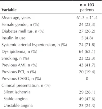
A total of 103 patients were enrolled between December, 2008 and November, 2009. The baseline characteristics are shown in Table 1. Overall, the mean age was 61.3 ± 11.4 years, 26.2% were diabetics, and 41.7% had a previous myocardial infarction. Regard-ing the clinical presentation, approximately half of the patients had stable angina.
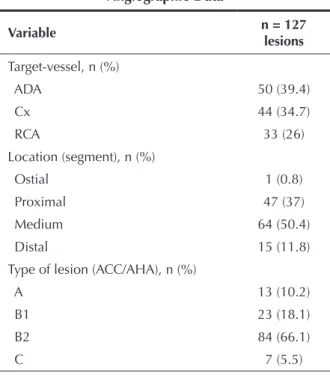
Angiographic data are shown in Table 2. In to-tal, 127 lesions were included (1.24 ± 0.43 lesions per patient) and the anterior descending artery was the target vessel most often treated (39.4%). Ad-ditionally, high-complexity lesions (type B2/C) were found in most cases (71.6%). During the procedure,
radial access was used in 46.6% of the cases, and a second stent in the target lesion was implanted in 4.7% of the cases (Table 3). At the end of the PCI, both angiographic success and device success were achieved in 100% of the cases.
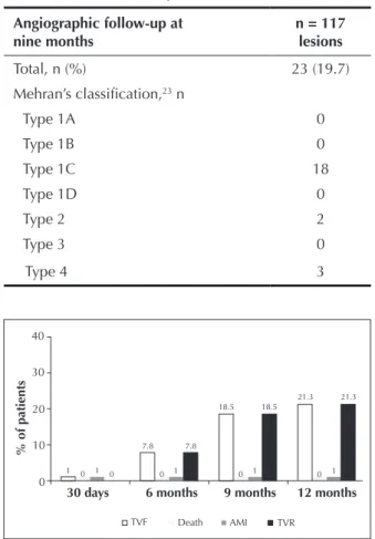
Table 4 shows the pre- and post-procedure results of the quantitative coronary angiography analysis. At the nine-month angiographic follow-up (92.1% of the lesions), the medians of the late intra-segment and in-stent lumen loss were 0.64 [0.32; 0.92] mm and 0.91 [0.50; 1.21] mm, respectively (Table 5). The rate of restenosis was 19.7%, and most recurrent cases were of focal in-stent type (Table 6).
Regarding clinical events while in the hospital, four patients (3.9%) had periprocedural myocardial infarctions (all without Q-wave), and one of these events led to TLR. In this case, the patient returned to the haemodynamics laboratory on the same day of the index procedure since he exhibited ischaemic symptoms after the procedure. At the angiographic assessment, a dissection was observed at the border of the previously implanted stent, which was treated with an additional stent implantation, overlapping with the struts of the other stent. Between hospital discharge and 30 days after the procedure, there was one instance of new myocardial infarction (non-Q). Figure 1 shows the cumulative rates of adverse events after hospital discharge up to the 12-month follow-up
TABLE 1
Baseline Clinical Characteristics
Variable
n = 103 patients
Mean age, years Female gender, n (%) Diabetes mellitus, n (%) Insulin in use
Systemic arterial hypertension, n (%) Dyslipidemia, n (%)
Smoking, n (%) Previous AMI, n (%) Previous PCI, n (%) Previous CABG, n (%) Clinical presentation, n (%)
Silent ischemia Stable angina Unstable angina
61.3 ± 11.4 24 (23,3) 27 (26.2) 5 (4.8) 74 (71.8) 64 (62.1) 23 (22.3) 43 (41.7) 20 (19.4)
0
29 (28.1) 49 (47.6) 25 (24.3)
TABLE 2 Angiographic Data
Variable n = 127
lesions
Target-vessel, n (%) ADA
Cx RCA
Location (segment), n (%) Ostial
Proximal Medium Distal
Type of lesion (ACC/AHA), n (%) A
B1 B2 C
50 (39.4) 44 (34.7) 33 (26)
1 (0.8) 47 (37) 64 (50.4) 15 (11.8)
13 (10.2) 23 (18.1) 84 (66.1) 7 (5.5)
ACC = American College of Cardiology; AHA = American Heart Association; RCA = right coronary artery; Cx = circumlex artery; ADA = anterior descending artery.
TABLE 3 Procedure
Variable n = 103 patients
(127 lesions)
Radial access, n (%) Pre-dilation, n (%)
Nominal balloon extension, mm Nominal balloon diameter, mm Balloon insuflation pressure, atm Implanted study stent, n (%) Nominal stent extension, mm Nominal stent diameter, mm Stent release pressure, atm Additional implanted stent, n (%) Post-dilation, n (%)
Nominal balloon extension, mm Nominal balloon diameter, mm Balloon insuflation pressure, atm Final TIMI low grade 3, n (%)
48/103 (46.6) 54/127 (42.5) 13.9 ± 3.2 2.68 ± 0.88
10.6 ± 2.9 127/127 (100)
16.9 ± 3.9 3.02 ± 0.37
13.5 ± 3.5 6/127 (4.7) 73/127 (57.5)
11.5 ± 2.6 3.21 ± 0.41
16.9 ± 3.8 127/127 (100)
TIMI = thrombolysis in myocardial infarction.
TABLE 4
Pre- and Post-procedural Quantitative Angiography
Variable n = 127
lesions
Pre-procedure Lesion extension, mm RVD, mm
MLD, mm SD, %
Post-procedure RVD, mm Intra-segment MLD, mm SD, %
Immediate gain, mm Intrastent
MLD, mm SD, %
Immediate gain, mm Baloon:artery ratio
12.1 [9.9; 15.8] 2.92 [2.64; 3.21] 0.70 [0.43; 1.00] 74.8 [64.8; 86.0]
2.95 [2.70; 3.30]
2.46 [2.22; 2.77] 15 [10.3; 21.1] 1.74 [1.40; 2.06]
2.79 [2.53; 3.11] 5.7 [3.6.; 8.5] 2 [1.72; 2.40] 1.10 [1.06; 1.16]
MLD = minimal lumen diameter; RVD = reference vessel diameter; SD = stenosis diameter.
TABLE 5
Angiographic follow-up at nine months
Variable n = 117 lesions
RVD, mm Intra-segment MLD, mm SD, %
Late lumen loss, mm Intrastent
MLD, mm SD, %
Late lumen loss, mm
2.81 [2.41; 3.07]
1.84 [1.40; 2.25] 31.7 [21.5; 44.7] 0.64 [0.32; 0.92]
1.94 [1.41; 2.30] 27.3 [16.4; 41.8] 0.91 [0.50; 1.21]
MLD = minimal lumen diameter; RVD = reference vessel diameter; SD = stenosis diameter.
DISCUSSION
In this prospective, multicentre, international registry, the non-polymeric paclitaxel-eluting Amazonia PAX® stent
showed excellent immediate results (device/angiographic success of 100% and procedural success of 96.1%), as well as sustained safety after one year. However, its clinical effectiveness was lower than that reported for other DES, with and without polymers.1–3,7–11,24–28
Notably, the angiographic indings in the long-term follow-up of the PAX-B study are similar to results reported with other previously tested non-polymeric paclitaxel-releasing stents,6 which also conirms that the
Amazonia PAX® stent is less effective than the Taxus®
Liberté paclitaxel-releasing stent with durable polymer at suppressing neointimal proliferation.19
In general, first-generation DES, such as Cypher®
(Cordis – Miami Lakes, USA) and Taxus®,
demon-strated remarkable and sustained clinical
effective-ness.29 However, the durable polymeric components
used as drug carriers in these devices were shown
to cause an intense local inflammatory response,30,31
which has been associated with recurrence and late
thrombotic phenomena.15,16,32,33 Thus, the second
generation of DES introduced more biocompatible polymers and systems with biodegradable poly-mers.7,8,10,11 Recent studies suggest that these devices
have an improved safety profile compared to the first generation of DES, without compromising their clinical effectiveness.34,35 Nonetheless, the
develop-ment of DES without polymers (third-generation DES) could, at least in theory, offer additional advantages, such as a) the prevention of adverse effects due to the prolonged or temporary presence of polymeric residues; b) the optimisation of vascular healing; c) the sustained integrity of the stent surface; and d) the maintenance of dual antiplatelet therapy for shorter
periods.36 However, the removal of the polymer from
the DES may overly compromise its effectiveness, as the polymer is responsible not only for carrying the drug, but also for controlling release kinetics.6 Previous
studies have shown that using a non-polymeric stent coating is safe and may lead to a dose-dependent decrease in restenosis rates; however, these studies have also demonstrated that the critical aspect is the elution profile of the antiproliferative drug.6,17,18 In
the current analysis, the relatively small sample size (n = 103) notwithstanding, impairment in safety was not observed, despite the high prevalence of complex cases and lesions, including an absence of death or stent thrombosis up to the 12-month follow-up. However, the unexpectedly high rates of recurrence indicate the technological challenge of maintaining effectiveness in the absence of a polymer.
In the prospective, randomised, multicentre study DELIVER, which included 1,043 patients, the
non-polymeric, paclitaxel-releasing DES Achieve® (Guidant
Corp. – Santa Clara, CA, USA) presented a mean late in-stent lumen loss of 0.81 ± 0.60 mm at the eight-month follow-up (vs. 0.98 ± 0.57 mm in the control group, treated with BMS, P = 0.003); however, this difference did not translate into signiicant decreases in the rates of binary restenosis or TLR. In this case, the device in the active group (Achieve® stent) received
a coating of paclitaxel at a dose of 3 µg/mm2 of the
stent surface area.6 Similar to the irst generation Taxus®
Liberté DES, which incorporates a durable polymer, the paclitaxel dose is rather low (1 µg/mm2 of the stent surface
area).3 Nevertheless, preclinical studies have estimated a
signiicant loss of the antiproliferative agent (up to 40% of the total) during stent delivery with the nonpolymeric device when compared to < 10% of drug elution in 30 days with the Taxus® stent.3,6 With the Amazonia PAX® stent,
tested in the PAX-A and PAX-B studies, the medians of late in-stent lumen loss were 0.77 [0.47; 1.05] mm (four months) and 0.91 [0.50; 1.21] mm (nine months), respectively. It is noteworthy that the paclitaxel dose (or
concen-tration) was 0.67 µg/cm2 of the stent surface area,
TABLE 6 Binary restenosis
Angiographic follow-up at nine months
n = 117 lesions
Total, n (%) 23 (19.7)
Mehran’s classiication,23 n
Type 1A 0
Type 1B 0
Type 1C 18
Type 1D 0
Type 2 2
Type 3 0
Type 4 3
TVR
% of patients
30 days
40 30 20 10 0
6 months 9 months 12 months 18.5 18.5
21.3 21.3
7.8 7.8
1 0 1 0 0 1 0 1 0 1
TVF Death AMI
with 33%–52% of the drug being released within eight hours; 55%–75%, within a week; and 100%, in 45 days.19 Thus, the kinetics of drug release appear to
explain, at least in part, the greater efficacy observed with polymeric vs. non-polymeric DES, despite the use of the same drug (in this case, paclitaxel) and
a reduced dose.6,19 Greater efficacy has also been
observed with stents releasing drugs from the ‘limus’ class.1,2,7,11,24,25 Late angiographic results with the
non-polymeric sirolimus-releasing DES Yukon®
(Trans-lumina GmbH, Hechingen, Germany) demonstrated relatively lower efficacy when compared with re-sults of the first generation sirolimus-releasing DES Cypher®. In the prospective, randomised study,
Intracoro-nary Stenting and Angiographic Restenosis (ISAR-TEST), which included 450 patients, the mean late in-stent
lumen loss was 0.48 ± 0.61 mm with the Yukon® stent
(vs. 0.48 ± 0.58 mm with the Taxus® stent).24 Similarly,
in the ISAR-TEST 3, which compared three sirolimus-eluting stents, including non-polymeric,
biodegrad-able polymer and durbiodegrad-able polymer systems (Cypher®),
the late in-stent lumen loss was 0.47 ± 0.56 mm vs. 0.17 ± 0.45 mm vs. 0.23 ± 0.46 mm, respectively.37
Alternatives tested in non-polymeric DES have included the incorporation of nonpolymeric carrier components and the structural modification of metal surfaces. The VESTAsync system (MIV Therapeutics, Atlanta, GA, USA) has a microporous hydroxyapatite coating that carries the drug sirolimus at a reduced dose (approximately 40% of the dose used in the Cypher® stent). In the initial study in humans (n = 15),
the late in-stent lumen loss was 0.30 ± 0.25 mm at four months and 0.36 ± 0.23 mm at nine months.
Corres pondingly, the obstruction volume percentage
with intracoronary ultrasound was 2.8 ± 2.2% at four months and 4 ± 2.2% at nine months. It is important to highlight that the VESTAsync stent had a drug-elution profile similar to that of the Cypher® stent, with total
release attained at 90 days.25,28 Additionally, the results
of a new nonpolymeric system using active carrier substances released from a ‘bioinductor’ surface (Cre8, CID – Saluggia, Italy) have been recently reported. In this prospective randomised study (n = 323), the non-polymeric stent Cre8, which releases a ‘limus’ drug, showed a late in-stent lumen loss at six months of
0.14 ± 0.36 mm vs. 0.34 ± 0.40 mm for the Taxus®
Liberté stent (P < 0.0001).26 Finally, the BioFreedom®
stent (Biosensors International – Singapore) incorporates a technology that uses a modified abluminal metal surface and does not use a polymer, resulting in a microporous structure that allows adhesion and the release of the antiproliferative agent Biolimus A9. In the prospective, randomised, BIOFREEDOM study, 182
patients were randomised into three groups and divided into two cohorts according to an angiographic restudy at four months (n = 75) or 12 months (n = 107). At four months, the median values of late in-stent lumen loss was 0.08 [0.02; 0.14] mm, 0.12 [0.07; 0.25] mm vs. 0.37 [0.14; 0.50] mm for the BioFreedom standard
dose, the BioFreedom reduced dose, and the Taxus®
Liberté, respectively; at 12 months, the values were 0.17 [0.09; 0.39] mm, 0.22 [0.17; 0.66] mm vs. 0.35 [0.22; 0.57] mm, respectively (P = 0.001 for non-inferiority in the comparison of the standard dose
BioFreedom group vs. Taxus®).27 Notably, both the
Cre8 and the BioFreedom stents showed efficacy that was relatively superior to that of other reported
non-polymeric DES.26,27 Possible reasons for the improved
efficacy include the use of the ‘limus’ drug family, which has demonstrated superiority in inhibiting neointimal hyperplasia in relation to other classes
of drugs, including paclitaxel.38 Moreover, the Cre8
system has drug release kine tics that are similar to
the first- and second-generations polymeric DES.26 In
contrast, approximately 90% of the drug is released
within 48 hours after BioFreedom implantation;27
how-ever, the drug release kinetics do not appear to have an impact on efficacy, as observed in other systems. It is speculated that this effect is probably due to the high lipophilicity of the drug Biolimus A9 (ten times greater than that of sirolimus), thus ensuring optimal drug absorption and a longer-lasting antiproliferative
effect. In the case of the Amazonia PAX® stent tested
in the present study, a new model, which utilizes a new carrier and release technology for the drug sirolimus based on nanoparticle encapsulation techno-logy (‘LEX’), has been developed (preclinical studies are in progress). This new model should increase the effectiveness in inhibiting neointimal hyperplasia for that system (R. Costa, personal communication, TCT 2011. San Francisco, CA, USA).
CONCLUSIONS
In this prospective, multicentre, international registry, the new drug-eluting, non-polymeric
paclita-xel-releasing Amazonia® PAX stent showed excellent
immediate results and a high safety proile, including the absence of death or stent thrombosis after one year of follow-up. However, recurrence rates were relatively high because of the low effectiveness of neointimal hyperplasia inhibition.
CONFLICTS OF INTEREST
REFERENCES
1. Sousa JE, Costa MA, Abizaid A, Abizaid AS, Feres F, Pinto IM, et al. Lack of neointimal proliferation after implantation of sirolimus-coated stents in human coronary arteries: a quantita-tive coronary angiography and three-dimensional intravascular ultrasound study. Circulation. 2001;103(2):192-5.
2. Moses JW, Leon MB, Popma JJ, Fitzgerald PJ, Holmes DR, O’Shaughnessy C, et al. Sirolimus-eluting stents versus standard stents in patients with stenosis in a native coronary artery. N Engl J Med. 2003;349(14):1315-23.
3. Stone GW, Ellis SG, Cox DA, Hermiller J, O’Shaughnessy C, Mann JT, et al. A polymer-based, paclitaxel-eluting stent in patients with coronary artery disease. N Engl J Med. 2004; 350(3):221-31.
4. Kirtane AJ, Gupta A, Iyengar S, Moses JW, Leon MB, Applegate R, et al. Safety and eficacy of drug-eluting and bare metal stents: comprehensive meta-analysis of randomized trials and observational studies. Circulation. 2009;119(25):3198-206. 5. Daemen J, Wenaweser P, Tsuchida K, Abrecht L, Vaina S,
Morger C, et al. Early and late coronary stent thrombosis of sirolimus-eluting and paclitaxel-eluting stents in routine clini-cal practice: data from a large two-institutional cohort study. Lancet. 2007;369(9562):667-78.
6. Lansky AJ, Costa RA, Mintz GS, Tsuchiya Y, Midei M, Cox DA, et al. Non-polymer-based paclitaxel-coated coronary stents for the treatment of patients with de novo coronary lesions: angiographic follow-up of the DELIVER clinical trial. Circula-tion. 2004;109(16):1948-54.
7. Meredith IT, Ormiston J, Whitbourn R, Kay IP, Muller D, Bonan R, et al. First-in-human study of the endeavor ABT-578-eluting phosphorylcoline-encapsulated stent system in de novo native coronary artery lesions: ENDEAVOR I Trial. EuroIntervention. 2005;1(2):157-64.
8. Serruys PW, Ong AT, Piek JJ, Neumann FJ, van der Giessen WJ, Wiemer M, et al. A randomized comparison of a durable polymer Everolimus-eluting stent with a bare metal coronary stent: the SPIRIT irst trial. EuroIntervention. 2005;1(1):58-65. 9. Costa RA, Lansky AJ, Mintz GS, Mehran R, Tsuchiya Y, Negoita M,
et al. Angiographic results of the irst human experience with everolimus-eluting stents for the treatment of coronary lesions (the Future I trial). Am J Cardiol. 2005;95(1):113-6.
10. Costa RA, Lansky AJ, Abizaid A, Mueller R, Tsuchiya Y, Mori K, et al. Angiographic results of the irst human experience with the Biolimus A9 drug-eluting stent for de novo coronary le-sions. Am J Cardiol. 2006;98(4):443-6.
11. Meredith IT, Worthley S, Whitbourn R, Walters D, Popma J, Cutlip D, et al. The next-generation endeavor resolute stent: 4-month clinical and angiographic results from the endeavor resolute irst-in-man trial. EuroIntervention. 2007;3(1):50-3. 12. Iakovou I, Schmidt T, Bonizzoni E, Ge L, Sangiorgi GM,
Stankovic G, et al. Incidence, predictors, and outcome of thrombosis after successful implantation of drug-eluting stents. JAMA. 2005;293(17):2126-30.
13. Costa RA, Sousa AGMR, Moreira AC, Costa JR Jr, Maldona-do G, Cano M, et al. Trombose de stent farmacológico no “mundo-real”: análise crítica do Registro DESIRE (Drug-Elu ting Stent in the Real World). Rev Bras Cardiol Invasiva. 2008; 16(2):144-54. 14. Joner M, Finn AV, Farb A, Mont EK, Kolodgie FD, Ladich E,
et al. Pathology of drug-eluting stents in humans: delayed healing and late thrombotic risk. J Am Coll Cardiol. 2006;48(1):193-202. 15. Finn AV, Nakazawa G, Joner M, Kolodgie FD, Mont EK,
Gold HK, et al. Vascular responses to drug eluting stents: Importance of delayed healing. Arterioscler Thromb Vasc Biol. 2007;27(7):1500-10.
16. Virmani R, Guagliumi G, Farb A, Musumeci G, Grieco N, Motta T, et al. Localized hypersensitivity and late coronary thrombosis secondary to a sirolimus-eluting stent: should we be cautious? Circulation. 2004;109(6):701-5.
17. Park SJ, Shim WH, Ho DS, Raizner AE, Park SW, Hong MK, et al. A paclitaxel-eluting stent for the prevention of coronary restenosis. N Engl J Med. 2003;348(16):1537-45.
18. Gershlick A, De Scheerder I, Chevalier B, Stephens-Lloyd A, Camenzind E, Vrints C, et al. Inhibition of restenosis with a paclitaxel-eluting, polymer-free coronary stent: the European evaluation of pacliTaxel Eluting Stent (ELUTES) trial. Circula-tion. 2004;109(4):487-93.
19. Chamié D, Costa JR Jr, Abizaid A, Costa RA, Feres F, Staico R, et al. Comparação randomizada entre o stent eluidor de paclitaxel de nova geração sem polímero e o stent eluidor de paclitaxel com polímero durável em pacientes com doen-ça arterial coronária: resultados da análise angiográica e ultrassonográica seriada do Estudo PAX-A. Rev Bras Cardiol Invasiva. 2011;19(4):379-91.
20. King SB 3rd, Smith SC Jr, Hirshfeld JW Jr, Jacobs AK, Morrison DA, Williams DO, et al. 2007 Focused Update of the ACC/AHA/SCAI 2005 Guideline Update for Percutane-ous Coronary Intervention: a report of the American College of Cardiology/American Heart Association Task Force on Practice Guidelines: 2007 Writing Group to Review new Evidence and Update the ACC/AHA/SCAI 2005 Guideline Update for Percutaneous Coronary Intervention, Writing on Behalf of the 2005 Writing Committee. Circulation. 2008;117(2):261-95.
21. Cutlip DE, Windecker S, Mehran R, Boam A, Cohen DJ, van Es GA, et al. Clinical end points in coronary stent trials: a case for standardized deinitions. Circulation. 2007;115(17):2344-51. 22. Popma JJ, Gibson CM. Qualitative and quantitative
angiogra-phy. In: Topol EJ, ed. Textbook of interventional cardiology. Philadelphia: Saunders; 2003.
23. Mehran R, Dangas G, Abizaid AS, Mintz GS, Lansky AJ, Satler LF, et al. Angiographic patterns of in-stent restenosis: classi-ication and implclassi-ications for long-term outcome. Circulation. 1999;100(18):1872-8.
24. Mehilli J, Kastrati A, Wessely R, Dibra A, Hausleiter J, Jaschke B, et al. Randomized trial of a nonpolymer-based rapamycin-elu ting stent versus a polymer-based paclitaxel-rapamycin-eluting stent for the reduction of late lumen loss. Circulation. 2006; 113(2):273-9.
25. Costa JR Jr, Abizaid A, Costa R, Feres F, Tanajura LF, Maldo-nado G, et al. 1-year results of the hydroxyapatite polymer-free sirolimus-eluting stent for the treatment of single de novo coronary lesions: the VESTASYNC I trial. JACC Car-diovasc Interv. 2009;2(5):422-7.
26. Carriè D, Berland J, Verheye S, Hauptmann KE, Vrolix M, Violini R, et al. A multicenter randomized trial compar-ing amphilimus- with paclitaxel-elutcompar-ing stents in de novo native coronary artery lesions. J Am Coll Cardiol. 2012; 59(15):1371-6.
27. Costa RA, Abizaid A, Mehran R, Magalhaes MA, Gambone L, Parise H, et al. De novo coronary lesions treated with the novel polymer-free biolimus-a9 coated stents: 12-month angio-graphic results from the prospective, randomized, multicenter biofreedom clinical trial. J Am Coll Cardiol. 2011;57:E1645. 28. Costa JR Jr, Abizaid A, Costa R, Feres F, Tanajura LF, Mattos LA,
29. Stone GW, Moses JW, Ellis SG, Schofer J, Dawkins KD, Morice MC, et al. Safety and eficacy of sirolimus-and paclitaxel-eluting coronary stents. N Engl J Med. 2007;356(10):998-1008. 30. van der Giessen WJ, Lincoff AM, Schwartz RS, van Beusekom
HM, Serruys PW, Holmes DR Jr, et al. Marked inlammatory sequelae to implantation of biodegradable and nonbiodegrada-ble polymers in porcine coronary arteries. Circulation. 1996; 94(7):1690-7.
31. Suzuki T, Kopia G, Hayashi S, Bailey LR, Llanos G, Wilensky R, et al. Stent-based delivery of sirolimus reduces neointimal formation in a porcine coronary model. Circulation. 2001; 104(10):1188-93.
32. Cook S, Ladich E, Nakazawa G, Eshtehardi P, Neidhart M, Vogel R, et al. Correlation of intravascular ultrasound indings with histopathological analysis of thrombus aspirates in pa-tients with very late drug-eluting stent thrombosis. Circulation. 2009;120(5):391-9.
33. Cook S, Wenaweser P, Togni M, Billinger M, Morger C, Sei-ler C, et al. Incomplete stent apposition and very late stent throm-bosis after drug-eluting stent implantation. Circulation. 2007;115(18):2426-34.
34. Stefanini GG, Kalesan B, Serruys PW, Heg D, Buszman P, Linke A, et al. Long-term clinical outcomes of biodegra dable polymer biolimus-eluting stents versus durable polymer sirolimus-eluting stents in patients with coronary artery disease (LEADERS): 4 year follow-up of a randomised non-inferiority trial. Lancet. 2011;378(9807):1940-8.
35. Bangalore S, Kumar S, Fusaro M, Amoroso N, Attubato MJ, Feit F, et al. Short- and long-term outcomes with drug-eluting and bare-metal coronary stents: a mixed-treatment comparison analysis of 117 762 patient-years of follow-up from random-ized trials. Circulation. 2012;125(23):2873-91.
36. Abizaid A, Costa JR Jr. New drug-eluting stents: an overview on biodegradable and polymer-free next-generation stent systems. Circ Cardiovasc Interv. 2010;3(4):384-93.
37. Mehilli J, Byrne RA, Wieczorek A, Iijima R, Schulz S, Bruskina O, et al. Randomized trial of three rapamycin-eluting stents with different coating strategies for the reduction of coronary restenosis. Eur Heart J. 2008;29(16):1975-82.