Neotropical Entomology
journal homepage: www.scielo.br/ne ISSN: 1519-566X
SyStEmatIcS, morphology aNd phySIology
taxonomic review of the genus
Tachardiella
cockerell (hemiptera: Kerriidae),
with a Key to Species of lac Insects recorded from the New World
T Kondo
1, PJ Gullan
2 1corpoIca, palmira, Valle, colombia2department of Entomology, Univ of california, davis, ca, USa
Keywords
argentina, brea, Parkinsonia praecox, taxonomy
Correspondence
Takumasa Kondo, corpoIca – corporación
Colombiana de Investigación Agropecuaria,
centro de Investigación palmira, calle 23,
Carrera 37, Continuo al Penal Palmira, Valle,
colombia; takumasa.kondo@gmail.com
Edited by roberto a Zucchi – ESalQ/USp
received 03 November 2010 and accepted 29 december 2010
Abstract
The lac insect genus Tachardiella Cockerell is reviewed and 17 species are recognized, including one new species. The adult female of Tachardiella palobrea Kondo & Gullan sp. n. is described and illustrated based on material collected in Argentina on ‘brea’, Parkinsonia praecox (Fabaceae). The adult female of Tachardiella argentina (Dominguez) is redescribed and a lectotype is designated based on newly discovered syntype material in the Bohart Museum of Entomology, University of California, Davis (BME). Diagnoses and notes are provided for the other species of Tachardiella studied and a lectotype is designated for Tachardiella ferrisi Chamberlin. A taxonomic key to separate all known species of lac insects in the New World is provided.
Introduction
Very little has been published on lac insects, the Kerriidae, in the New World since the work of Chamberlin (1923, 1925). Matile-Ferrero & Couturier (1993) described a new species of Austrotachardiella Chamberlin from Peru. Later, Kondo & Gullan (2005) added a new species of Austrotachardiella from Colombia, and gave an updated key to species of that genus. More recently, Kondo & Gullan (2007) reviewed the genus Paratachardina Balachowsky and described the invasive species Paratachardina pseudolobata Kondo & Gullan, which in the New World has been reported from USA (Florida), Bahamas and Cuba. For lac insects in North America, Gill (1993) illustrated those species of Tachardiella Cockerell occurring in California, and provided a key to separate them by adapting Chamberlin´s (1923) key to species of the genus. Gill (1993) mentioned the existence of various forms (perhaps putative species) occurring in California, including those cited by Chamberlin (1923), and suggested that the genus Tachardiella needed critical revision.
Tachardiella species occur in the Nearctic and Neotropical regions, but usually in the drier parts of southwest North America and Mexico and areas of Argentina and Brazil (Ben-Dov 2006). Recently, Kondo & Gullan (2010) transferred the species Coccus resinatum Kieffer & Herbst, previously classified in the family Coccidae, to the lac insect family Kerriidae as Tachardiella resinata (Kieffer & Herbst) after translation of the original description written in German. The total number of named species in the genus is now sixteen.
The present study is not intended to be a revision of Tachardiella, but should be treated as a brief review of the genus. We describe and illustrate the adult female of a new species of Tachardiella fromArgentina collected on Parkinsonia praecox (Fabaceae), a tree locally known as ‘brea’, and we also redescribe Tachardiella argentina
(Dominguez), which is described for the first time on
a lectotype for Tachardiella ferrisi Chamberlin from BME syntype material. To facilitate further study, we make information from older literature available to a wider audience by providing a diagnosis, mostly based on Chamberlin (1923), and notes for each of the other 15 species of Tachardiella, and by presenting a key to the adult females of all species of lac insects known to occur in the New World. The key is based partly on the works of Chamberlin (1923, 1925), partly on translations of
original descriptions by the first author, and partly on
the study of museum specimens, including the types of some species.
Material and Methods
The description of the adult female of the new species and the redescribed species are based on multiple specimens that were slide-mounted recently by the second author. Museum specimens of other species in the genus Tachardiella were also examined for comparison and additional specimens were slide-mounted from dry material in the BME. Slide-mounting techniques follow those of Williams & Granara de Willink (1992) except that xylene was used instead of clove oil. Collection data, number of slides with the total number of specimens, and the depository in parentheses are given for each lot of material studied.
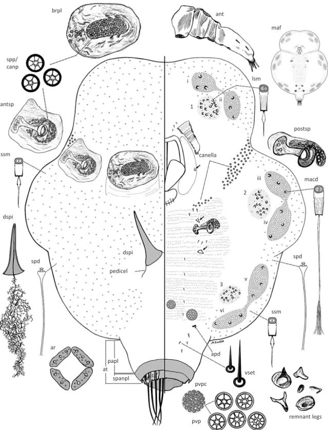
Measurements of adult female lac insects were made using an ocular micrometer in an Olympus compound microscope. In the two descriptions, the body shape of the adult female is described both when unmounted and when mounted on a microscope slide. An “unmounted” adult female refers to the insect’s resinous test, either alive, preserved dry or in ethanol. Body length and width of the adult female are measured in mm as mounted on the slide; other measurements are in microns. Length is measured from the apex of the head to the posterior end of the body. Width is measured as the greatest width. The length of each spiracle is the length of the spiracular apodeme plus the peritreme. The drawing is a generalization of several specimens and was made with the assistance of a camera lucida attached to an
Olympus compound microscope. The figure shows an
entire insect with the venter depicted on the right side of the illustration and the dorsum shown on the left. Special features of the specimen are enlarged to the side of the main illustration, although enlargements are not in direct proportion to each other.
The terms used to describe the lac insects follow those of Kondo & Gullan (2007) and Gullan & Kondo (2009). Marginal duct clusters (mdc) in the New World genera Austrotachardiella and Tachardiella can be separated into simplex, duplex and triplex types. A simplex type mdc is composed of one type of duct, either composed
solely of spermatoid ducts or, in some cases, of just macroducts. This latter case appears to apply in cases in which spermatoid ducts are scarce or hard to detect due to the condition of the specimen. High quality
slide-mounts are essential for lac insect identification.
Duplex mdc are found in Tachardiella, and we believe that Chamberlin’s suggestion that some species might
have simplex mdc was due to the difficulty of seeing
spermatoid ducts in poor slide-mounts. Duplex mdc are composed of macroducts and spermatoid ducts only, whereas Austrotachardiella species have triplex mdc composed of macroducts, spermatoid ducts and an outer narrow band of microducts (Kondo & Gullan 2005, 2007, Gullan & Kondo 2009).
A key to the adult females of New World kerriids was compiled using mostly published keys and descriptions, i.e., Kondo & Gullan (2005) for Austrotachardiella, Gullan & Kondo (2009) for Kerria lacca (Kerr), Kondo & Gullan (2007) for Paratachardina pseudolobata and mostly Chamberlin (1923) for Tachardiella. Specimens of Tachardiella species deposited at the Bohart Museum of Entomology were studied for comparison and for testing of the key (see individual species entries for details of specimens studied). For those Tachardiella species that were not included in Chamberlin’s (1923) key to Tachardiella, original descriptions were used here for construction of the key couplets, as follows: Hempel (1921) for T. artocarpi, Leonardi (1911) for T. cordaliae and T. lycii, Fonseca (1975) for T. nigra, Hempel (1937) for T. ourinhensis, Hempel (1900) for T. parva, and Kieffer & Herbst (1909) for T. resinata. These descriptions had to be translated from Portuguese, Italian or German. We did not examine specimens of any of the above seven species.
This review collates as much of the previously published information as possible for each named species. As far as possible, brief diagnoses of the species, based on the adult female only, are provided. For nine species, these diagnoses are based on those of Chamberlin (1923), as acknowledged in the text for the species. This important publication is not readily or freely available, although it may be purchased from the publisher. In his descriptions, Chamberlin (1923) provides counts of pores and ducts for each species, but these meristic data appear in most cases to have been based on a very few or perhaps sometimes one individual of each species and thus should not be
regarded as definitive of the species. For five species,
the diagnosis is based on a translation of the original description, as noted above for the construction of the key. In each diagnosis we have inserted [in square brackets] our interpretation of the various features.
lectotype has been designated for two speciesto provide stability of nomenclature, and designation is done in a revisionary context in agreement with the amended Recommendation 74G of Article 74.7.3.
Abbreviations for the depositories are as follows: BME (the Bohart Museum of Entomology, Department of Entomology, University of California, Davis, California, U.S.A.); CSCA (California State Collection of Arthropods, the California Department of Food and Agriculture, Sacramento, California); IMLA (Fundación e Instituto Miguel Lillo, Universidad Nacional de Tucumán, Tucumán, Argentina); USNM (United States National Collection of Coccoidea of the National Museum of Natural History, Smithsonian Institution, housed at the United States Department of Agriculture (USDA), Beltsville, Maryland).
Key Based on Adult Females to Separate Species of the Family Kerriidae Known to Occur in the New World
1. Without perivulvar pore clusters. Insect test (resin cover) letter-X shaped. Highly polyphagous. Reported from the Bahamas, Christmas Island (Australia), Cuba, and the USA (Florida) ...………… Paratachardina pseudolobata Kondo & Gullan (Fig 1b)
− With perivulvar pore clusters. Insect test not letter-X
shaped ... 2 2. With more than three pairs of perivulvar pore clusters,
usually with 18-50. Marginal duct clusters composed of large-sized microducts only and arranged in linear serpentine groups. Canellae absent. Polyphagous. Fig 1 Some lac insects known from the New World. a) Kerria lacca [on Albizzia sp., Peradeniya, Sri Lanka; inset showing close-up of adult female]; b) Paratachardina pseudolobata [paratypes, on Myrica cerifera, University of Florida, Fort Lauderdale Research Education Center, Davie, Broward Co., Florida, U.S.A.]; c) Austrotachardiella colombiana [paratypes, on Psidium guajava, Santander de Quilichao, Cauca, Colombia]; d) Tachardiella ferrisi [paralectotypes, on Acacia flexicaulis, La Paz, Lower California, Mexico]; e) Tachardiella fulgens [on
Coursetia microphylla, Sabino Canyon, Catalina Mts, Arizona]; f) Tachardiella glomerella [on Gutierrezia sp., Pecos River, near Sheffield,
Texas]; g) Tachardiella ingae [syntypes, Mogy-Guassu (= Mogi Guaçu), Brazil]; h) Tachardiella larreae [on Larrea tridentata, near Rice, Arizona, USA]; i) Tachardiella mexicana (Comstock) [on Myrica cerifera, Lake Buena Vista, Orange County, Florida]. Photographs by TK except that of Tachardiella mexicana by Lyle Buss, University of Florida].
a
d
g
b
e
h
c
f
Reported from Neotropic (Guyana), Indo-Malayan and Palearctic Regions ... Kerria lacca (Kerr) (Fig 1a)
− With two (rarely three) pairs of perivulvar pore clusters.
Marginal duct clusters composed of macroducts and spermatoid ducts, with some taxa having an outer row of large-sized microducts; clusters not arranged in linear serpentine groups. Canellae generally present. Monophagous or oligophagous. Known from the New World .…………...……. 3
3. Marginal duct clusters triplex; with two setae on last antennal segment (Austrotachardiella Chamberlin) ... 4
− Marginal duct clusters duplex; with 3-8 setae on last
antennal segment (Tachardiella Cockerell) ... 12 4. Marginal duct clusters not paired (six clusters in total),
never with a deep constriction subdividing clusters ... 5
− Marginal duct clusters paired (12 clusters in total),
completely separated or each pair connected with at most a narrow isthmus of microducts (e.g., as in Figs 3, 4) .... 7 5. Posterior marginal duct cluster (mdc-iii) with two
macroducts. Known from Brazil (São Paulo); on Cydonia
and Rosa (Rosaceae) ………..…... A. cydoniae (Hempel)
− Posterior marginal duct cluster (mdc-iii) with 3-5
macroducts ...……. 6
6. Canellae well developed, each composed of 50-60 pores, each pore about size of a spiracular pore, extending in a line from area mesad to anterior spiracles towards area near mouthparts. Known from Brazil (São Paulo); on Croton (Euphorbiaceae) ... A. rubra (Hempel)
− Canellae poorly developed, each composed of a linear
group of 5-10 pores, each pore much smaller than a spiracular pore, present on each side of mouthparts. Known from Mexico (Jalisco and Veracruz); on Acacia (Fabaceae) ... A. nigra (Townsend & Cockerell)
7. Test of live insect with three elevated lobes on mid-dorsum. Known from Brazil (Rio de Janeiro); on
unidentified genus of Myrtaceae ...
... A. trilobata (Mendes)
− Test of live insect with one or no elevated lobe on
mid-dorsum ...………... 8
8. Always with some marginal duct clusters with two macroducts, occasionally a few clusters may have more than three macroducts per cluster. Known from Mexico (Jalisco); on plants locally known as “zicna” and “guasima”, latter probably Guazuma ulmifolia (Malvaceae) ... A. rotundata (Cockerell & Cockerell)
− Marginal duct clusters never with just two macroducts,
each with three or more macroducts ………..…9
9. Number of microducts in each anterior ventral duct cluster (vdc-1) 35-70 (mostly <60) ……... 10
− Number of microducts in each anterior ventral duct
cluster (vdc-1) 75-150 (mostly >80) ……... 11
10. Marginal duct clusters each with three or four (mostly three) macroducts; test crimson red. Known from Jamaica; on Chrysobalanus (Chrysobalanaceae) ….………. …...….... A. gemmifera (Cockerell)
− Marginal duct clusters each with four or five macroducts;
test orange-red to faintly orange-ruby. Known from Guyana; on Sapium (Euphorbiaceae) and Ficus (Moraceae) ………... A. bodkini (Newstead) 11. Each anterior marginal duct cluster (mdc-i) with
50-70 microducts; most marginal duct clusters with
three or four (rarely with four or five) macroducts;
test of sticky texture. Known from Peru; on Myrciaria (Myrtaceae) ... A. sexcordata Matile-Ferrero
− Each anterior marginal duct cluster (mdc-i) with 80-115
microducts; each marginal duct cluster always with four
or five macroducts; test of hard texture. Known from
Colombia; on Psidium (Myrtaceae) …... ....……….... A. colombiana Kondo & Gullan (Fig 1c)
12. Test of golden color. Known from Chile (Concepcion); on Baccharis (Asteraceae) ... Fig 2 a) Tachardiella argentina on twig of Acacia caven (Fabaceae),
with arrow pointing to nipple-like elevation; b) Tachardiella palobrea tended by Camponotus sp. cf. rosariensis, on Parkinsonia praecox (Fabaceae). Photograph A by TK; B by PJG.
a
... T. resinata (Kieffer & Herbst)
− Not with above combination ..……….…….. 13
13. Test of adult female light yellowish in color, shiny, sticky. Known from Brazil (Ourinhos, São Paulo); on a
cultivated plant of unidentified genus of Myrtaceae ...
...……… T. ourinhensis Hempel
− Not with above combination ……….… 14
14. Test of individual adult female, round, oval, dark purple in color, with two conspicuous humps on dorsum. Known from Argentina (Cacheuta, Mendoza); on Condalia (Rhamnaceae) …...….. T. cordaliae (Leonardi)
− Not with above combination …...……….. 15
15. Test of individual adult female, round, oval, dark purple in color, with one conspicuous mammiform hump on dorsum. Known from Argentina (Cacheuta, Mendoza); on Lycium (Solanaceae) …... T. lycii (Leonardi)
− Not with above combination ………...……….. 16
16. Test of mature female globose, with pointed elevation on mid dorsum, dilated margins and shallow sulci at base; resin reddish-brown in color, slightly yellowish, darker towards margins; young individuals with test red, star-shaped, with elevation on dorsum, becoming pyramidal with increase in size. Known from Brazil (Rio de Janeiro); on Anacardium (Anacardiaceae), Artocarpus (Moraceae) and Terminalia (Combretaceae) ... T. artocarpi (Hempel)
− Not with above combination ……...……….. 17
17. Leg remnants with three segments, basal segment tubercle-shaped, middle segment about half width and as long as basal segment, apical segment ending in an acute tip. Test globular, 3-4 mm in length, black, shiny, with consistency somewhat soft and
flexible, dorsal region with three white curved filaments. Known from Brazil (Morumbi, São Paulo);
on native forest plant (Myrtaceae) ... ... T. nigra Fonseca – Leg remnants not as above. Test generally of hard and
brittle texture ... 18
18. Marginal duct clusters duplex, each with no more than two macroducts. Large-sized microducts on posterior ventral duct cluster (vdc-3) spread over an area several times larger than area of anterior spiracle. Known from
Brazil (Ypiranga and Cachoeira); on unidentified genus of
Myrtaceae ……….………… T. parva (Hempel)
− Not with above combination ……….. 19
19. Macrotubular ducts absent on dorsum ....………….. 20 – Macrotubular ducts present on dorsum, although
sometimes not very conspicuous ……....…. 23 20. Maximum dimension of brachial plate 200-250 µm. Canellar pores near anterior spiracle forming clusters involving 10-30 fused pores,
often fused in two or several groups; each pore
tubular in profile, usually longer than wide ... 21 − Maximum dimension of brachial plate 130-200 µm.
Canellar pores near anterior spiracle either not forming clusters of fused pores or with just a few small clusters of
only 2-4 fused pores; in profile each pore usually about
as long as wide ...……….. 22 21. Crater of brachial plate ovate-oblong with one end
having distinct sub-depressions; rim around crater mostly much narrower than width of crater itself. Canellae well developed and very long (at least half maximum body width), with 115-160 or more canellar pores. Known from Brazil (São Paulo); on Inga (Fabaceae) ………. T. ingae (Hempel) (Fig 1g)
− Crater of brachial plate ovate, usually obviously
tapering at one end, without distinct sub-depressions in crater; rim around crater broader, at least in some places, than width of crater itself. Canellae well-developed but rarely as long as half maximum body width, generally with 100 or fewer canellar pores. Known from Mexico (Oaxaca and Tamaulipas) and USA (Florida and Texas); on Acacia and Mimosa (Fabaceae) ………... T. mexicana (Cockerell)
22. Lac test often with a nipple-like elevation (Fig 2a). When seen from above, brachial crater almost central, edge of crater not touching outer rim of brachial plate. Macroducts in marginal duct clusters rather small, each 7.5-10 µm wide. Second marginal duct cluster (mdc-ii) with 1-3 macroducts. Antennae four segmented. Known from Argentina (Córdoba Province); on Acacia (Fabaceae) ………….…... T. argentina (Dominguez) – Lac test without a nipple-like elevation. When seen from
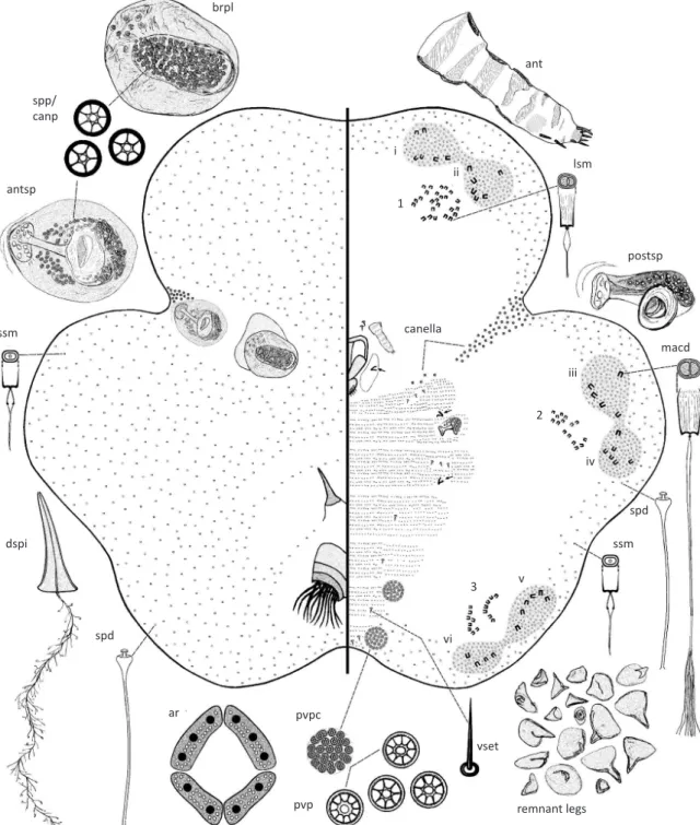
above, brachial crater not central, touching outer rim of brachial plate. Macroducts in marginal duct clusters larger, each 10-16 µm wide. Second marginal duct cluster (mdc-ii) with 4-6 macroducts. Antennae 5-7 segmented. Known from Argentina (Salta Province); on Parkinsonia (Fabaceae) .…….... T. palobrea Kondo & Gullan sp. n. 23. Posterior ventral duct clusters (vdc-3) always present
and well marked ...… 24 – Posterior ventral duct clusters (vdc-3) absent
or represented by only two or three scattered large-sized microducts ... 25
24. Dorsal macroducts conspicuous, 22 (mostly 12-17) µm long, aperture surrounded by circular area of light sclerotization. Posterior spiracles each with 10-20 (usually 12-18) associated pores. Known from Mexico and USA (Arizona); on Sesbania and Coursetia (Fabaceae) ……….. T. fulgens (Cockerell) (Fig 1e) – Dorsal macroducts not so conspicuous, mostly
(only Baja California Sur); on Acacia (Fabaceae) ………... T. ferrisi Chamberlin (Fig 1d) 25. Dorsal macroducts broad (10-13 µm wide), and very prominent, always more than twice diameter of a canellar pore and larger than a perivulvar p o re . K n o w n f ro m M e x i c o a n d U S A ( N e w Mexico and Texas); on Parthenium and Viguiera (Asteraceae) ….……….. T. cornuta (Cockerell) – Dorsal macroducts never more than 7.0-7.5 µm wide (often narrower) and not prominent …... Subgroup of T. larreae sensu Chamberlin (1923), c o n s i s t i n g o f T. g l o m e r e l l a ( C o c ke re l l ) , T. larreae (Comstock) and T. pustulata (Cockerell)
Note. At present, it is not possible to adequately distinguish the putative species included in the subgroup of T. larreae sensu Chamberlin (1923); refer to discussion below the synonymy for the genus.
Tachardiella Cockerell
Tachardiella Cockerell, 1901: 249.
Tachardiella (Tachardiella); Chamberlin, 1923: 174. Change of status to subgenus.
Tachardiella Chamberlin; Chamberlin 1925: 39. Change of status: elevation of subgenus to genus.
Type species. Tachardiacornuta Cockerell, by monotypy and original designation.
Chamberlin (1923) used a broad concept for Tachardiella and recognized two subgenera, Tachardiella (Tachardiella) and Tachardiella (Austrotachardiella). Two years later, Chamberlin (1925) elevated his former subgenera to genera as Tachardiella and Austrotachardiella (he did not combine them into a single genus as stated by Ben-Dov
(2006). Chamberlin’s (1925) classification has been used
by all subsequent authors, including in this review. The diagnosis below is based on what Chamberlin (1923) referred to as “subgenus typicus” of Tachardiella. With the description of one new species below, 17 species are recognized currently in Tachardiella.
There are seven North American species of Tachardiella and, with the exception of T. mexicana, the adult females of all of these specieshave an often diffuse cluster or
more often a pair of clusters of macroducts on the mid dorsum, with the number of ducts in each cluster ranging from two to 15 or more, depending on population and species. Chamberlin (1923) placed six of the North American species, namely T. cornuta, T. ferrisi, T. fulgens, T. glomerella,T. larreae and T. pustulata, in his informal ‘Group of T. cornuta’, which he said was extremely homogeneous and required further study. He recognized this group by the adult female having a brachial plate with “an ovate, non-subdepressed crater, without a crest
at one end and with a rim or collar which is never so wide as breadth of crater, usually no more than half as wide” (Chamberlin 1923: 177), but did not mention that these six species all shared the possession of dorsal macroducts. He further split this group into two subgroups: (i) ‘Subgroup of T. larreae’ containing T. cornuta, T. glomerella, T. larreae and T. pustulata, for which adult females can be diagnosed most readily by having a very narrow collar around the brachial crater and lacking the posterior ventral duct clusters (vdc-3), and (ii) ‘Subgroup of T. fulgens’ containing T. ferrisi and T. fulgens, for which adult females are diagnosed by having a brachial collar that is wider than half the width of the crater and a distinct vdc-3. Gill (1993) suggested that there may be a species complex involving T. glomerella, T. larrea and T. pustulata because he found that the features used by Chamberlin to separate these species can vary within the Californian populations that he studied and thus species characteristics overlapped. Furthermore, Gill (1993) suggested that there might be an association with host plants, i.e., T. larrea with Larrea (Zygophyllaceae), T. glomerella with Adenostoma (Rosaceae) and T. pustulans with Asteraceae. However, he added that more data are needed to substantiate his hypothesis. We concur with Gill (1993) and also suggest that T. ferrisi and T. fulgens are part of a species complex. We treat all of these six species separately below, pending detailed study, preferably with molecular data.
Note.The synonymy listed in Ben-Dov (2006, 2010) is partially incorrect for this genus and has been corrected above.
Generic diagnosis (modified from Chamberlin 1923). Dorsum. Dorsal setae generally absent. Dorsal macroducts present or absent. Microducts and spermatoid ducts present. Brachial tube either short or long. Brachial plate sclerotized, with a shallow, usually ovate to elliptical crater
surrounded by a collar or flat rim sometimes as wide as
in size and shape to those on dorsum. Ventral duct clusters in three pairs, posterior pair sometimes reduced or absent, as in T. cornuta. Microducts present. Perivulvar pore clusters usually present and four (rarely six) in number; in T. lycii clusters greatly reduced, often represented by no more than a single pore or absent in part.
Tachardiella argentina (Dominguez) (Figs 2a, 3)
Tachardiaargentina Dominguez, 1906: 219-222; Autran 1907: 148, 150 & 156.
Tachardiellaargentina; MacGillivray, 1921: 154. Change of combination.
Proposed common names. Spanish: Insecto laca de la Tusca; English: Tusca lac insect.
Dominguez (1906) described the live appearance and chemical composition of tests of this lac insect species from a collection made on tusca trees, Acacia cavenia (now A. caven), in the surroundings of Totoral in Córdoba Province, Argentina. We believe that dry material in the BME labeled as T. argentina is part of Dominguez’s original collection because it was collected on the type host from the type locality and by the author of the species. Also written on the label is the name of the entomologist Mr. Lizer y Trelles, who appears to have obtained the specimens described by Dominguez (1906) and probably gave them to T.D.A. Cockerell, who must have sent them either to G. F. Ferris or to Ferris’ student, Joseph Conrad Chamberlin, a world authority on the taxonomy of lac insects at the time (Judson & Chamberlin 1998). The scale insect collections of both Ferris and Chamberlin are housed now in the BME. Chamberlin did not include a description based on specimens of T. argentina in his publications on Kerriidae (Chamberlin 1923, 1925), so it is likely that he acquired the material after the publication of his papers. Chamberlin (1923) considered T. argentina to be a nomen nudum because he thought that no valid description of the species had been published. However, Chamberlin (1923) added
that because of the existence of a figure associated with the first mention of the species, this figure could render
the name valid. In his supplement to the monograph of lac insects, Chamberlin (1925: 40) later considered the species name as valid, writing as follows: “Prof. T.D.A. Cockerell has kindly furnished me with some notes concerning this species. From these it is at once apparent that the species must be regarded as valid. We cannot, however, as yet, say whether it will fall in Tachardiella or Austrotachardiella.”
The present study has confirmed that T. argentina does belong in Tachardiella.
Adult female (Figs 2a, 3)
Unmounted material.The original description written in French by Dominguez (1906: 219) translates as follows:
“The production [lac] that we have encountered on the branches of tusca: – form masses of a resinous aspect, of a deep red color; isolated [individuals], more or less voluminous, round or ovoid in shape, 5-8 mm in diameter, 2.5-3.0 mm thick, surface smooth, convex, and interiorly
flattened, or more or less slightly concave, somewhat in
the form of a crude crust, 4-5 mm thick, which wraps the branch entirely, often a considerable surface. This crust [the resinous mass made up from the fusion of numerous individuals] is rugose, unequal, covered in nipples [see arrow on Fig 2a] which correspond to the cells of the insects that remain underneath; some individuals are in contact with the exterior throughout the year through
small orifices, whereas in others these [orifices] are much
smaller, or missing. The material of which constitutes this
product is odorless, without flavor, denser than water, hard and easily breakable, but difficult to pulverise.”
Mounted adult female. Insects oval to elongate oval, margin 6-lobed in youngest specimens, however, becoming 3-lobed in older specimens, and unlobed and ovoid in much older specimens. Body 0.9-2.4 (lectotype 1.9) mm long, 0.9-2.5 (lectotype 1.5) mm wide (Fig 2) (n = 13 adult females).
Dorsum. Derm membranous. Dorsal setae and macroducts absent. Microducts numerous, but absent from around brachial plates and anterior spiracles, from around anal tubercle, and from dorsal spine; diameter of duct rim 3 µm. Spermatoid ducts each 6 µm wide, scattered throughout dorsum, less abundant on areas devoid of microducts. Brachia very short and membranous in just molted specimens, probably about 10 µm long; becoming up to 430 µm long and 280 µm wide at base and slightly sclerotized at maturity. Brachial plates oval to broadly oval, each 130-175 long, 118-145 µm wide; brachial crater elongate oval, central, with two or three (usually three) setae on anterior margin and one or two (usually two)
setae on posterior margin (high magnification needed to
detect); brachial crater 5-10 pores wide, 10-16 pores long. Brachial pores with 5-7 loculi, each 5 µm wide. Anterior spiracles present on dorsum, large, surrounded by a spiracular sclerotization, 130-188 µm long, 118-188 µm wide; width of anterior spiracular peritremes 60-68 µm; spiracular pores of similar structure to brachial pores,
with 5-7 (mostly five) loculi, each 5 µm wide, numerous
around each spiracle. Anal tubercle tapering, highly sclerotized; pre-anal plate longer than supra-anal plate, with four pairs of setae; no setae observed on supra-anal plates (not illustrated). Dorsal spine well-developed, length 140-180 µm, width at base 53-75 µm; dorsal spine duct of dendritic type. Anal fringe entire, each anal fringe plate ligulate, 13-20 µm long. Anal ring 58-78 µm wide, divided into four separate sections, with 10 setae, tip of setae surpassing anal fringe. Eyespots absent.
Fig 3 Tachardiella argentina adult female. Abbreviations: ant, antenna; antsp, anterior spiracle; apd, apodeme; ar, anal ring; at, anal tubercle; brpl, brachial plate; dspi, dorsal spine; lsm, large-sized microduct; maf, mature adult female; macd, macroduct; papl, pre-anal plate; postsp, posterior spiracle; pvp, perivulvar pore; pvpc, perivulvar pore cluster; ssm, small-sized microduct; spanpl, supra-anal plate; spd, spermatoid duct; spp/canp, spiracular pore/canellar pore; vset, ventral setae. Marginal duct clusters labeled as i, ii, iii, iv, v and vi. Ventral duct clusters labeled as 1, 2 and 3. Variation in size and shape of leg remnants drawn from various individuals.
brpl
ssm
dspi
dspi
pedicel
canella
spd
ar spp/
canp
antsp
ssm spd
macd postsp lsm
ant
maf
remnant legs pvpc
papl at
spanpl
apd
vset
pvp i
1
2
vi
3 v
ii
iii
microtrichia. Antennae 70-115 µm long, segmentation
poorly defined, 4-5 segmented; first three segments
showing signs of sclerotization, fourth segment
membranous, with a fleshy seta about mid area, and another near apex, with two fleshy setae and two or three
slender setae at apex of terminal segment. Clypeolabral shield 153-185 µm long, 118-135 µm wide. Labium one segmented, 65-85 µm long, 55-75 µm wide; with four pairs of setae. Legs each reduced to a remnant claw (arrowed on Fig 3), each claw 8-30 µm long, prothoracic claw remnant smallest, often absent, metathoracic claw remnant largest. Canella composed of a linear group of 45-85 pores extending from dorsal spiracles ventrally towards mouthparts, with a smaller group of 4-12 pores present lateral to mouthparts; canellar pores each 4-6 µm wide, each with 3-7 loculi, pore group near mouthparts generally smaller, 3-5 µm wide, with fewer (2-6) loculi. Ventral setae slender, each 7.5-15.0 µm long, present in groups of three just anterior to each meso- and metathoracic leg, and also medially, a pair per abdominal segment, and ventral to the anal tubercle, setae absent elsewhere. Posterior spiracles much smaller than anterior spiracles, spiracular peritreme 30-38 µm wide; with 10-28 spiracular pores present within a spiracular pocket
anterior to each spiracle, each pore 5 µm wide, with five
or six loculi, similar in structure to those on anterior peritreme. Marginal duct clusters distinct, elongate oval, of the duplex type, composed of macroducts surrounded by spermatoid ducts; six pairs of double clusters, with one pair of clusters present near margin of each body lobe, pair on each lobe generally connected by a area rich in spermatoid ducts; number of macroducts in each marginal duct cluster as follows: mdc-i: 2-4, mdc-ii: 1-3, mdc-iii: 2-3, mdc-iv: 2-4, mdc-v: 2-4, mdc-vi: 2-4; rim of macroducts 7.5-10.0 µm wide. Spermatoid ducts similar in size and shape to those on dorsum, present around body margin, numerous in marginal duct clusters, absent from mid-ventral area. Ventral duct clusters subcircular to elongate oval, composed of large-sized microducts present medial to each pair of marginal duct clusters; number of large-sized microducts in each ventral duct cluster as follows: vdc-1: 12-23, vdc-2: 13-21, and vdc-3: 11-18; duct rim of large-sized microducts each 6.0-7.0 µm wide. Microducts outside ventral and marginal duct clusters smallest, each with rim 5.0 µm wide, present around body margin. Rest of ventral derm completely devoid of microducts. Perivulvar pore clusters: two pairs present around vulva, each cluster 52-80 µm in diameter, each perivulvar pore cluster with 25-60 pores, each pore with 4-11 (mostly 10) loculi and 6-8 µm wide.
Morphological variation.The dimensions of the brachia, the pedicel of the dorsal spine and the pre-anal plate of
the anal tubercle are affected mostly by age. Features
such as the number of macroducts in the marginal duct clusters, number of large-sized microducts in the ventral
duct clusters, antennal segmentation, number of setae on the antennae, number of canellar pores in the canella, the number of loculi in the spiracular pores and the number of pores should be considered more stable features.
Host plants. Acacia caven (Fabaceae).
Distribution. Neotropical region: Argentina.
Notes. The adult female most closely resembles that of T. palobrea but can be distinguished readily by the features given in the key.
Etymology.The species was likely named by Dominguez (1906) after its country of origin, namely Argentina.
Material studied. LECTOTYPE: Adult female, here
designated, 1 slide (1 specimen) (BME Type # 1788). ARGENTINA, Province of Córdoba, date not given, coll. Dominguez, ex Acacia caven,slide mounted by PJG in 2004 from dry material labeled: “Tachardiella argentina (Domínguez) / on Acacia cavenia Hook. & Arn. / Province of Córdoba / Domínguez coll. –Lizer y Trelles leg.”.
PARALECTOTYPES: Same data as lectotype: 11 slides (11
adult females), 2 slides (3 immature females, including one on same slide as a small adult female), 2 slides (2 embryos) (BME, except 2 adult females to IMLA); one twig with dry insects in their tests (BME).
tachardiella artocarpi (Hempel)
Tachardiaartocarpi Hempel, 1921: 145-146.
Tachardiellaartocarpi; Chamberlin, 1923: 194. Change of combination.
is about 190 microns in length. The anal tubercle is black [in color], conical shaped, about 400 microns long. On the dorsal surface, near the base of the anal tubercle, there are four oval-shaped groups [perivulvar pore clusters] of round glands [perivulvar pores], each group having 170 or more pores. The two lac glands [brachial plates] are conical in shape, with the height being same as the diameter of the base. Next to these [the brachial plates] there are two large spiracles, and next to the antennae there are two more spiracles, but small. The antennae are about 125 microns long, and each with about three segments. Legs were not observed.” (Hempel 1921).
“Habitat – Rio de Janeiro, on branches of jack tree and
cashew tree, being the first samples received from Mr.
Luiz de Azevedo Marques. Later, samples were received from Mr. Carlos Moreira, collected from almond tree (Terminalia catappa)” (Hempel 1921: 146).
Host plants. Anacardium occidentale (Anacardiaceae), Terminalia catappa (Combretaceae) and Artocarpus heterophyllus (Moraceae) (Hempel 1921).
Distribution. Neotropical region: Brazil (Hempel 1921).
Notes.The description of the adult female lac test (resin cover) as having a pointed elevation on the mid dorsum with dilated margins and with shallow sulci at the base, of the young individuals being star-shaped with an elevation on dorsum, and medium-sized specimens having a pyramid form, suggests the possibility that T. artocarpi might be a member of the genus Austrotachardiella, because the tests of adult females and young individuals of A. colombiana and A. sexcordata closely match the above description. Specimens of T. artocarpi were not available during the present study, so we were not able to verify the morphology or the generic placement of this species.
tachardiella cordaliae (Leonardi)
Tachardia cordaliae Leonardi, 1911: 258-259.
Tachardiella cordaliae; MacGillivray, 1921: 154. Change of combination.
Tachardiella condaliae; Lizer Y Trelles, 1939: 184.
Unjustified emendation.
Tachardiella condaliae; Sharma & Ramani, 1999: 439. Misspelling of name.
Tachardiella condaliae; Varshney, 1997: 29, 2009: 7, 2010: 118. Misspelling of name.
Diagnosis. The original description written in Italian translates as follows: “Adult female. – The body, like the preceding species [referring to T. lycii], after laying its eggs, is more or less deformed. Antennae short, of three segments, of which the apical segment bears two to three short setae. Legs rudimentary, represented by a chitinous
dentiform process. Anterior spiracles much larger than posterior ones, with disc pores extending in a line that goes from one spiracle to the other; posterior spiracles smaller, each with six disc pores. Anal process highly developed and highly sclerotized towards the apex. Anal ring with 10 long and robust setae located on the four chitinized parts [of the anal ring], which are surrounded by the other chitinous process. Dorsal spine robust and very long. Derm rich in tubular glands. Color of body rose purple. Body length: about 5 mm long. Antennal length: 90 µm long. Dorsal spine: 160 µm long. Test. – The test is very similar to that of the preceding species [referring to T. lycii], except that, on the dorsum, instead of having a single hump, this one has two conspicuous humps. With a large aperture corresponding to the anal opening. Walls of the test thick. In cases where the lac of individuals are close to each other, their lac merges to form a protective envelope, forming an encrustation, but never reaching the size that can be attained by some entities. Color of the test dark purple. Length of test 6-9 mm. Habitat. Collected at Cacheuta on Cordalia lineata” (Leonardi 1911: 259).
Host plants. Condalia lineata (Rhamnaceae) (Leonardi 1911).
Distribution. Neotropical region: Argentina (Leonardi 1911).
Notes.The host plant of T. cordaliae was given as “Cordalia lineata”in the original description (see translation above under diagnosis). Leonardi (1911) apparently named the species after its host, but spelt the name incorrectly, since there is no plant genus “Cordalia”. Lizer y Trelles (1939) apparently noticed the error, and emended the name to T. condaliae (Leonardi), however, this was an unjustified emendation of the name (see articles 32.2 and 32.5 of the Code (ICZN 1999)). Leonardi (1911) wrote the host genus of his new lac insect as “Cordalia” and the lac insect name “cordaliae” thus is consistent with the original published
name for the host plant. The unjustified emendation to
“condaliae” was used by subsequent authors (Sharma & Ramani 1999; Varshney 1997, 2009, 2010) but was treated as a misspelling of the species name by Ben-Dov (2006). Morrison (1919) suggested that this species was a junior synonym of T. lycii, but this is unlikely. No material of the species was available during the present study.
tachardiella cornuta (Cockerell)
Tachardiacornuta Cockerell, 1894: 284-285.
Tachardiella cornuta; Cockerell, 1901: 249. Change of combination.
description by Cockerell (1894: 284). Female scales “crowded on the stems of the plant, lively red-brown in color, smooth and rather shiny, sub-translucent; elevated so as to form in outline a triangle, the base of which is greater than either side viewed from one side, but with the sides greater than the base when the scale is viewed from one end. In a lateral view the two sides are about equal and meet each other at a right angle; all the other
angles of the profile, whether taken from the side or
from the end, are necessarily less than right angles. A more minute inspection shows that the apex of the scale is not a simple pyramid, but consists of a horn or tooth inclined backwards, so that a small but distinct notch appears in the lateral outline on the posterior side. This horn gives the whole scale somewhat the shape of certain teeth of sharks. Viewed from above, the scale is roughly oval in outline, but presents on each side a slight bulging, before and behind which is a groove or constriction.” The dimensions given for the female test are 2 mm high, 2 mm wide and 2.7 mm long. Cockerell’s (1894) description of the adult female is so brief and general that it does not distinguish it from other members of the genus.
According to the redescription of T. cornuta by Chamberlin (1923: 182), the species is: “Solitary or lightly massed on stem of host; solitary individuals show distinct lobations; lac somewhat variable in color, between a burnt sienna and brown ochre.” Chamberlin’s description of the adult female can be summarized as follows: Body length 2 mm. Dorsal duct cluster [composed of macroducts] prominent, of 7-8 (rarely up to 16 or 17) very large macroducts [about 15 µm long and 10-13 µm wide], always measurably larger than a canellar pore. Brachia long, with a constriction behind brachial plate; brachial plates typical for group but with a narrower collar than in other species. Anterior spiracles distinctly smaller than brachial plate, longer than broad and bearing 25-30 pores. Anal tubercle of the usual short subquadrate type, with pre-anal plate large and distinctly elongate. Dorsal spine slightly more than 1.5 times as long as diameter of brachial plate. Antennae of four sclerotized segments. Legs represented by minute claws arising from circular, convex, nipple-like patches. Canella well developed, consisting of a single more or less convoluted line of 35-45 canellar pores, with canella of each side almost meeting a little posterior of mouthparts. Posterior spiracles with 4-5 pores. Marginal duct clusters consisting of a more or less lunate line of 4-7 ducts, clusters apparently of an obscure duplex type [spermatoid ducts hard to detect]. Perivulvar pore clusters small, often smaller than half breadth of anal tubercle at fringe, and rarely, if ever, larger than this, in some cases a cluster may be reduced to as few as 4-5 pores. Anterior ventral duct cluster [vdc-1] loosely organized, composed of 9-12 microducts, median ventral duct clusters [vdc-2] compact with about six microducts, posterior ventral duct cluster [vdc-3] apparently absent
or occasionally represented by one or two isolated large-sized microducts.
Host plants.Partheniumincanum (Asteraceae) (Cockerell 1894), Viguiera sp. (Asteraceae) (Schroer et al 2008).
Distribution. Nearctic region: Mexico (Cockerell 1902); USA (New Mexico [Cockerell 1894], Texas [Schroer et al 2008]).
Notes. The synonymy listed in Ben-Dov (2006, 2010) is incorrect for this species and has been corrected above.We examined the material listed by Chamberlin (1923) and now held by the BME. The adult females are most readily distinguished from those of closely related Tachardiella species found in the southwest USA by having prominent dorsal ducts that are nearly as broad as long.
tachardiella ferrisi Chamberlin (Fig 1d)
Tachardia sp. Ferris 1921: 86.
Tachardiella (Tachardiella) ferrisi Chamberlin, 1923: 183-184.
Diagnosis.The following notes on the adult female and its
test are modified from Chamberlin (1923: 184). Loosely
massed upon twigs of host; tests smooth and gently lobed [may be an artifact of the test having melted – see Notes below]; lac smooth with color between that of dragon’s blood and burnt sienna. Body length 2 mm. Dorsal duct cluster composed of eight macroducts, much less conspicuous than in T. cornuta or T. fulgens and ducts lacking a heavily sclerotized rim. Brachia typical of group; brachial plates ovate to subcircular with rim or collar about half width of crater. Anterior spiracles subequal in size to brachial plate, with 50-60 pores. Anal tubercle similar to that of T. cornuta. Dorsal spine as long as diameter of brachial plate, often basally bent
or distorted. Antenna of five evident segments. Legs very
with 15-16 large-sized microducts very closely grouped. Perivulvar pore clusters with diameter about half width of anal tubercle at fringe.
Host plants. Acacia flexicaulis (Fabaceae) (Chamberlin 1923).
Distribution. Nearctic region: Mexico (Baja California Sur) (Chamberlin 1923); note that records of this species from USA (California) by Gill (1993) are incorrect [see below].
Notes. We agree with Chamberlin (1923) that T. ferrisi is closest to T. fulgens. Material of T. ferrisi in the BME includes four original slides (with 8 adult females) used by J.C. Chamberlin and associated dry material consisting of two pieces of twig covered in resinous tests containing dry insects (Fig 1d). The resin of these tests was partially melted
and thus their shape may not be an accurate reflection their
natural shape. The dry material has the following label data:
“ferrisi, Tachardiella Type Material / On Acacia flexicaulis
/ La Paz, Lower Calif / Summer 1919, G.F. Ferris”. Twenty-four new slide-mounts with adult females, an immature
female, adult males and first-instar nymphs were prepared
for this study by PJG from the dry material (details given below). Two adult parasitoid wasps taken from the tests of the type collection also were mounted on one slide. All are part of the G.F. Ferris Collection in BME. Chamberlin (1923: 183) listed the type host and locality as “Mexico: Lower California, La Paz, on Acacia flexicaulis, July 1919 (G. F. Ferris)” but his original description does not refer to a holotype or paratypes, even though he labeled the BME slides with these type designations. Therefore, Chamberlin’s labeled type specimens must be regarded as syntypes and below we designate a lectotype to avoid future nomenclatural confusion.
Gill (1993) provided a color photograph of the female tests of T. ferrisi (ex Adenostoma sparsifolium, Jacumba, San Diego County, California, coll. 6 March 1983, R. J. Gill) and illustrated the putative adult female of T. ferrisi based on specimens from A. sparsifolium (Rosaceae) from Temecula in southern California (belonging to BME). However the morphology depicted (Gill 1993, Fig 48) is not consistent with that of the types of T. ferrisi; for example, the females from Temecula have many more dorsal, submarginal and marginal macroducts. We believe that Gill illustrated an unnamed species of the group, but further study is required to verify this hypothesis.
Material studied. LECTOTYPE: Adult female, here
designated, on slide with two other adult females, lectotype clearly marked, apparently in Chamberlin’s handwriting, with sketch of the three females and an arrow to the primary type but incorrectly labeled as “Holotype”, slide labeled: “Tachardiella / ferrisi Chamberlin / Holotype [with sketch of position of specimens] / On Acacia / flexicaulis / La Paz,
Lower Calif. / G. F. Ferris, col. / Entomological Laboratory / Stanford University [4 printed words] / JCC G.F.F.” (BME). The lectotype is rather opaque due to aging of the mountant, but we believe that it is desirable to maintain Chamberlin’s choice of primary type specimen. PARALECTOTYPES:
Original Ferris slides with same data as lectotype: 3 slides (5 adult females including two on same slide as lectotype) (BME); new slides prepared from Ferris dry material by PJG, same data as lectotype: 22 slides (24 adult females and 1 immature female on same slide as small adult females), 1
slide (4 first-instar nymphs), 1 slide (2 adult males), two
twigs with dry insects in their tests (BME).
Other material. USA: 1 slide (2 adult females), Baja California, District Sur, San Pedro, July 1913, G.F. Ferris, on Acacia flexicaulis (BME). Although this slide bears Chamberlin’s label “Paratypes”, the locality is not the type locality and thus the specimens have no type status. The host A. flexicaulis and the localities La Paz, San Pedro and San Bortolo all were listed for this lac insect species by Ferris (1921) and these towns are near each other at the southern end of Baja California.
Note that on the four original Ferris slides listed above, the name of the species, the name of the author of the species (i.e., Chamberlin), the type names (either ‘Holotype’
or ‘Paratypes’) and the initials ‘JCC’ are in a different
handwriting (apparently that of Chamberlin) and darker ink from the writing of the original Ferris label.
tachardiella fulgens (Cockerell) (Fig 1e)
Tachardiafulgens Cockerell, 1895: 1-2.
Tachardiellafulgens; MacGillivray, 1921: 154. Change of combination.
Tachardiella (Tachardiella) fulgens; Chamberlin, 1923: 183.
Diagnosis.Female scales “usually massed together, more or less surrounding the twig, forming an irregular nodulose bright reddish-orange coating about 4 mm. thick. A single scale is about 5 mm. long and 4 broad, and presents a conspicuous somewhat curved, blunt, shining, dorsal hump; also a tail-like projection, sometimes directed upwards, and two or three irregular projections on the side.” (Cockerell 1895: 1) (Fig 1e). The following diagnosis of the adult female is adapted from Chamberlin (1923: 183).
consisting of 16-25 canellar pores. Posterior spiracles with about 13 spiracular pores [10-20 pores counted on 20 females examined]. Marginal duct clusters of a duplex type, anterior marginal duct cluster [vdc-1] with 4-6 macroducts and with a few scattered microducts, median marginal duct cluster (mdc-ii) as for anterior marginal duct clusters, posterior marginal duct cluster [mdc-iii] with 8-9 macroducts and closely associated with posterior ventral duct cluster, with 30-40 small microducts scattered closely around both posterior marginal and ventral duct clusters. Anterior ventral duct cluster [vdc-1] composed of 16-18 large-sized microducts, median ventral duct cluster [vdc-2] more closely grouped but large-sized microducts numbering about same as for vdc-i, posterior ventral duct cluster [vdc-3] closely grouped and with 8-10 ducts and very closely associated with marginal duct cluster [mdc-iii]. Perivulvar pore clusters well developed, somewhat greater in diameter than width of anal tubercle at fringe.
Host plants. Possibly Sesbania sp. (Fabaceae) [Cockerell 1895], Mimosa or Prosopis and Coursetia spp. (Fabaceae) (Chamberlin 1923).
Distribution. Nearctic region: Mexico (Chamberlin 1923); USA (Arizona) [Cockerell 1895; Chamberlin 1923].
Notes.According to Chamberlin (1923), T. fulgens is most close to T. ferrisi. The identity of the original host plant was uncertain (see quote below) and Chamberlin suggested that the type host might have been Coursetia axillaris (Fabaceae). This lac insect species has been considered to have medicinal properties since Cockerell (1895: 1-2) wrote as follows: “Hab. Arizona, received from Prof. J. W. Toumey, who gives the following interesting particulars. He got it from a Mexican, and has seen only the stem of the food-plant, but thinks it is a Sesbania. He was told that this lac was used quite extensively by the Mexicans as a medicine for stomach troubles, under the name of “Gomea”. It is kept in the drug shops at Tucson, and meets quite a sale. It is also used to some extent in mending pottery, etc. Finally, he adds, the Mexicans make a marked distinction between this and T. larreae, the latter not being considered to have any medicinal qualities.”
Material studied. 25 slides (36 adult females), mounted from dry material by PJG in 2010, USA: Arizona, Pima County, Catalina Mts, Sabino Canyon, 27 Feb. 1949, R.B. Marlatt, ex Coursetia microphylla, Wehrle Collection No. 673 (BME). The photograph of the female tests (Fig 1e) is from this collection; 1 slide (2 adult females), USA: Arizona, Tucson, 1919, C.T. Vorhies, ex Coursetia axillaris (BME); 1 slide (2 adult females), MEXICO: Sonora, hills near Huasihuas, from Cockerell, on legume (BME); 2 slides (4 adult females), MEXICO: Sonora, O.E. Bremner Collection,
615 (BME). The last three collections represent three of the four lots of material examined by Chamberlin (1923).
tachardiella glomerella (Cockerell) (Fig 1f)
Tachardiaglomerella Cockerell, 1905: 52.
Tachardiellaglomerella; MacGillivray, 1921: 153. Change of combination.
Tachardiella (Tachardiella) glomerella; Chamberlin, 1923: 180-181.
Tachardiella (Tachardiella) glomerella baccharidis Chamberlin, 1923: 181-182. Synonymy by Ferris 1955: 221.
Diagnosis. “Tests of females crowded on stems with
individuals coalescing, color very dark, with translucent shining orange-red bosses; scales smooth and rounded, without (even in young females) any distinct projection such as seen in T. cornuta” (Cockerell 1905). “Solitary or massed on twigs; lobation distinct; lac clear and translucent, amber-like; near dragon’s blood in color” (Chamberlin 1923). The following morphology of the adult female is summarized based on Chamberlin (1923: 181). Body length 2-3 mm. Dorsal duct clusters present. Brachia and brachial plates rather small but typical in structure. Anterior spiracle typical in shape for group and subequal [in size] to brachial plate, with 30-35 pores. Dorsal tubercle typical. Dorsal spine 1.3 times as long as width of brachial plate, often basally bent. Antennae of 5 segments. Canella distinct, composed of 27-30 canellar pores. Posterior spiracles very close to ends of canellae and almost connected with them, with 9-12 pores. Marginal duct clusters distinctly duplex; anterior marginal duct clusters [mdc-i] with 4-5 macroducts, median marginal duct clusters [mdc-ii] with 2-4 macroducts, posterior marginal duct clusters [mdc-iii] with 4-5 macroducts. Anterior ventral duct clusters [vdc-1] prominent with about 25 conspicuous large-sized microducts loosely grouped into two parts, median ventral duct clusters [vdc-2] very compact and consisting of 6-8 ducts, posterior ventral duct clusters [vdc-3] absent; small-sized microducts scattered sparingly around the marginal and ventral duct clusters. Perivulvar pore clusters about subequal in diameter to width of anal tubercle at fringe. Posterior perivulvar pore clusters distinctly a little larger than anterior perivulvar pore clusters.
Host plants. Gutierreziaglomerella (Asteraceae) (Cockerell 1905), Baccharis sp. (Asteraceae) and Adenostoma sp. (?) (Rosaceae) (Chamberlin 1923).
Distribution. Nearctic region: USA (New Mexico) (Cockerell 1905).
and Glenn Springs, Texas, by Ferris (1921) differ from
typical specimens of T. glomerella by having many more microducts in vdc-2. Chamberlin erected a new form, T. glomerella f. baccharidis, based on this population. Ferris (1955) stated that ‘forms’ had no nomenclatural standing and thus he placed Chamberlin’s ‘form’ from Baccharis in synonymy with T. glomerella. However, according to Article 45.6.4 (International Commission on Zoological
Nomenclature 1999), a name is to be treated as subspecific
[and thus regulated by the International Code of Zoological
Nomenclature] if first published before 1961 and its
author expressly used either the term “variety” or “form”, including the terms “var.”, “v.”, “forma” or “f.” (the latter was used by Chamberlin 1923). We examined the type material of Chamberlin’s ‘form’ and the other material of T. glomerella listed by Chamberlin (1923), all now in the BME. The specimens from Baccharis at Tornillo Creek
do differ from adult females from other populations of T. glomerella in the features listed by Chamberlin, including by having more microducts in vdc-2. However, this is not
a host-correlated difference because adult females from
Baccharis at Dawson Canyon, Riverside County, California (housed in CSCA) resemble the New Mexico and other Texas populations and not those from Tornillo Creek. More extensive collecting and detailed study based on both morphological and molecular data are required to understand this variation.
tachardiella ingae (Hempel) (Fig 1g)
Tachardiaingae Hempel, 1900: 415-416.
Tachardiellaingae; MacGillivray, 1921: 154. Change of combination.
Diagnosis.The original description ofT. ingae by Hempel (1900) was in Portuguese but the description was republished a year later in English (Hempel 1901: 124-125), as follows: “Adult female scale subglobular, dorsum
slightly flattened, with aperture in the centre. The lac
[resin] is dull, shiny when the surface becomes rubbed, semitransparent, thick, brittle, light green with brown
stripes. Some fine white filaments usually protrude from the dorsal orifice. The lac of many individuals usually unites
to form a confused mass. Diameter 5.25 millim.; height 3.75 millim. Denuded of lac the insect is three-lobed.” Hempel (1900, 1901) also described the cuticular morphology of the adult female but his description is not very informative. The following morphology of the adult female is summarized based on Chamberlin (1923: 177) with some additions. Body length 3.0-3.5 mm. Brachia lightly chitinised and fairly distinct; brachial plate almost circular, crater without a crest broadly ovate-oblong, half as wide as long and much wider than surrounding collar, one longitudinal half of crater subdepressed into pseudodimples. Anterior spiracles large, about two-thirds size of brachial plate and bearing numerous pores. Dorsal spine straight and shorter
than width of brachial plate. Anal tubercle and supra-anal plate more or less typical of genus, but a little broader at base than usual. Antennae distinctly seven segmented. Legs comparatively large, almost as large as posterior spiracles. Canella prominent and very long [with 115-160 or more canellar pores, including 15-30 pores forming one or more distinct clusters of fused and sclerotized pores just near anterior spiracles]. Posterior spiracles with 7-8 [9-18 on eight females examined] pores. Marginal duct
clusters apparently of simplex type [definitely duplex] with
about six macroducts [4-8 in each double cluster]. Ventral duct clusters distinct, anterior ventral cluster [vdc-1] of about 25-30 loosely clustered large-sized microducts, median ventral cluster [vdc-2] of about 35 loosely grouped microducts, posterior ventral cluster [vdc-3] of about 25 loosely grouped microducts. Perivulvar pore clusters large, measuring considerably more in diameter than half width of anal tubercle at fringe and with more pores [40-55 per cluster] than typical of genus.
Host plants. Inga sp. (Fabaceae) (Hempel 1900).
Distribution. Neotropical region: Brazil (Hempel 1900).
Notes.This species was described based on specimens collected from “the margins of the river Mogy-Guassú, near the town of Mogy-Guassú” (Hempel 1900: 416). This town also is known by the name Mogi Guaçu and is a municipality in the state of São Paulo in Brazil. The BME has one slide with three adult females and the label: “N. Y. Agr. Exp. Sta. [printed] / Tachardia / ingae / Hempel / Type / Mogy-guassu / J.C.C.”, with the “J.C.C.” in Chamberlin’s handwriting. We consider these adult females to be syntypes; the other syntypes are supposed to be housed in the Museu de Zoologia da Universidade de São Paulo, Brazil (Ben-Dov 2006). We have not designated a lectotype because one should be selected from the syntype specimens in Brazil that we have not seen. The BME also has one short piece of dry twig with a single dry pale orange, eaten-out test of an adult female with the labels: “28. / Tachardia ingae Hempel. Type / Mogy-Guassu.” [typed] and “ingae, Tachardiella / On ? TYPE”. There is another dry collection in the BME with label: “Tachardiella ingae Hempel. / On Ingá sp. / Bethania, State of São Paulo, / Brazil, 26-X-1928. / At the margin of the Tieté River. / Adolph Hempel, Coll.”. PJG prepared slide-mounts of the Bethania collection (four adult females and
six slides of numerous first-instar nymphs) and compared
the rim around crater is mostly much narrower than the width of the crater itself. Chamberlin (1923) considered this species to be so distinctive that he placed it in its own group, separate from all other Tachardiella species.
tachardiella larreae (Comstock) (Fig 1h)
Carterialarreae Comstock, 1882: 211-212.
Tachardiellalarreae; MacGillivray, 1921: 154. Change of combination.
Tachardiella (Tachardiella) larreae; Chamberlin, 1923: 177-179.
Tachardiella (Tachardiella) larreaecalifornica Chamberlin, 1923: 179. Synonymy by Ferris, 1955: 223.
Lakshadialarreae; Mahdihassan, 1923: 53. Change of combination.
Tachardiellalarrea; Colton, 1943: 21-32; Colton, 1944: 1-24. Misspelling of species name.
Tachardiellalarrae; Stacey et al 1998: 53. Misspelling of species name.
Diagnosis.Tests of adult females aggregated but each test more-or-less globular in form, with tendency to be 6-lobed if isolated (Comstock 1882); resin dark grey-brown in color with tinges of crimson or orange (Gill 1993, photograph 44) to reddish-brown with tinges of orange (Fig 1h).
The following morphology of the adult female is summarized based on Chamberlin (1923: 178). Body length 2.5-3.5 mm. Dorsal duct cluster present but macroducts hard to see [see below under Notes]. Brachia and brachial plates typical of group. Anterior spiracles normal in shape and subequal [in size] to brachial plate; anterior spiracles with 40-50 pores. Anal tubercle heavily sclerotized; supra-anal plate typical of group; pre-anal plate staining [in acid fuchsin] rather deeply. Dorsal spine as long as diameter of brachial plate. Antennae of six obvious segments. Legs greatly reduced, represented by tip of vestigial claw. Canella not conspicuous, consisting from 35-40 scaterred canellar pores. Posterior spiracle with 9-10 pores. Marginal duct clusters with 5 6 macroducts. Anterior ventral duct cluster [vdc-1] loosely grouped, more or less annular, with 25-30 ducts, median ventral duct clusters [vdc-2] similar and with 20-22 ducts; posterior ventral duct clusters [vdc-3] absent. Perivulvar pore clusters small, varying in diameter from half to more than half width of anal tubercle at fringe; anterior and posterior perivulvar pore clusters subequal.
Host plants. Larrea tridentata (as L. mexicana) (Zygophyllaceae) (Comstock 1882]); T. larreae f. californica from Peucephyllum schottii (Fabaceae) (Chamberlin 1923).
Distribution. Nearctic region: Mexico (Comstock 1882), USA (Arizona [Cockerell 1893], California [Chamberlin 1923, 1925]).
Notes.The species was named after its host-plant genus, Larrea, and occurs on creosote bush, L. tridentata, which has a number of synonyms (including Larrea mexicana and Covillea glutinosa). Chamberlin’s taxon T. larreae forma californica was described from insects collected by G.F. Ferris on the shrub Peucephyllum schottii, commonly called pygmy-cedar, which has resinous foliage and apparently closely resembles the creosote bush. Ferris (1955) stated that forms have no nomenclatural standing and thus he placed Chamberlin’s ‘form’ in synonymy with T. larreae. However, according to Article 45.6.4 (International Commission on Zoological Nomenclature 1999), a name
is to be treated as subspecific [and thus regulated by the International Code of Zoological Nomenclature] if first
published before 1961 and its author expressly used either the term “variety” or “form”, including the terms “var.”, “v.”, “forma” or “f.” [the latter was used by Chamberlin (1923)].
Furthermore, the differences noted by Chamberlin might be sufficient to warrant species status for this ‘form’,
which he described as having 23-25 pores associated with the posterior spiracles (9-10 pores in T. larreae), 10-11 macroducts in each marginal duct cluster (5-6 ducts in T. larreae), 42-43 large-sized microducts in vdc-1 (25-30 in T. larreae), 14-15 large-sized microducts in vdc-2 (20-22 in T. larreae), and distinct dorsal ‘collared’ ducts (not distinct in T. larreae). In his redescription of T. larreae, Chamberlin (1923) was not sure about the presence or absence of dorsal macroducts. However, we have studied the slides examined by Chamberlin of T. larreae from Arizona and California and the dorsal macroducts are present but hard to see because they are small (5-7 µm long, 2.5-3.0 µm wide at inner sclerotized end). However, Gill (1993) in his redescription of the species, clearly illustrates distinct dorsal macroducts. Gill (1993)
prepared the drawing (figure 50, page 94) and took a color
![Fig 1 Some lac insects known from the New World. a) Kerria lacca [on Albizzia sp., Peradeniya, Sri Lanka; inset showing close-up of adult female]; b) Paratachardina pseudolobata [paratypes, on Myrica cerifera, University of Florida, Fort Lauderdale Resear](https://thumb-eu.123doks.com/thumbv2/123dok_br/15572645.604246/3.892.138.768.107.641/albizzia-peradeniya-paratachardina-pseudolobata-paratypes-cerifera-university-lauderdale.webp)