Predictors of Hospital Mortality in Hemodynamically Stable Patients
with Pulmonary Embolism
André Volschan
1, Denilson Albuquerque
2, Bernardo Rangel Tura
2, Marcos Knibel
8, José Péricles Esteves
3, Luiz
Carlos Bodanese
5, Francisco Silveira
9, João Pantoja
4, Paulo Cesar Pereira da Silva e Souza
7, João Mansur
6, Evandro
Tinoco Mesquita
1**on behalf of the EMEP (Estudo Multicêntrico de Embolia Pulmonar) investigators
Hospital Pró-Cardíaco1; Universidade do Estado do Rio de Janeiro2, Rio de Janeiro, RJ; Hospital Português3, Salvador, BA; Hospital Copa D’Or4, Rio
de Janeiro, RJ; Hospital São Lucas5, Porto Alegre, RS; Hospital Samaritano6, Rio de Janeiro, RJ; Hospital de Clínicas de Niterói7, Niterói, RJ; Hospital
CardioTrauma8, Rio de Janeiro, RJ; Hospital Prontocor9, Belo Horizonte, MG – Brazil
Summary
Background: Pulmonary embolism is associated with high mortality in patients with hypotension or circulatory shock. However, the association between some clinical variables and mortality is still unclear in hemodynamically stable patients.
Objective: To derive an in-hospital mortality risk stratification model in hemodynamically stable patients with pulmonary embolism.
Methods: This is a prospective multicenter cohort study of 582 consecutive patients admitted in emergency units or intensive care units with clinically suspected pulmonary embolism and whose diagnosis was confirmed by one or more of the following tests: pulmonary arteriography, spiral CT angiography, magnetic resonance angiography, Doppler echocardiography, pulmonary scintigraphy, or venous duplex scan. Data on demographics, comorbidities and clinical manifestations were collected and included in a logistic regression analysis so as to build the prediction model.
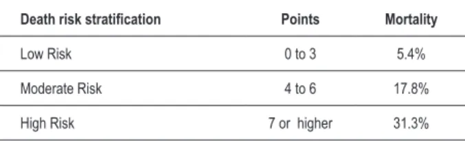
Results: Overall mortality was 14.1%. The following parameters were identified as independent death risk variables: age > 65 years, bed rest > 72h, chronic cor pulmonale, sinus tachycardia, and tachypnea. After risk stratification, mortalities of 5.4%, 17.8%, and 31.3% were found in the low, moderate and high-risk subgroups, respectively. The model showed 65.5% sensitivity and 80% specificity, with a 0.77 area under the curve.
Conclusion: In hemodynamically stable patients with pulmonary embolism, age > 65 years, bed rest > 72h, chronic cor pulmonale, sinus tachycardia andtachypnea were independent predictors of in-hospital mortality. However, further validation of the prediction model in other populations is required so that it can be incorporated into the clinical practice. (Arq Bras Cardiol 2009; 93(2):128-132)
Key Words: Hospital mortality; pulmonary embolism; pulmonary heart disease; tachycardia, sinus.
Mailing Address: André Volschan •
Rua Baronesa De Pocone, 137/201 – Lagoa - 22471-270 - Rio de Janeiro, RJ, Brazil
E-mail: [email protected], [email protected]
Manuscript received August 12, 2008; revised manuscript received September 12, 2008; accepted September 19, 2008.
Introduction
Pulmonary embolism (PE) is the third cardiovascular cause of hospital admission1, following acute coronary syndrome
and stroke. It has a broad spectrum of clinical manifestations, especially when associated with decompensated heart failure2 and chronic obstructive pulmonary disease. Massive
PE characterized by hemodynamic instability defines the subgroup of more severely ill patients among whom high mortality rates are observed3,4, and who can benefit from a
more aggressive therapeutic approach. On the other hand, clinically stable patients considered at low risk require a shorter
length of hospital stay and, in some cases, can be treated as outpatients with low-molecular-weight heparin5,6. Thus,
prognostic assessment becomes useful to guide the therapeutic strategy and other care.
If, on one hand, hypotension characterizes patients with PE at a higher risk of death, on the other hand echocardiogram, troponin, and brain natriuretic peptide levels help identify cases with a worse prognosis in hemodynamically stable patients. Since the early 1990’s, the presence of right ventricular dysfunction as detected in the echocardiogram of normotensive patients classified as having submassive PE has been related to higher mortality7,8. Despite its ability to
stratify patients at a higher risk, echocardiography is not always available 24 hours a day, and this limits its use.
Troponin and brain natriuretic peptide are parameters that indirectly express ventricular involvement9,10 and help select
progressively increased, they are still uncommonly used in less complex health centers that also admit patients with PE.
Prognostic clinical markers in stable patients that may contribute to death risk stratification in patients with PE have not been frequently studied13-15. Demographic data,
risk factors, signs and symptoms comprise a group of low-complexity variables that can be assessed at any hospital environment, regardless of ancillary tests, and this makes this strategy universally usable.
Objective
The objective of this study was to elaborate a model based on clinical stratification markers of in-hospital mortality risk in hemodynamically stable patients with PE.
Methods
This is a prospective multicenter cohort study conducted in 24 investigation centers of 20 Brazilian tertiary-care hospitals from January 1988 to May 2003. From an initial group of 727 consecutive patients with suspected PE, thosepresenting hemodynamic instability were excluded; thus a sample of 582 consecutive patients (42.1% males, median age of 73 years, ranging from 18 to 102) admitted in emergency or intensive care units was analyzed. PE was clinically suspected by the physician who evaluated the patient, based on risk factors,
signs and symptoms of the disease. Systolic blood pressure ≥
90 mmHg was considered a criterion of hemodynamic stability. In addition to clinical suspicion, PE had to be documented using one of the following ancillary methods:
1. Pulmonary arteriography with visualization of the thrombus in pulmonary artery.
2. Spiral CT angiography with visualization of the thrombus in pulmonary artery.
3. Magnetic resonance angiography with visualization of the thrombus in pulmonary artery.
4. Echocardiography with visualization of the thrombus in pulmonary artery.
5. Ventilation/perfusion pulmonary scintigraphy with a high probability of PE.
6. Duplex scan with visualization of the thrombus and reduced compressibility in the deep venous system.
The following variables were considered for the univariate analysis: age, gender, previous history of venous thromboembolism, hip or lower limb fracture in the past 90 days, abdominal or pelvic surgery in the past 30 days, neoplasia, bed rest > 72h, chronic cor pulmonale, cigarette smoking, heart failure, stroke, chest pain, sinus tachycardia (heart rate > 100 bpm), syncope, dyspnea, tachypnea (respiratory rate > 20 bpm), fever (axillary temperature > 37 ºC), cough, cyanosis and hemoptisis or bloody sputum.
The information collected was included in a standardized form by investigators of each study center and later sent to the chief investigator, to be stored in a CSV-format database and exported to the R statistical package version 2.6.0, where the analyses were carried out16.
Statistical analysis
The model used demographic variables, comorbidities and clinical manifestations that could be easily collected during the baseline visit of patients with PE.
Data were described as percentages, means and standard deviation or median and interquartile deviation, according to the type of variable (categorical, normal, and nonparametric). Dichotomization of continuous variables was performed using the ROC (Receiver Operating Characteristic) curve. Student’s t, Mann-Whitney, chi square or Fisher’s exact tests were used to measure the association between clinical variables and the endpoint. P values < 0.20 or the existence of a strong clinical association with mortality were used as selection criteria for inclusion in the multivariate model17.
The multivariate model was adjusted using logistic regression with the selection of variables guided by the likelihood ratio, using the least possible number of variables without loss of the predictive ability; its accuracy was assessed by the C statistics. After the model was created, a score was elaborated based on the odds ratio value approximate to the unit of each variable. The risk score was analyzed using the chi square test of linear trend, and the death risk was further evaluated in each stratum.
Survival analysis was carried out in the three subgroups using the Kaplan-Meyer estimator.
Results
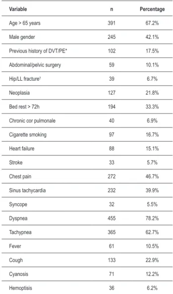
The overall mortality of this study was 14.1%; demographic and clinical characteristics are described in Table 1.
The median age was 73 years, ranging from 18 to 102 years, and the best cut-off point was the age of 65 years, as determined using the ROC curve16, with an area under the
curve of 0.60 (Figure 1), sensitivity of 81.8%, specificity of 88.3%, and positive and negative likelihood ratios of 6.99 and 0.21, respectively. In addition to age > 65 years, the model identified bed rest, chronic cor pulmonale, sinus tachycardia, and tachypnea as variables able to independently predict death risk, using logistic regression (Table 2).
From the prediction model, the expected death risk was calculated for each patient, and an ROC curve was constructed18
(Figure 2).The area under the curve – also known as C statistics – was 0.77, which corresponds to the model accuracy, and operating characteristics with 65.5% sensitivity, 80.0% specificity, positive likelihood ratio of 3.3, and negative likelihood ratio of 0.4. Figure 3 shows the relationship between pre-test probability (14.1%), which corresponds to theoverall mortality in the study, and the post-test probability of death in the case of a positive test (35.1%) and of a negative test (6.2%). Comparison of mortality curves (Figure 4) showed a significant difference between the risk groups, with p value = 0.000004.
Based on the predictive power of each variable, as guided by the respective odds ratio, the model showed a linear association for prediction of death (p>0.001).
Table 1 - Demographic and clinical characteristics.
Variable n Percentage
Age > 65 years 391 67.2%
Male gender 245 42.1%
Previous history of DVT/PE* 102 17.5%
Abdominal/pelvic surgery 59 10.1%
Hip/LL fracture† 39 6.7%
Neoplasia 127 21.8%
Bed rest > 72h 194 33.3%
Chronic cor pulmonale 40 6.9%
Cigarette smoking 97 16.7%
Heart failure 88 15.1%
Stroke 33 5.7%
Chest pain 272 46.7%
Sinus tachycardia 232 39.9%
Syncope 32 5.5%
Dyspnea 455 78.2%
Tachypnea 365 62.7%
Fever 61 10.5%
Cough 133 22.9%
Cyanosis 71 12.2%
Hemoptisis 36 6.2%
DVT/OE - deep-vein thrombosis/pulmonary embolism, †LL - lower limbs
Figure 1 - ROC curve for age cut-off point
S
ens
iti
vi
ty
1-Specificity 1.0
0.8
age=65
Sens: 81.8% Spec: 38.3%
Area under the curve: 0.604 0.6
0.4
0.2
0.0
1.0 0.8 0.6 0.4 0.2 0.0
Table 2 - Multivariate logistic regression analysis.
Variable OR p
Age > 65 years (1.6 a 2.4)1.9 0.0061
Bed rest 2.1
(1.8 a 2.3) 0.0002
CCP* 2.5
(1.8 a 2.7) 0.0080
Tachycardia 1.7
(1.2 a 2.1) 0.0099
Tachypnea 1.8
(1.1 a 2.1) 0.0135
* CCP – Chronic cor pulmonale
Figure 2 - Operating characteristics of the model.
S
ens
iti
vi
ty
1-Specificity 1.0
0.8
0.6
0.4
0.2
0.0
1.0 0.8 0.6 0.4 0.2 0.0
p=0.163
Sens: 65.5% Spec: 80.0%
Area under the curve: 0.773
Figure 3 - Relationship between pre and post-test probability
P
os
t-tes
t probabi
lity
Pre-test probability 1.0
0.8
0.6
0.4
0.2
0.0
Figure 4 - Mortality curves (Kaplan-Meyer)
1.0
0.8
0.6
0.4
0.2
0.0
40 30
20 Low Risk Moderate Risk High Risk
10 0
Table 3 - Prediction score.
Variable Points
Age > 65 years 3
Bed rest > 72h 2
Chronic cor pulmonale 4
Sinus tachycardia 2
Tachypnea 2
Table 4 - Death risk stratiication.
Death risk stratiication Points Mortality
Low Risk 0 to 3 5.4%
Moderate Risk 4 to 6 17.8%
High Risk 7 or higher 31.3%
Discussion
Most of the studies on the prognosis of PE use echocardiographic data of right ventricular dysfunction19,20
or laboratory data such as troponin21,22 and brain natriuretic
peptide23-25 elevation to quantify the risk of a worse outcome
for the patients. Despite their good prognostic accuracy, these tests are not broadly available.
In this first analysis of our study, we chose to use a model comprised of clinical variables only, based on the recommendations that by using a simpler prediction rule its application becomes more encompassing26. Hemodynamically
unstable patients were excluded so that we could evaluate a more homogeneous subgroup at a lower death risk. Despite the fact that this population was less severely ill in comparison
to patients with hypotension, the overall mortality of 14.1% was higher than those found in related publications.
Wicki et al27 studied 296 consecutive patients and
constructed a prediction model, the Geneva prognostic score, and observed 10.1% of adverse events, with an overall mortality of 8.4%. The predictors of adverse events used in this model were cancer, hypotension, hypoxemia, heart failure, history of previous deep-vein thrombosis, and documented deep-vein thrombosis on duplex scan.
In the PESI study28, Aujesky et al derived a risk stratification
model in a total of 10,534 patients distributed into five subgroups using 11 clinical variables, and observed an overall mortality of 9.2%, ranging from 1.1% (Class I, very low risk) to 24.5% (Class V, very high risk).
These two models were later compared as to their ability to select patients for treatment as outpatients, and demonstrated a significant difference for the prediction of 30-day mortality (PESI 0.9%; 95% CI, 0.3 to 2.2; vs Geneva, 5.6%; 95% CI, 3.6 to 7.6 - p < 0.0001)5.
The higher mortality in our case series suggests that, despite hemodynamic stability, the population studied, which was comprised of patients admitted in intensive care units, could be at a higher risk of complications. For the same reason, it is possible to understand the higher death risk in each stratum assessed in comparison with those of other studies mentioned27,28.
The identification of a subgroup that could be treated on an outpatient basis was not possible in this analysis, since the 5.4% mortality risk attributed to the low-risk stratum is too high to permit the choice of this treatment modality. On the other hand, the 31.3% mortality in the high-risk subgroup indicates the need for more complex interventions and surveillance of these patients.
Derivation of a prediction model is the starting point of a strategy to obtain a rule for clinical decision making. Thus, internal and external validation phases are fundamental for the model to be universally applied26. Similar databases are
being collected in other investigation centers, so that we can validate the model in other populations.
Conclusions
In hemodynamically stable patients with PE, age > 65 years, bed rest, chronic cor pulmonale, sinus tachycardia, and tachypnea were able to independently predict death risk using the logistic regression model.
Acknowledge
We acknowledge the participation of the following EMEP (Estudo Multicêntrico de Embolia Pulmonar – Multicenter Study on Pulmonary Embolism) investigators, for data collection:
References
1. Giuntini C, Di Ricco G, Marini C, Melillo E, Palla A. Pulmonary embolism: epidemiology. Chest. 1995; 107: 3S-9S.
2. Terzi CB, Lage SG, Dragosavac D, Terzi RGG. Insuficiência cardíaca grave em unidade de terapia intensiva: existe um índice prognóstico ideal? Arq Bras Cardiol. 2006; 87: 344-51.
3. Goldhaber SZ, Visani L, De Rosa M. Acute pulmonary embolism: clinical outcomes in the International Cooperative Pulmonary Embolism Registry. Lancet. 1999; 353: 1386-9.
4. Kurkciyan I, Meron G, Sterz F, Janata K, Domanovits H, Holzer M, et al. Pulmonary embolism as a cause of cardiac arrest: presentation and outcome. Arch Intern Med. 2000; 160: 1529-35.
5. Jiménez D, Yusen RD, Otero R, Uresandi F, Nauffal D, Laserna E, et al. Prognostic models for selecting patients with acute pulmonary embolism for initial outpatient therapy. Chest. 2007; 132 (1): 7-8.
6. Wells PS. Outpatient treatment of patients with deep-vein thrombosis or pulmonary embolism. Curr Opin Pulm Med. 2001; 7: 360-4.
7. Ribeiro A, Lindmarker P, Juhlin-Dannfelt A, Johnsson H, Jorfeldt L. Echocardiography Doppler in pulmonary embolism: right ventricular dysfunction as a predictor of mortality rate. Am Heart J. 1997; 134: 479-87.
8. Grifoni S, Olivotto I, Cecchini P, Pieralli F, Camaiti A, Santoro G, et al. Short-term clinical outcome of patients with pulmonary embolism, normal blood pressure, and echocardiographic right ventricular dysfunction. Circulation. 2000; 101: 2817-22.
9. Meyer T, Binder L, Hruska N, Luthe H, Buchwald AB. Cardiac troponin I elevation in acute pulmonary embolism is associated with right ventricular dysfunction. J Am Coll Cardiol. 2000; 36: 1632-6.
10. Tulevski II, ten Wolde M, van Veldhuisen DJ, Mulder JW, van der Wall EE, Buller HR, et al. Combined utility of brain natriuretic peptide and cardiac troponin T may improve rapid triage and risk stratification in normotensive patients with pulmonary embolism. Int J Cardiol. 2007; 116: 161-6.
11. Becattini C, Vedovati MC, Agnelli G. Prognostic value of troponins in acute pulmonary embolism: a meta-analysis. Circulation. 2007; 116: 427-33.
12. Douketis JD, Leeuwenkamp O, Grobara P, Johnston M, Sohne M, Tem Wolde M, et al. The incidence and prognostic significance of elevated cardiac troponins in patients with submassive pulmonary embolism. J Thromb Haemost. 2005; 3: 508-13.
13. Aujesky D, Roy PM, Le Manach CP, Verschuren F, Meyer G, Obrosky DS, et al. Validation of a model to predict adverse outcomes in patients with pulmonary embolism. Eur Heart J. 2006; 27: 476-81.
14. Aujesky D, Obrosky DS, Stone RA, Auble TE, Perrier A, Cornuz J, et al. A prediction rule to identify low-risk patients with pulmonary embolism. Arch Intern Med. 2006; 166 (2): 169-75.
15. Jiménez D, Díaz G, Valle M, Martí D, Escobar C, Vidal R, et al. Prognostic value of syncope in the presentation of pulmonary embolism. Arch Bronconeumol. 2005; 41 (7): 385-8.
16. R Development Core Team (2007) R: a language and environment for statistical computing. R Foundation for Statistical Computing, Vienna, Austria; 2007.
17. Greeland, S. Modeling and variable selection in epidemiology analysis. Am J Public Health. 1989; 79 (3): 340-9.
18. Hanley JA, McNeil BJ. The meaning and use of the area under the receiver operating characteristic (ROC) curve. Radiology. 1982; 143: 29-36.
19. Grifoni S, Vanni S, Magazzini S, Olivotto I, Conti A, Zanobetti M, et al. Association of persistent right ventricular dysfunction at hospital discharge after acute pulmonary embolism with recurrent thromboembolic events.. Arch Intern Med. 2006; 166: 2151-6.
20. Sociedade Brasileira de Cardiologia. Diretriz de embolia pulmonar. Arq Bras Cardiol. 2004; 83 (supl 1): 1-8.
21. Konstantinides S, Geibel A, Olschewski M, Kasper W, Hruska N, Jäckle S, et al. Importance of cardiac troponins I and T in risk stratification of patients with acute pulmonary embolism. Circulation. 2002; 106: 1263-8.
22. Giannitsis E, Müller-Bardorff M, Kurowski V, Weidtmann B, Wiegand U, Kampmann M, et al. Independent prognostic value of cardiac troponin T in patients with confirmed pulmonary embolism. Circulation. 2000; 102 (2): 211-7.
23. Pruszczyk P, Kostrubiec M, Bochowicz A, Styczynski G, Szulc M, Kurzyna M, et al. N-terminal pro-brain natriuretic peptide in patients with acute pulmonary embolism. Eur Respir J. 2003; 22: 649-53.
24. Binder L, Pieske B, Olschewski M, Geibel A, Klostermann B, Reiner C, et al. N-terminal pro-brain natriuretic peptide or troponin testing followedN-terminal pro-brain natriuretic peptide or troponin testing followed by echocardiography for risk stratification of acute pulmonary embolism. Circulation. 2005; 112: 1573-9.
25. Sanchez O, Trinquart L, Colombet I, Durieux P, Huisman MV, Chatellier G, et al. Prognostic value of right ventricular dysfunction in patients with haemodynamically stable pulmonary embolism: a systematic review. Eur Heart J. 2008; 29 (12): 1569-77.
26. Thomas G, McGinn TG, Guyatt GH, Wyer PC, Naylor CD, Stiell IG, et al. for the Evidence-Based Medicine Working Group. Users’ Guides to the Medical Literature XXII: How to Use Articles About Clinical Decision Rules. JAMA. 2000; 284: 79-84.
27. Wicki J, Perrier A, Perneger TV, Bounameaux H, Junod AF. Predicting adverse outcome in patients with acute pulmonary embolism: a risk score. Thromb Haemost. 2000; 84 (4): 548-52.
28. Aujesky D, Obrosky DS, Stone RA, Auble TE, Perrier A, Cornuz J, et al. Derivation and validation of a prognostic model for pulmonary embolism. Am J Resp Crit Care Med. 2005; 172: 1041-6.
de Janeiro RJ, Gustavo Gouvêa, Alexandre Drumond, Eduardo Eiras, Emanuel Salgueiro (Casa de Saúde São José) Rio de Janeiro RJ, Mônica Callafiori (Hospital CardioTrauma) Rio de Janeiro RJ, Paulo César Pereira da Silva e Souza, Marcelo Magalhães (Hospital de Clinicas de Niterói) Niterói RJ, Ana Paula Lacerda (Hospital Barra D`Or) Rio de Janeiro RJ, Fernando Pacheco (Hospital Copa D`Or) Rio de Janeiro RJ, Marcelo Vieira Gomes, Luis Felipe Camille (Clinica São Vicente) Rio de Janeiro RJ, Guilherme Aguiar (Hospital São Vicente de Paulo) Rio de Janeiro RJ, Maria Luiza Toscano, Roberto Caetano (Instituto Nacional do Cancer) Rio de Janeiro RJ, José Péricles Esteves, Carolina Barbosa (Hospital Português) Salvador BA, Francisco Silveira (Hospital Prontocor) Belo Horizonte MG, Alex Macedo (Hospital Santa Casa) Santos SP, Luis Carlos Bodanese (Hospital São Lucas) Porto Alegre RS.
Potential Conflict of Interest
No potential conflict of interest relevant to this article was reported.
Sources of Funding
This study was partially funded by Sanofi-Aventis.
Study Association