Emergency EEG
Study of survival
Moacir Alves Borges1, Harethusa Junia Botós2, Ricardo Funes Bastos2,
Moacir Fernandes Godoy3, Nely Silvia Aragão de Marchi1
ABSTRACT
Objective: To determine the survival rate according to the main findings of emergency electroencephalography (EEGs) of patients treated in a tertiary hospital. Method: In this prospective study, the findings of consecutive emergency EEGs performed on inpatients in Hospital de Base in São José do Rio Preto, Brazil were correlated with survival utilizing Kaplan-Meyer survival curves. Results: A total of 681 patients with an average age of 42 years old (1 day to 96 years) were evaluated, of which 406 were male. The main reasons for EEGs were epileptic seizures (221 cases), hepatic encephalopathy [116 cases of which 85 (73.3%) were men, p-value=0.001], status epilepticus (104 cases) and impaired consciousness (78 cases). The underlying disease was confirmed in 578 (84.3%) cases with 119 (17.5%) having liver disease [91 (76.0%) were men, p-value=0.001], 105 (15.4%) suffering strokes, 67 (9.9%) having metabolic disorders, 51 (7.5%) central nervous system infections and 49 (7.2%) epilepsy. In the three months following EEG, a survival rate of 75% was found in patients with normal, discreet slow activity or intermittent rhythmic delta activity EEGs, of 50% for those with continuous delta activity and generalized epileptiform discharges, and of 25% for those with burst-suppression, diffuse depression, and in alpha/theta-pattern coma. Death was pronounced immediately in patients with isoelectric EEGs. Conclusion: The main findings of EEGs, differentiated different survival rates and are thus a good prognostic tool for patients examined in emergencies. Key words: EEG, emergency, prognosis, probability of survival.
EEG de urgência: taxa de sobrevivência
RESUMO
Objetivo: Determinar a taxa de sobrevivência (TS), segundo os principais achados de eletrencefalograma de urgência (E-EEG), dos pacientes atendidos nas emergências de hospital de alta complexidade. Método: Estudo prospectivo, por ordem de chegada, da correlação entre os achados de E-EEG, feitos nos pacientes à beira do leito, com TS, utilizando-se as curvas de sobrevidas de Kaplan Meyer no Hospital de Base de São José do Rio Preto, São Paulo/Brasil. Resultados: Foram estudados 681 pacientes, dos quais 406 (59,6%) masculinos, com idade média de 42 anos (1 dia a 96 anos). As principais motivações para o E-EEG foram crises epilépticas (221 casos), encefalopatia hepática [(116 casos, dos quais 85 masculinos (73,3%), p= 0,001]; estado de mal epiléptico 104 e rebaixamento de consciência 78. O diagnóstico da doença de base foi confirmado em 578 (84,3%), sendo 119 (17,5%) hepatopatia, dos quais 91 (76,%) masculinos, p= 0,001; 105 (15,4%) acidente vascular encefálico; 67 (9,9%) distúrbio metabólico; 51 (7,5%) infecção do sistema nervoso central e 49 (7,2%) epilepsia. TS de 75% nos três primeiros meses foi encontrada nos pacientes com E-EEG com alentecimento discreto ou com atividade delta rítmica intermitente. TS por volta de 50% nos três meses foi encontrado nos pacientes com E-EEG com delta contínuo, crítico e com descargas periódicas. A TS foi menor que 25% nos dois primeiros meses após E-EEG, nos pacientes com E-EEG com surto/supressão, com depressão difusa e com comas alfa/teta e 0% nos E-EEG iselétricos. Conclusão: O E-EEG, com seus principais achados, foi capaz de diferenciar as diversas taxas de sobrevivências na amostra estudada, constituindo-se, portanto, bom instrumento de prognóstico para pacientes atendidos nas unidades de emergência hospitalar. Palavras-chave: EEG, emergência, probabilidade de sobrevida.
Correspondence
Moacir Alves Borges Rua Nair do Santos Lima 110 15090-290 São José do Rio Preto SP - Brasil
E-mail [email protected]
Received 17 February 2009 Received in final form 19 October 2009 Accepted 30 October 2009
1 PhD, Professor, Neuroscience Department of the Medicine School in São José do Rio Preto, São José do Rio Preto SP, Brazil;
2MD, Resident, Neuroscience Department of the Medicine School in São José do Rio Preto, São José do Rio Preto SP, Brazil;
Emergency departments and intensive care units (ICU) are becoming increasingly more important in hos-pitals. Within the enormous range of tools available in these sectors, emergency electroencephalography (EEG), due to its low cost, the time required to carry out the ex-amination, the lack of risk and personnel requirements, contributes greatly to the treatment of patients with acute diseases, in particular those with changes in conscious-ness1-3. he diferent patterns of EEG obtained in emer-gency patients, although nonspeciic, can often be corre-lated with the etiology of diseases of the central nervous system such as trauma4, vascular injury and anoxic-isch-emic injury due to cardiorespiratory arrest5. In hepatic encephalopathy6,7, Creutzfeldt-Jakob disease8, non-con-vulsive status epilepticus9-15 and herpes simplex enceph-alitis16, EEG is decisive for diagnosis and thus guides ther-apy and gives an indication of the prognosis.
EEG also contributes by characterizing the state of consciousness in exogen intoxication and metabolic encephalopathy17,18. Hence, EEG has gained importance in the identiication of cases with persistently reduced con-sciousness but with normal imaging examinations4,18.
his study aimed at correlating survival to emergency EEG indings in patients examined in a tertiary hospital.
Method
his is a prospective study of emergency EEGs ob-tained from patients submitted to urgent examinations in the Emergency Department, on the ward or in inten-sive care units (ICUs) of Hospital de Base in São José do Rio Preto. Hospital de Base is a tertiary hospital and re-gional reference center with 716 beds, 155 of which are in ICUs. he Emergency Department attends an average of 9,700 patients per month.
The emergency EEGs were performed in the labo-ratory of neurophysiology or at the bedside. Electrodes were placed according to the 10/20 international system utilizing two Berg analogue apparatuses (time constant: 0.3; ilter: 70 Hz; notch ilter: 60 Hz; paper speed: 3 cm/ second) with eight channels and two Nihon digital ap-paratuses (12-bit) with 22 channels. Only the irst emer-gency EEG examination of each patient studied between 1/7/06 and 1/8/07 was included in this work, although some patients were submitted to as many as 10 exami-nations. he results were interpreted by two experienced neurophysiologists (MAB, NSAM) assisted by two train-ees (HJB, FRB), with the inal decision being by consen-sus. Patients with diagnoses from previous hospitaliza-tions, those with technically low quality examinations and patients for whom consent to participate was refused by the next of kin were excluded from the study.
For study purposes, only the most signiicant altera-tion, whether related to the baseline rhythm or
paroxis-tic disorders, was considered in each EEG; some exami-nations had more than one evident alteration. hus, only one emergency EEG result was analyzed for each patient. he following indings were standardized according to the modiied criteria of Husain3: 1) intermittent rhyth-mic delta activity (IRDA); 2) critical activity epileptiform according to Treiman et al.19 with modiications by Gar-zon et al.20; 3) normal; 4) burst-suppression; 5) continu-ous high-voltage delta activity according to Amodio et al.7; 6) low-voltage, slow, nonreactive EEG of less than 10 mV; 7) difuse periodic epileptiform disorder (PED) and lateralized (PLED); 8) coma with speciic rhythms (alpha, theta and spindle); 9) electrocerebral inactivity (according to the minimum technical requirements proposed by the American Clinical Neurophysiology Society and 10) in-tercritical epileptiform disorder. he data were obtained and stored in an Excel spreadsheet.
Diagnosis of the underlying disease was made by in-vestigating the clinical history, even if precarious, comple-mentary biochemical examinations, imaging examinations such as computed tomography and magnetic resonance, as well as the diagnosis conirmation service, that is, autopsy.
Epilepsy was diagnosed only when epileptic seizures occurred, without being caused by any acute disorder, on more than two occasions with an interval of more than 24 hours. Generalized convulsive status epilepticus (GCSE) was deined according to the Commission of Classiica-tion and Terminology of the InternaClassiica-tional League against Epilepsy21, with modiications by Lowenstein et al.22 and Brenner14.
he Pearson chi-square or Fisher exact tests were uti-lized in the statistical analysis of the gender, age and sam-ple data. Kaplan-Meyer curves23 were employed to esti-mate survival probabilities. To compare survival curves, the Log-rank test and empirical Hazard function were used. he event of death was considered after conirma-tion by autopsy, the data of which was registered in the patient’s hospital records or was obtained by telephoning the relatives of the deceased. Surviving patients, who did not return to the outpatient’s clinic within three months after release from hospital, were also contacted by tele-phone to check on their health. he level of statistical sig-niicance was set at 0.05.
Results
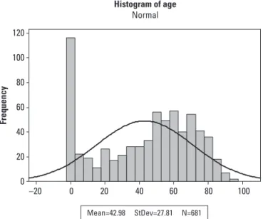
A total of 712 cases were considered however only 681 illed the inclusion criteria of this study. Of these, 406 (59.6%) were men (p-value=0.001). he ages had a non-Gaussian bimodal distribution with peaks for patients within the irst year of life and for those between 50 to 70 years old. he average age was 42 years old (range: 1 day to 96 years old – Fig 1).
patients records or by telephone) in the three months fol-lowing the last day for patient enrollment (1/8/2007).
he reasons to perform emergency EEGs are shown in Table.
he underlying disease was elucidated in 578 (84.3%) of the participating patients whose main diseases are shown in Figure 2. Among the 119 (17.5%) patients who sufered hepatic encephalopathy, 26 (21.9%) presented with triphasic waves.
he survival curves of the ten most common electro-encephalographic indings are shown in Figure 3.
discussion
he current study was designed to evaluate survival in respect to emergency EEG indings of patients
exam-ined in a tertiary general hospital. Performing emergen-cy EEGs is feasible24 however subject to many artifacts as was commented in the excellent review article published by Kaplan25. To reduce artifacts, ICUs have been adapted to local speciications with adequate earthing being pro-vided for the apparatuses. Even so, whenever possible, when there was no risk of life, the patients were trans-ferred either in wheel chairs or on stretchers to the neu-rophysiology laboratory.
he irst interesting data found in this study was that
–20 0 20 40 60 80 100 120
100
80
60
40
20
0
Frequency
Mean=42.98 StDev=27.81 N=681
Histogram of age
Normal
Fig 1. Distribution of the patients who underwent E-EEG.
0 20 40 60 80 100 Hepatic encephalopathy
Unknow Stroke Central nervous system infection Epilepsy Newborn disorder Heart disease Head-brain injury Brain tumor Renal failure Diabetes Alcoholism Chronic respiratory insufficiency Alzheimer’s disease Acute abdomen Central nervous system malformation Psychogenic non-epileptic seizures Drowning Poisoning Creutzfeld-Jakob disease
F M
Fig 2. The main diseases found correlated with gender;*p- value= 0.001
Fig 3. Survival curve of patients attended in emergency units of Hospital de Base according to the ten most common elec-troencephalographic inding. Critical: critical activity epilepcti-cus; IRDA: intermittent rhythmic delta activity; PED: period-ic epileptperiod-ic form disorder; SS: burst-suppression. Relationship among curves p-value=0.001.
400 300
200 100
0 100
80
60
40
20
0
Time to death (days)
Percent
the percentage of male patients was signiicantly higher than female patients. he explanation for this is that the study sample included a high number of patients with encephalopathy due to alcoholic cirrhosis; men are more prone to alcoholism. Another reason for there being more men in this study was the high rate of brain-encephalic injuries caused by motorbike accidents, which, in our re-gion, are generally driven by young men.
In the studies by Hesdorfer et al.10 in Rochester, USA and by Coeytaux et al.26 in Switzerland, more men than women were attended with status epilepticus (SE), how-ever in the current study the percentages of men and women with this condition are practically equal. here was no statistical diference between the genders of pa-tients who underwent emergency EEG for impaired con-sciousness to conirm non-convulsive status epilepticus. he distribution of the ages presented a bimodal as-pect with the irst peak being within the irst year of life and the second between 50 and 70 years old. he peaks occur exactly at ages when individuals have greater vul-nerability due either to the severe diseases of childhood or the fatal illnesses that afect adults in Brazil including heart and brain diseases. In this study, the relatively high number of severely ill children of up to one year old is due to the hospital being a regional reference center for sur-geries to correct congenital heart disease.
All patients with isoelectric EEGs were declared dead on the day of the examination, as this is the last test in the brain death protocol for organ transplantation. Of the 21 patients with requests to certify brain death, 6 deaths
were conirmed by EEG, while the others, who demon-strated low-voltage activity without the electroencephalo-graphic criteria of brain death, were referred for brain ar-teriography or scintigraphy. One patient with an isoelec-tric EEG underwent this examination because of coma.
Considering the order of the EEG indings in decreasing severity, the group with the highest death rate comprised patients with burst-suppression, of which three quarters died before the end of the irst month. his electroenceph-alographic inding occurred in particular as a consequence of anesthetics (midazolam, fentanil and barbiturates) dur-ing the treatment of SE and of anoxia caused by cardio-respiratory arrest, although in this study there were some cases due to other serious lesions of the central nervous system such as head-brain injury and intra-parenchyma-tous hemorrhages. In respect to the dismal prognosis, the data reported here are similar to other publications1,27 with only 15% surviving after the third month.
he low-voltage, slow, nonreactive EEG group also presented low survival rates, with approximately three quarters having died by the second month after the exam-ination. Similar to the indings of Celesia et al.28, patients with cardiorespiratory arrest and patients with suspicion of brain death were the most common with this pattern. Additionally, as was described by Young1 and Püttgen and Geocadin29, this electroencephalographic pattern may oc-cur within the irst hours after anoxia owing to prolonged cardiorespiratory arrest and after evolving to a burst-sup-pression pattern30.
he current study, unlike that by Young1, found a low survival rate for patients in alpha, theta and spindle co-mas; by the end of the second month, almost two thirds of the cases in this group had already died. hese results are in agreement with the indings of Kaplan31. In the cur-rent study the majority of alpha and theta comas originat-ed in patients with cardiorespiratory arrest, while spin-dle comas originated in cases of intoxication by poison or medications. Comparable to the results found in oth-er publications6,32, spindle comas had a better prognosis than alpha and theta comas.
Continuing in order of decreasing severity, the PED-type electroencephalographic pattern was found in several pa-tients, principally those with myoclonus encephalopathies after anoxia, prion diseases (three Creutzfeldt-Jakob cases)32 and encephalitis. After the second month a few more than two ifths survived. Triphasic waves were not considered PEDs, although, sometimes, the distinction is difficult.
Patients with the ictal electroencephalographic pat-tern presented with intermediate survival among the studied groups, but even so, mortality was considerably high, as by the end of the second month, half the patients had died. he mortality rate is high due to the underlying disease and the seriousness inherent in SE because of the Table. Reasons to perform the E-EEG correlated to gender (n=681).
Reason
M* F**
n % n %
Seizure 131 59.3 90 40.7
Hepatic encephalopathy# 85 73.3 31 26.7
Reduction in awareness 49 62.8 29 37.2
CSE1 45 46.4 52 53.6
Coma 21 53.8 18 46.2
CRA2 25 60 10 40
Brain death 14 66.7 7 33.3
Stroke 6 64.7 11 35.3
Mental confusion 6 37.5 10 62.5
Miscellanea3 7 50 7 50
Encephalitis 7 63.6 4 36.4
HBI4 7 63.6 4 36.4
PNEE5 3 60 2 40
Total# 406 59.6 275 40.4
1convulsive status epilepticus; 2cardiorespiratory arrest; 3apnea, Parkinson
disease, abstinence, tremor, poisoning; 4head-brain injury; 5psychogenic
excessive release of catecholamine which is the mecha-nism responsible for death33. In the current work, EEGs with PLED were grouped with the ictal pattern EEGs, in particular when there were clinical indications of sei-zures, although there is still controversy about the epi-leptiform nature of this discharge34. Triphasic waves were also analyzed with much care as they may appear in com-mon clinical situations such as epileptic seizures or in en-cephalopathies, a situation that was explained well in the report of Kaplan and Birbeck35. hese authors showed the diiculties in distinguishing between encephalopathy and non-convulsive status epilepticus in a series of seven pa-tients intoxicated by lithium and individuals with myo-clonic SEs after cerebral anoxia. EEGs critical to com-plex partial non-convulsive status epilepticus are diicult to identify given the variability in presentation, as was shown by Kaplan36 and Markand37. In the current study, this diiculty was minimized by studying clinical indings and the behavioral phenomenology of the patient.
Another group in which half the patients died up to the end of the second month in this study is that of con-tinuous delta rhythm. his group basically comprises pa-tients with hepatic encephalopathy due to cirrhosis and who are waiting for liver transplantation. he EEGs of one ifth of these patients, apart from having slow activ-ity, presented triphasic waves6,29.
he group of patients with normal baseline rhythm EEGs and intercritical epileptiform disorders, and those with IRDA, which are the irst manifestations of involve-ment of the central nervous system, have similar survival rates and correspond to patients with encephalopathies associated to underlying diseases that have good evolu-tions, including epilepsy, meningitis and head injuries.
As was reported in review articles by Young1, Mar-kand37, Kaplan2 and Nishisaki et al.38 the prognosis and the survival rate of patients submitted to emergency EEGs depends on the underlying disease. Hence, the main ind-ings of emergency EEGs were capable of diferentiating the diferent survival rates in the sample studied and is thus a good prognostic tool for patients attended in hos-pital emergency units.
acknowlEdGMEnTs– he authors wish to thank the statistician
José Antonio Cordeiro, PhD, of the Statistics Department of the Medi-cine School in São José do Rio Preto, for help with statistical analysis.
RefeRences
1. Young GB. The EEG in coma. J Clin Neurophysiol 2000;17:473-485. 2. Kaplan PW. The EEG in metabolic encephalopathy and coma. J Clin
Neuro-physiol 2004;21:307-318.
3. Husain AM. Electroencephalographic assessment of coma. J Clin Neurophys-iol 2006;23:208-220.
4. Peets AD, Berthiaume LR, Bagshw SM, et al. Prolonged refractory status ep-ilepticus following acute traumatic brain injury: a case report of excellent neurological recovery. Critical Care 2005;9:725-728.
5. Krumholz A, Stern BJ, Weiss HD. Outcome from coma after cardiopulmonary resuscitation: relation to seizures and myoclonus. Neurology 1988;38:401-405. 6. Bickford RG, Butt AR. Hepatic coma: the electroencephalographic patterns.
J Clin Invest 1955:34:790-799.
7. Amodio P, Marchetti P, Del Piccolo F, et al. Spectral versus visual EEG analy-sis in mild hepatic encephalopathy. Clin Neurophysiol 1999;110:1334-1344. 8. Bortone E, Bettoni L, Giorgi C, Terzano MG, Trabattoni GR, Mancia D. Reliabil-ity of EEG in the diagnosis of Creutzfeldt-Jakob disease. Electroencephalogr Clin Neurophysiol 1994;90:323-330.
9. Tomson T, Lindbom U, Nilsson BY. Nonconvulsive status epilepticus in adults: thirty-two consecutive patients from a general hospital population. Epilep-sia 1992;33:829-835.
10. Hesdorfer DC, Logroscino G, Cascino G, Annegers JF, Hauser WA. Incidence of status epilepticus in Rochester, Minnesota, 1965-1984. Neurology 1998; 50:735-741.
11. Fountain NB, Waldman WA. Efects of benzodiazepines on triphasic waves: implications for nonconvulsive status epilepticus. J Clin Neurophysiol 2001; 18:345-352.
12. Towne AR, Waterhouse EJ, Boggs JG, et al. Prevalence of nonconvulsive sta-tus epilepticus in comatose patients. Neurology 2000;54:340-349. 13. Husain AM, Horn GJ, Jacobson MP. Non-convulsive status epilepticus:
useful-ness of clinical features in selecting patients for urgent. EEG. J Neurol Neuro-surg Psychiatry 2003;74:189-191.
14. Brenner RP. EEG in convulsive and nonconvulsive status epilepticus. J Clin Neuroisiol 2004;21:319-331.
15. Holtkamp M, Masuhr F, Harms L, Einhäupl KM, Meierhord H, Buchheim K. The management of refractory generalized convulsive and complex partial status epilepticus in three European countries: a survey among epileptologists and critical care neurologists. J Neurol Neurosurg Psychiatry 2003;74:1095-1099. 16. Roches JC, Probst A, Scollo-Lavizzari G. How speciic are periodic complexes
in the diagnosis of herpes simplex encephalitis? Eur Neurol 1984;23:466-471. 17. Husain AM. Electroencephalographic assessment of coma. J Clin
Neurophys-iol 2006;23:208-220.
18. Kolls BJ, Husain AM. Assessment of hairline EEG as a screening tool for non-convulsive status epilepticus. Epilepsia 2007;48:959-965.
19. Treiman DM, Walton NY, Kendrick C. A progressive sequence of electroen-cephalographic changes during generalized convulsive status epilepticus. Epilepsia 1990;5:49-60.
20. Garzon E, Fernandes RMF, Sakamoto AC. Serial EEG during human status epi-lepticus: evidence for PLED as an ictal pattern. Neurolology 2001;57:1175-1178. 21. Commission on Classification and Terminology of International League
against Epilepsy Liga International. Epilepsia 1981;22:489-501.
22. Lowenstain DH, Bieck T, Macionald RL. It’s time to revise the deinition of sta-tus epilepticus. Epilepsia 1999;40:120-122.
23. Lee EP. Statistical methods for survival data analysis. New York: LifeTime Learning Publication,1980:75-154.
24. Quigg M, Sheel B, Domer. Current practice in administration and clinical cri-teria of emergent EEG. J Clin Neurophysiol 2001:18:162-165.
25. Kaplan PW. EEG monitoring in the intensive care unit. Am J Technol 2006;46: 81-97. 26. Coeytaux A, Jallon P, Galobardes B, MOrabia A. Incidence of status epilepti-cus in French-speaking Switzerland: Epistar. Neurology 2000;55:693-697. 27. Hockadaday JM, Potts F, Epstein E, Bonazzi A, Schwabb RS.
Electroenceph-alographic changes in acute cerebral anoxia from cardiac or respiratory ar-rest. Electroencephagr Clin Neurophysiol 1965;18:575-586.
28. Celesia GG, Grigg MM, Ross E. Generalized status myoclonicus in acute an-oxic and tan-oxic-metabolic encephalopathies. Arch Neurol 1988;45:781-784. 29. Püttgen HA, Geocadin R. Predicting neurological outcome follow cardiac
ar-rest. J Clin Neurophysiol 2007;261:108-117.
30. Rossetti AO, Logroscino G, Liauder L, et al. Status epilepticus: an indepen-dent outcome predictor after cerebral anoxia. Neurology 2007;69:255-60. 31. Kaplan PW, Genoud D, Ho TW, Jallon P. Etiology, neurology correlations, and
prognosis in alpha coma. Clin Neurophysiology 1999;110:205-213. 32. Au WJ, Gabor AJ, Viyan N, Markand ON. Periodic lateralized epileptiform
com-plexes (PLEDs) in Creutzfeldt-Jakob disease. Neurology 1980;30:611-617. 33. Parson-Smith BG, Summerkil WHJ, Dawson AM, Sherlock S. The
electroen-cephalograph in liver disease. Lancet 1957;2:867-871.
34. Brenner RP, Schauí N. Periodic EEG patterns: classiication, clinical correlation, and pathophysilogy. J Clin Neurophysiol 1990;7:249-288.
35. Kaplan PW, Birbeck. Lithium-induced confusional state: Nonconvulsive sta-tus epilepticus or triphasic encephalopathy? Epilepsia 2006;47:2071-2074. 36. Kaplan PW. The EEG of status epilepticus. J Clin Neurophysiol 2006;32:221-229. 37. Markand ON. Pearls, perils, and pitfalls in the use of the
electroencephalo-gram. Semin Neurol 2003;23:1-46.