Radiofrequency Catheter Ablation of Premature Ventricular
Contractions Originating in the Right Ventricular Outflow Tract
Francisco C. C. Darrieux, Maurício I. Scanavacca, Denise T. Hachul, Sissy L. Melo, André B. D’Ávilla, César J. Gruppi,
Paulo J. Moffa, Eduardo A. Sosa
Instituto do Coração do Hospital das Clínicas – FMUSP - São Paulo, SP - Brazil
Summary
Objectives: To evaluate if radiofrequency catheter ablation is an effective procedure for the treatment of right ventricular outflow tract premature ventricular contraction (RVOT-PVC) and ascertain if it results in an improvement of symptoms.
Methods: A prospective study with 30 consecutive patients (mean age 40 ± 13 years, 25 females), with no apparent structural cardiopathy, with very frequent (mean density of 1,263 ± 593/h) RVOT-PVC, symptomatic for more than one year (mean = 74 months) and resistant to antiarrhythmic drugs (3 ± 1.7, including beta-blockers), who underwent radiofrequency catheter ablation.
Results: After the first procedure, there were 23 initial successful cases (76.6%) and 7 initial failures (23.4%). Four patients experienced relapses, two of whom did not undergo the second procedure. The second procedure was carried out in 9 patients (7 initial failures and 2 relapses), and there was success in 5 additional patients, one of them by epicardial access. The final success rate was 80% (24/30), and there were no major complications. After a mean follow-up of 14 ± 6 months, in the successful group there was a reduction greater than 90% in density of premature ventricular contractions (PVC) (24/24; p<0.0001) and a resulting absence of symptoms in the majority of patients (23/24; p<0.001).
Conclusion: Radiofrequency catheter ablation is a safe and effective treatment for patients with persistent and symptomatic PVC with RVOT morphology.
Key words: Catheter ablation; premature ventricular contractions; arrhythmia; heart ventricles.
Mailing address: Francisco C. C. Darrieux •
InCor - Clinical Unit of Arrhythmia and Pacemaker - Francisco C. C. Darrieux Av. Dr. Enéas de Carvalho Aguiar, 44 Bl. II – Andar AB
05403-000 – São Paulo, SP
E-mail: [email protected]
Manuscript received April 9, 2006; revised manuscript received June 2, 2006; accepted July 21st, 2006
Introduction
Premature ventricular contractions (PVC) are very common arrhythmias in clinical practice1-3. They may occur in healthy
individuals with no evidence of structural cardiopathy 2-5 and
most originate in the right ventricular outflow tract (RVOT)1-3.
Many patients with RVOT premature ventricular contractions (RVOT-PVC) may experience a symptomatic progression with complaints of palpitations frequently associated with other clinical manifestations such as dyspnea, atypical precordial pain, dizziness, pre-syncope or syncope that may be incapacitating2. Conventional treatment with antiarrhythmic
drugs and psychological support may not be helpful in some cases. Therefore, a therapeutic approach seeking complete remission of these arrhythmias and associated symptoms may be an option in this subgroup of patients.
RVOT-PVC originate in an automatic point of origin similar to that responsible for RVOT idiopathic tachycardia, and therefore constitute a legitimate target for radiofrequency catheter ablation (RFA)6. Even though radiofrequency catheter
ablation is a safe and effective treatment for idiopathic
ventricular tachycardias7,8, its use in RVOT-PVC is still not
clearly defined. The objectives of this study were to evaluate the efficacy of RFA for treatment of persistent RVOT-PVC and ascertain if the success of this RFA results in an improvement of symptoms.
Methods
sustained ventricular tachycardia (NSVT) during the exercise stress test.
All patients were resistant to an average number of 3.0 ± 1.7 antiarrhythmic drugs, including beta-blockers, in all cases. Other medications used were sotalol (21/30), amiodarone (18/30), propafenone (10/30), quinidine (4/30), mexiletine (1/30) and disopyramide (1/30).
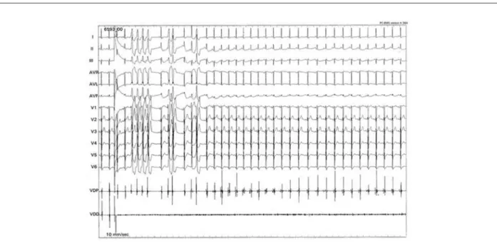
Electrophysiological study and radiofrequency catheter ablation - The electrophysiological study (EPS) and RFA were performed by the same team of electrophysiologists, with patients in a fasting condition for at least 8 hours and sedated with propofol. All anti-arrhythmic medications were stopped for at least 5 half-lives before the procedure. Two catheters with multipolar electrodes were positioned in the right ventricle, one for programmed stimulation, and the other for mapping and ablation. The ventricular stimulation catheter was positioned at the apex of the RVOT. Programmed ventricular stimulation was repeated after an additional stimulation using 1 to 5 µg/min of isoproterenol with the intent of documenting RVOT-PVC before and after ablation and eliminating the possibility of sustained ventricular tachycardia secondary to the reentry mechanism as in cases of arrhythmogenic right ventricular dysplasia.
Mapping and ablation of PVC were carried out using conventional catheters with a 4 mm deflecting tip (EP Technologies - Mountain View, CA, USA). Positioning of the catheter was guided by fluoroscopy (Fig. 2).
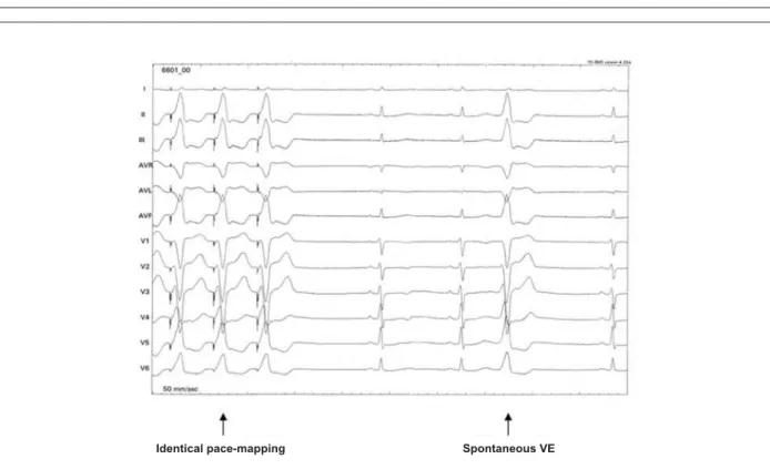
Radiofrequency (RF) application was guided by criteria of earliest endocardial activation (EEA) during spontaneous ectopic ventricular activity and the pace-mapping technique (Fig. 3). Unipolar RF pulses were applied with maximum controlled temperature of 70°C, maximum energy potency of 50W, and duration of up to 60 seconds.
End-of-procedure criterion was the complete disappearance of RVOT-PVC (Fig. 4), even after 30 minutes of isoproterenol infusion (immediate success). For simultaneous 12-lead ECG tracings and endocavitary electrograms, a digital computerized system was used for storage on an optic disk (Electrophysiological Measurement System – EMS - version 4.3 - University of Limburg -The Netherlands). The intracardial bipolar electrograms were filtered with a cut-off frequency of 30 to 500 Hz.
In case of failure of the first procedure (P1) and of symptomatic persistence of RVOT-PVC, patients underwent a second procedure (P2) with the same protocol or using different approach techniques such as the epicardial transthoracic approach9 or access of the left ventricular outflow
tract. In cases of failure after P2, patients were then classified as the final failure group.
Follow-up - Within the first 24 hours, all patients underwent 12-lead ECGs and 24-hour Holter tests. In cases of the epicardial approach, a bidimensional echocardiogram was taken. After 24 hours, patients were discharged from the hospital.
All patients were submitted to clinical evaluations in outpatient clinic visits at 30, 60, and 120 days, and for up to 1 year of follow-up. The following ancillary tests were done: 12-lead ECGs, 24-hour Holter test (at least 3 tests) and exercise stress test (at least 2). During the medical evaluation,
Fig. 1 - 12-lead ECG. Example of a patient with bigeminate premature ventricular contractions extrasystole with probable right ventricle outflow tract origin (inferior axis and morphology of left bundle branch block).
by the Ethics Committee of the Hospital das Clínicas, Medical School, Universidade de Sao Paulo.
Clinical characteristics - The clinical characteristics are listed on Table 1. Thirteen (13/30) patients presented no associated disease or comorbidity (43.5%), and the mean ejection fraction of the left ventricle was 73 ± 4%.
The symptom of palpitation was present in all patients (100%) and its mean duration was 74±66 months (median of 53 months). The RVOT-PVC were present during the stress test and were more frequent (17/30; 56.7%) during the periods of rest and recovery. Eight patients (26.7%) presented
non-Table 1 - Clinical characteristics of patients undergoing radiofrequency catheter ablation of right ventricle
outflow tract extrasystoles
Mean age (years) 40±13
Sex (F/M) 25/5
Absence of structural cardiopathy (n/%) 30(100%)
Associated conditions (n/%)
Hypertension 10(33.3%)
Hypothyroidism 3(10 %)
Morbid obesity 1(3.3%)
Mitral valve prolapse 1(3.3%)
Asthma 1(3.3%)
Smoking 1(3.3%)
LV Ejection fraction (%) 73±4
Nº. of Antiarrhythmic drugs 3.0±1.7
Symptoms (n/%)
Palpitations 30(100%)
Dizziness 19(63.3%)
Dyspnea 15(50 %)
Syncope 4(13.3%)
Duration of symptoms (months) 74±66
PVC/h density (Holter) 1,263±593
a specific questionnaire on symptoms was completed. A greater than 90% reduction in the density of PVC/hour in at least three consecutive Holter tests was considered a criterion of persistent success.
Statistical analysis - At the end of the study, patients were classified in two groups: Group 1 (final success) and Group 2 (final
failure). According to the RFA results, the percentage of reduction of PVC/hour on the Holter was analyzed using Wilcoxon’s test for independent samples and Fisher’s exact test, where reduction
ZDVGHILQHGDV7KHFRUUHODWLRQRIWKH5)$UHVXOWVRU
both) with the symptoms (yes or no) was made by Fisher’s exact
WHVW$SYDOXHZDVFRQVLGHUHGVLJQLILFDQW
Fig. 2 -Radiofrequency catheter ablation of right ventricle outflow tract (RVOT). Fluoroscopic aspect in right anterior oblique (A) and left anterior oblique projections (B). The arrows indicate the catheter in the RVOT and His bundle. The mapping and RF application start at RVOT, septal region, right below the pulmonary valves.
A B
Fig. 3 - An example of an adequate signal obtained during stimulation of the probable point of origin of premature ventricular contractions (PVC). Observe the agreement on the 12 leads, suggesting that the ablation catheter is close to the PVC point.
Results
Ablation procedure - Thirty-nine electrophysiological procedures were performed. In 9 patients, 2 procedures were necessary. The mean total time was 98 ± 37 minutes, and the mean time of radiation was 3 ± 1.7 minutes. The mean number of RF applications was 8 ± 5.5 pulses, with a mean temperature of 58 ± 7°C. The pace-mapping criterion was obtained in 24 patients (80%). The criterion of earliest endocardial activation (EEA) of less than zero milliseconds occurred in 11 patients (36.6%). Both criteria were utilized in 10 patients (33.3%). In 4 patients (16.6%), no signal adequate for ablation was attained.
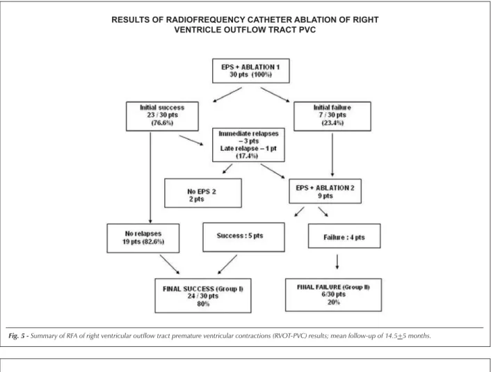
Regarding P1, the rate of immediate success (end of procedure, after 30 minutes of isoproterenol infusion) was 76.7% (23/30 patients), and immediate failure was evident in 7 patients (23.3%). Four patients (17.4%) presented relapses, 3 during the 24-hour Holter monitoring (13.05%) and 1 during late 24-hour Holter follow-up (4.35%). In two of these four patients with relapses, the second procedure was not performed - in one patient because of pregnancy and in the other because she became asymptomatic in spite of maintaining high density RVOT-PVC.
The second procedure (P2) was done in 9 patients, attaining success in 5 of them. In 4 of these patients, success came via the conventional endocardial route. In one case, success was achieved by the epicardial approach. In the remaining 4 patients of the failure group, the origin of the PVC could not be determined, even accessing the LV and the epicardium, and the high-density of PVC was maintained according to the 24-hour Holter test in late follow-up.
At the end of the study, final success was obtained in 24 patients (group 1), who also showed an absence or minimal density of RVOT-PVC on the 24-hour Holter (24/30; 80%). With the exception of only one, all the remaining patients of
group 1 became asymptomatic (23/24; 95.8%).
Figure 5 summarizes the sequence of results obtained by RFA during this study.
Application site for RF pulses - Appropriate sites for RF applications could be determined in 26 patients. In 18 patients, the RF applications were made on the septal surface of the RVOT, right below the pulmonary valves. In 7 patients, the application site was the free wall of the RVOT and in one patient the epicardial surface. In 4 patients, no RF pulse could be applied because of a lack of adequate signals for ablation (Fig. 6).
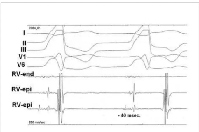
Epicardial access - The transthoracic epicardial approach was used in 5 patients in whom the endocardial approach failed. In 4 of them, no adequate signal was obtained. In one patient, however, one signal of earliest epicardial activation (-40 ms) was obtained. At this site, pace mapping was perfect (12/12) and two RF pulses resulted in the complete disappearance of the PVC (Fig. 7).
Reduction of RVOT-PVC density on the Holter test - The mean follow-up was 14.5 ± 5 months. In 24 patients (Group 1), there was an elimination or significant reduction of the PVC density per hour. In the other patients (Group 2), no significant PVC density reduction occurred. Thus, patients from Group 1 presented a significant reduction in RVOT-PVC density when compared to those of Group 2, both in the classified percent value (p<0.0001) and in the continuous percent value (p=0.0017) (Tables 2 and 3). The success rate (80%) of RFA of the RVOT-PVC persisted during the study in patients from Group 1; no spontaneous reduction was seen in RVOT-PVC density in patients from Group 2.
Symptoms - After one or two procedures, the final success rate in Group 1 patients was 95.8% (23/24). Only one patient from Group 1 reported persistence of palpitations
(1/24; 4.2%). On the other hand, one patient from Group 2 reported an improvement in symptoms. The maintenance of symptoms in Group 2 was 83.3% (5/6), which correlated with the persistence of high density RVOT-PVC seen on the Holter Test. Thus, comparing the two groups, a significant
difference was observed in improvement of symptoms in Group 1 patients (p<0.001), which correlated with the percentage of reduction of RVOT-PVC density on the Holter test (p<0.0001) (Table 4).
Complications - There were no significant complications.
Fig. 6 -Sites where RFA pulses were applied according to electrophysiological criteria for mapping premature ventricular contractions (PVC) point of origin. Note that most points of origin were on the septal face of right ventricular outflow tract (RVOT).
Fig. 5 -Summary of RFA of right ventricular outflow tract premature ventricular contractions (RVOT-PVC) results; mean follow-up of 14.5+5 months. RESULTS OF RADIOFREQUENCY CATHETER ABLATION OF RIGHT
Fig. 7 - An example of RF ablation RVOT-PVC by epicardial mapping technique. Note early epicardial activation at approximately 40msec, identifying the target for RFA.
Two patients presented minor complications (hematoma in the inguinal region, readily treated in a conservative manner).
Discussion
This study confirms that RVOT-PVC in deeply symptomatic patients without evidence of structural heart disease may be safely eliminated by RFA. In our study, success of RFA in RVOT-PVC ablation was attained in 80% of the patients during a mean follow-up of 14 months, and no significant complication occurred.
RVOT-PVC and RFA - RVOT-PVC not associated with structural cardiopathy generally present a good prognosis and treatment may not be necessary. Nevertheless, a rare subgroup of patients may experience a symptomatic progression, associated with the persistence of a high density of PVC (> 1,000 PVC/24 h.).
Even though RFA has become the treatment of choice for idiopathic ventricular tachycardias resistant to clinical treatment7,8,10,11, its role in treating monomorphic symptomatic
PVC is not yet well established. However, patients who satisfied the inclusion criteria in our study had been symptomatic for a long period and were not responsive to usual antiarrhythmic drugs. To date, the results of some successful clinical studies12-18 have left several questions unanswered regarding
radiofrequency catheter ablation of RVOT-PVC. Examples of these include the possibility of spontaneous remission of RVOT-PVC, RFA success predictor factors, the possibility
of ablation by the epicardial route in failure cases, and the possibility of immediate relapses seen on the Holter test in the first 24 hours of the RFA. Gursoy et al12, reported the first
case of successful RFA of monomorphic PVC; one patient had RVOT-PVC, and the other, PVC of the outflow tract of the left ventricle. Other authors reported recurrences of 10 to 20% of cases during follow-up14-17. Krittayaphong et al17, analyzed
the efficacy of RFA in 33 patients with RVOT-PVC resistant to clinical treatment, and obtained success in 32 cases (97%) and relapses in 8 (24%). Therefore, one important characteristic of these patients who undergo RVOT-PVC is the considerable possibility of relapses. In our study with 30 patients, there were 4 relapses, most of which (3/4; 75%) a result of the inclusion of the Holter test in the first 24 hours of RFA.
In some cases of initial failure of RFA, the site of origin of monomorphic PVC may be the intramyocardial region, or even the epicardial surface, as was demonstrated in one patient from our group.
Reduction of PVC on the holter test - The RVOT-PVC density seen on the Holter test was very high before the RFA (mean = 1,262 ± 593 PVC/h). It is important to point out that all patients in our study had at least 3 consecutive Holter tests with a high density of RVOT-PVC in a period of less than 180 days before the RFA. This fact was important in order to avoid the effects of temporal variations of RVOT-PVC and false-positive readings. The final success rate was 80%. The percentage reduction in RVOT-PVC density reached statistical significance (p<0.0001) and was particularly related to the success of the RFA. There were no cases of spontaneous remission of the RVOT-PVC in Group 2 patients.
PVC and symptoms - Perception of the PVC varies among patients. All patients presented with complaints of palpitations. Other symptoms were more diverse among the patients. Intriguing symptoms were dyspnea and fatigue experienced by
Table 2 - Final results of VE/h reduction (Holter) – continuous variable
Variable Final Result n Minimum Maximum Mean Median SD P
PVC/h Failure 6 634 2,151 1,225 1,309 723 0.8183
Before Success 24 366 2,955 1,151 1,236 607
PVC/h Failure 6 246 2,020 636 885 809 0.0017
After Success 24 0 166 0.37 24 43
Final Reduction % Failure 6 -15 78 34 33 44 0.0017
PVC/h Success 24 90 100 99 98 3
Table 3 - Final results of PVC/h reduction (Holter) – categorical variable
Final reduction PVC/h
Final > 90%
Result n % n % p
Failure 0 0.00 6 100.00 < 0.0001
References
1. Hiss RG, Averill KH, Lamb LE. Electrocardiography findings in 67,375 asymptomatic subjects. III. Ventricular rhythms. Am J Cardiol. 1960; 6: 96-107.
2. Barrett PA, Peter CT, Swan HJ, Singh BN, Mandel WJ. The frequency and prognostic significance of electrocardiographic abnormalities in clinically normal individuals. Prog Cardiovasc Dis. 1981; 23: 299-319.
3. Kennedy HL, Underhill SJ. Frequent or complex ventricular ectopy in apparently healthy subjects: a clinical study of 25 cases. Am J Cardiol. 1976; 38: 141-8.
4. Gallavardin L. Extrasystole ventriculaire a paroxysmes tachycardiques prolongés. Arch Mal Coeur. 1922; 15: 298-312.
5. Hiss RG, Lamb LE. Electrocardiographic findings in 122,043 individuals. Circulation. 1962; 25: 947-61.
6. Gumbrielle T, Bourke JP, Furniss SS. Is ventricular ectopy a legitimate target for ablation? Br Heart J. 1994; 72: 492-4.
7. Morady F, Kadish AH, Dicarlo L, Kou WH, Winston S; de Buitlier M, et al. Long-term results of catheter ablation of idiopathic right ventricular tachycardia. Circulation. 1990; 82: 2093-9.
8. Klein LS, Shih HT, Hackett FK, Zipes DP, Miles WM. Radiofrequency catheter ablation of ventricular tachycardia in patients without structural heart disease. Circulation. 1992; 85:1666-74.
9. Sosa E, Scanavacca M, D’Avila A, Piccioni J, Sanchez O, Velarde JF, et al. Endocardial and epicardial ablation guided by nonsurgical transthoracic epicardial mapping to treat recurrent ventricular tachycardia. J Cardiovasc Electrophysiol. 1998; 9: 229-39.
10. Wilber DJ, Baerman J, Olshansky B, Kall J, Kopp D. Adenosine-sensitive ventricular tachycardia: clinical characteristics and response to catheter ablation. Circulation. 1993; 87: 126-34.
11. Rodriguez LM, Smeets JL, Timmermans C, Wellens HJ. Predictors for successful ablation of right- and left-sided idiopathic ventricular tachycardia. Am J Cardiol. 1997; 79: 309-14.
12. Gursoy S, Brugada J, Souza O, Steurer G, Andries E, Brugada P. Radiofrequency ablation of symptomatic but benign ventricular arrhythmias. Pacing Clin Electrophysiol. 1992; 15: 738-41.
13. Zhu DW, Maloney JD, Simmons TW, Nitta J, Fitzgerald DM, Trohman RG, et al. Radiofrequency catheter ablation for management of symptomatic
Table 4 – Study of symptoms final results (After ESP 1- 2)
Final results
Failure (II) Success (I)
Symptoms Category n %(Res) %(Symp) n %(Res) %( Symp)) P
Presence of symptoms No 1 17 0 23 96 100 < 0.001
Yes 5 83 80 1 4 20
these patients. Lauck et al14, by continuous invasive monitoring
during the period of severe bigeminate PVC, observed that the effective cardiac output was reduced to half during the ventricular bigeminism period, which could explain these symptoms and justify a more aggressive approach to PVC. The same aspect was observed by transthoracic continuous echo-Doppler, showing a loss of cardiac output in the left ventricular outflow tract14. Recently
Yarlagadda et al18 demonstrated cases of tachycardiomyopathy
secondary to RVOT-PVC, justifying intervention with RFA. In our series, we did not observe this characteristic of RVOT-PVC, which we believe to be quite uncommon.
All patients in this study were very symptomatic and resistant to conventional antiarrhythmic medications (an average of 3 drugs), including the use of beta-blockers. The association between the PVC and symptoms may be suggested since the reduction of PVC density coincided with the control of the symptoms. One issue would be the fact that the RVOT-PVC had been spontaneously suppressed. Gaita et al19
conducted a longitudinal study in 61 patients with RVOT-PVC and observed that the symptoms disappeared spontaneously in two-thirds of patients for an average period of 15 years. However, one third of the patients persisted with RVOT-PVC and continued symptomatic. The patients in our study were all very symptomatic and spontaneous resolution of these RVOT-PVC was considered improbable. Additionally, suppression and relapses of RVOT-PVC were directly correlated to the
suppression and recurrence of the symptoms.
Limitations - Our study has some limitations. There was no randomization with a control group. Nonetheless, the groups that presented relapses and the cases of failure served, in some way, as a control group, confirming that maintenance of the high density of RVOT-PVC and symptoms resulted from RFA failures. Because of the methodology of the study, no third procedure was performed in final cases of failure. Thus, the failure rate could have been modified if there had been an additional procedure or if other RFA strategies had been used. However, the objective of the study was met in most patients by means of the conventional RFA technique.
Conclusions
RFA is an effective and safe procedure for treatment of persistent and symptomatic RVOT-PVC, and the success of this approach results in an improvement of symptoms. Cases of RFA failure in RVOT-PVC may be related to the origin of the PVC at other sites, such as epicardial, intramyocardial or left ventricular regions.
Potential Conflict of Interest
ventricular ectopic activity. J Am Coll Cardiol. 1995; 26: 843-9.
14. Lauck G, Burkhardt D, Manz M. Radiofrequency catheter ablation of symptomatic ventricular ectopic beats originating in the right outflow tract. Pacing Clin Electrophysiol. 1999; 22: 5-16.
15. Lauribe P, Shah D, Jais P, Takahashi A, Haissaguerre M, Clementy J. Radiofrequency catheter ablation of drug refractory symptomatic ventricular ectopy: short- and long-term results. Pacing Clin Electrophysiol. 1999; 22 (5): 783-9.
16. Seidl K, Schumacher B, Hauer B, Jung W, Drogemuller A, Senges J, et al. Radiofrequency catheter ablation of frequent monomorphic ventricular activity. J Cardiovasc Electrophysiol. 1999; 10 (7): 924-34.
17. Krittayaphong R, Sriratanasathavorn C, Bhuripanyo K, Raungratanaamporn O, Soongsawang J, Khaosa-ard B, et al. One-year outcome after radiofrequency catheter ablation of symptomatic ventricular arrhythmia from right ventricular outflow tract. Am J Cardiol. 2002; 89 (11): 1269-74.
18. Yarlagadda RK, Iwai S, Stein KM, Markowitz SM, Shah BK, Cheung JW, et al. Reversal of cardiomyopathy in patients with repetitive monomorphic ventricular ectopy originating from the right ventricular outflow tract. Circulation. 2005; 112: 1092-7.