300 • Arq Gastroenterol • 2017. v. 54 nº 4 Out/Nov 300 • Arq Gastroenterol • 2017. v. 54 nº 4 Out/Nov
INTRODUCTION
Helicobacter pylori (H. pylori) is a gram-negative bacterium that causes one of the most common chronic infections in human. This microorganism is a known pathogen associated with chronic gastritis, peptic ulcer disease (PUD), gastric carcinoma and gas-tric lymphoma. However, the clinical effects of this pathogen is beyond of the stomach and the epidemiological studies suggested an association between H. pylori infection and atherosclerosis or vascular diseases. The role of H. pylori and its virulence factors involved in the pathogenesis of atherosclerosis has not been identi-ied so far(18). Atherosclerosis as a chronic inlammatory disease is
a multifactorial inlammatory phenomenon and it is known that some pathogens is associated with chronic infection can induce inlammation(12). In the case of H. pylori infection, the
organ-ism after colonization, by activating proinlammatory signaling pathways such as nuclear factor kappa B (NF-κB)(8), can cause
the production and release of the proinlammatory cytokines such
Flow mediated dilation and carotid intima media
thickness in patients with chronic gastritis
associated with
Helicobacter pylori
infection
Arezo
JUDAKI
1, Siros
NOROZI
2, Mohammad Reza Hafezi
AHMADI
3,Samira Mis
GHAVAM
2,
Khairollah
ASADOLLAHI
4and Asghar
RAHMANI
5Received 9/4/2017 Accepted 17/7/2017
ABSTRACT – Background – Endothelial dysfunction is one of the early stages of vascular diseases. Objective – The aim of this study was to investigate the endothelial dysfunction markers in patients with chronic gastritis associated with Helicobacter pylori (H. pylori) infection. Methods – By a cross sectional study, basic and clinical information of 120 participants (40 patients with positive H. pylori infection, 40 patients with negative H. pylori
infection and 40 healthy people) were analyzed. Carotid intima media thickness and flow-mediated dilation levels were measured in all patients and controls. Soluble vascular cell adhesion molecule-1 (sVCAM-1) and intercellular adhesion molecule-1 (ICAM-1) were measured with Elisa for all subjects. IgG level was assessed in chronic gastritis patients. Results – The flow-mediated dilation level in patients with positive H. pylori infection (0.17%±0.09) was signiicantly lower than those with negative H. pylori infection (0.21% ±0.10, P<0.05) and compared to the control group (0.27% ±0.11, P<0.05). Carotid intima media thickness level in patients with positive H. pylori infection (0.58±0.13 mm) was signiicantly higher than those with negative H. pylori infection (0.48±0.32 mm, P<0.05) and compared to the control group (0.36±0.44mm, P<0.05). The mean level of sICAM-1 in positive H. pylori infection group (352.16±7.54 pg/mL) was higher than negative H. pylori infection group (332.64±8.75 pg/mL =0.75) and com-pared to the control group (236.32±12.43 pg/mL, P<0.05). A direct relationship was revealed between flow-mediated dilation and carotid intima media thickness changes and between sICAM-1 and sVCAM-1 associated with the level of H. pylori IgG in chronic gastritis. Conclusion – The levels of flow-mediated dilation, carotid intima media thickness and sICAM-1 were higher among patients with positive H. pylori infection. Patients with chronic gastritis associated with H. pylori infection are at risk of endothelial dysfunction due to flow-mediated dilation and carotid intima media thickness abnormalities and increased level of sICAM-1 and sVCAM-1.
HEADINGS – Gastritis. Helicobacter pylori. Endothelial growth factors. Carotid intima-media thickness.
Declared conflict of interest of all authors: none
Funding/Support: Ilam University of Medical Sciences, Ilam, Iran, supported this study.
1 Department of Gastroenterology, Ilam University of Medical Sciences, Ilam, Iran; 2 Department of Cardiology, Faculty of Medicine, Ilam University of Medical Sciences, Ilam, Iran; 3 Department of Pathology, Faculty of Medicine, Ilam University of Medical Sciences, Ilam, Iran; 4 Department of Social Medicine, Faculty of Medicine, Ilam University of Medical Sciences, Ilam, IR Iran; 5 General physician, Faculty of Medicine, Ilam University of Medical Sciences, Ilam, Iran.
Correspondence: Asghar Rahmani. Student Research Committee, Faculty of Medicine, Ilam University of Medical Sciences, Ilam, Iran. Email: [email protected]
as CRP, mRNA and protein expression of tumor necrosis factor alpha (TNFα) and interleukin(17). Systemic effect of this
inlam-matory process is a basic hypothesis of the association between
H. pylori infection and extragastrointestinal (GI) diseases(15). The
inflammatory cytokines especially CRP are independent risk factor for CVD, so that it is known that inlammatory cytokines can induce function and structural vessel abnormalities in the arterial wall, by many mechanisms, such as changes in the serum lipid levels, and metabolism and production of oxidative stress. Endothelial dysfunction plays a key role in the pathogenesis of atherosclerosis, that refers to the lack of ability in endothelium to providing vascular homeostasis(4). The endothelial dysfunction is a
consequence of overproduction of vasoconstrictors and dysregula-tion of hemostasis, inlammadysregula-tion and thrombosis in endothelium of vascular wall(20). Activation of vascular endothelium causes an
atherosclerosis(16). In addition to these markers, some non-invasive
procedures such as low mediated dilation (FMD) and carotid inti-ma media thickness (CIMT) can be indicators of atherosclerosis(19).
As mentioned previously, inlammation is a common feature of
Helicobacter pylori and atherosclerosis(15); however, in reality there
is no study to indicate the atherogenic mechanisms of H. pylori
and evidences are available only based on epidemiological studies. Therefore, this study aimed to investigate the relationship between
H. pylori infection and serologic markers related to H. pylori with blood markers of endothelial dysfunction such as ICAM-1 and VCAM-1 and markers related to atherosclerosis such as CIMT and FMD in patients with chronic gastritis associated with
H. pylori infection.
METHODS
Study protocol
This case-control study was performed among 120 partici-pants who showed some gastrointestinal manifestations such as dyspepsia and attended to the gastroenterology clinics, for rou-tine checks or endoscopy, in Ilam city, West of Iran, during May and November 2015. For all of participants, a detailed history including the starting time of signs and symptoms, duration of disease, past medical history and predisposing factors such as smoking, alcoholism, medication used especially Nonsteroid Anti-inlammatory Drugs (NSAID) was obtained. Patients with chronic diseases such as cardiovascular, endocrine, hematologic and respiratory disorders, pregnancy or lactation and hormone replacement therapy (HRT), antibiotic use in the previous 6 weeks, previous attempts at H. pylori eradication, previous gastric surgery or a previous history of alcohol consumption or drug abuse were excluded from the study. For better comparison of the results, the control group was selected among subjects who had no history of atherosclerotic risk factors or conirmed diseases. Also, controls did not show any dyspepsia symptom, or did not report a his-tory of antibiotics, bismuth, proton pump inhibitors or NSAID application during the past 8 weeks. The Ethics Committee of Ilam University of Medical Sciences conirmed the study, and a written informed consent was obtained from each participant. All clinical procedures were performed according to the principles expressed in the Helsinki declaration.
Detection of H. pylori infection
H. pylori infection was detected via cultivation of two biopsy samples taken from antrum of stomach in a transport medium (Portagerm pylori, bioMe´rieux, Marc l’Etoile, France) and Urease Breath Test (UBT) viaa CLOtest (Tri-Med Specialities, Osborne Park, Western Australia). In addition, two biopsy samples were taken from antrum and corpus of stomach for histopathologic assessment. Three different tests including UBT, histology and cultivation of H. pylori were performed for conirmation of infec-tion and if the results of all tests were positive, the patient was considered as an infected individual for H. pylori.
Endoscopic evaluations
All participants were inspected by the same upper Olympus (GIF-V) endoscopic apparatus. Endoscopy was performed by a gastrointestinal subspecialist and the biopsy needle as well
as the apparatus were sterilized for each patient separately. All patients were sedated by 10% oral xylocaine spray and 5.5-7 mg midazolam intravenously and samples of biopsies were taken by the same needle and the same method. Endoscopy was done at a lying position on the left side and after macroscopic evaluations, the needed biopsy samples were taken from the different parts of the stomach.
Histology
In this study, histological assessment was performed ac-cording to the Sydney System for the classiication of gastritis. Hematoxylin-Eosin (H&E) staining was preformed for the H. pylori
diagnosis and histological indings. For conirming the presence of
H. pylori “Giemsa” stain was also done. Identiication of rods and curved gram-negative organisms in the mucosal surface in the pit of stomach with ×40 zoom of light microscope was precisely sought by expert individuals. The existence of at least five bacilli per microscopic ield was necessary for conirmation. Iniltration of lymphocytes and plasma cells in the lamina propria were con-sidered as signs of chronic gastritis and neutrophil iniltration was considered as sign of active gastritis. The presence of goblet cells and replacement of mucosal cylindrical cells by absorption cells was considered as a sign of intestinal metaplasia(17).
FMD assay
FMD measurement was preformed by ultrasound imaging with 18-MHz linear array transducer on M-mode (Acuson S3000; Siemens). All measurements were performed at the morning and all patients had 30 minutes rest and were inhibited to consume coffee or a special diet.
Carotid intima media thickness assay
CIMT assessment was performed for all participants using B-mode ultrasonography (Esaote, MylabTM 70 Co., Italy) using a high-resolution, 18-MHz linear array transducer. All measurements were performed by a radiologist who was blinded to the clinical conditions of patients. According to a previous study, the CIMT of both left and right common carotid arteries was measured only once, and the CIMT value was calculated by averaging the meas-urements of left and right common carotid IMT.
Laboratory assay
After a fasting of 12 hours, 10 mL of blood sample from brachi-al vein was collected from brachi-all participants. Samples were centrifuged for 5 minutes at 4000 rpm and then were frozen and stored at -70°C until the time of assessment. sICAM-1 and sVCAM-1 levels were measured with sandwich enzyme immunoassay technique (Elisa) kits (Bender medical systems GmbH Viena, Austria).
Statistical analysis
302 • Arq Gastroenterol • 2017. v. 54 nº 4 Out/Nov RESULTS
Basic information of subjects
Basic and clinical indings of subjects are showed in Table 1. In the control group, 21 (52.5%) participants were male and 19 (47.5) were female. In the H. pylori positive group, 18 (45%) pa-tients were male and 22 (55%) were female. Twenty three (57.5%) patients of the H. pylori negative group were male and 17 (42.5%) were female. There wasn’t any signiicant difference between three studied groups for gender (P=0.876). Mean ± standard deviation (SD) for age among control, negative and positive
H. pylori groups were 47.21±7.54, 46.52±5.52 and 45.64±8.32 years respectively (P=0.943). Five (12.5%) participants in the control group, six (15%) patients in the positive and seven (17.5%) patients in the negative H. pylori groups reported alcohol con-sumption (P=0.245). Also, four (10%) participants in the control group were current smokers and 10 (25%) others were passive smokers. In the positive H. pylori group, six (15%) patients and 12 (30%) patients and in the negative H. pylori group 8 (20%) patients and 9 (22.5%) patients were current and passive smokers respectively (P=0.032). NSAID was applied by 12 participants in the control group, 16 (40%) patients in the positive H. pylori and 14 (35%) patients in the negative H. pylori groups respectively (P=0.042). The frequencies of atrophy, metaplasia and dysplasia among the positive H. pylori group were 12 (30%), 10 (25%) and 14 (35%) respectively and among the negative H. pylori group these igures were 15 (37.5%), 7 (17.5%) and 13 (32.55%) respectively. Mean ±SD of IgG in the positive and negative H. pylori groups
were 87.54±12.32 IU/mL and 14.32±6.43 IU/mL respectively with a high signiicant difference (P=0.000).
Relationship between endothelial dysfunction and
H. pylori
The level of endothelial dysfunction among both negative and positive H. pylori groups is showed in Table 2. Mean ±SD of FMD in the control group (0.27% ±0.11) was signiicantly higher in comparison with the positive (0.17% ±0.09, P=0.034) and negative H. pylori groups (0.21% ±0.10, P=0.041). Also, the mean ±SD of CIMT in the positive H. pylori group (0.58±0.13 mm) was signiicantly higher than that in the control (0.36±0.44 mm,
P=0.021) and negative H. pylori groups (0.48±0.32 mm, P=0.040). Mean ±SD of CIMT in the chronic gastritis patients with nega-tive H. pylori was signiicantly higher than that for participants in the control group (P=0.037). Mean ±SD of sICAM-1 in the chronic gastritis patients with positive and negative H. pylori was signiicantly higher than that for participants in the control group (332.64±8.75 pg/mL, 352.16±7.54 pg/mL, 236.32±12.43 pg/mL,
P=0.000, respectively). Level of sICAM-1 in positive H. pylori
group was higher than that in the negative H. pylori group but this difference was not signiicant (P=0.053). Mean ±SD of sV-CAM-1 in patients with positive H. pylori (731.43±21.34 pg/mL) was signiicantly higher than that in the negative H. pylori patients (665.45±17.54 pg/mL, P=0.001) as well as those in the control group (456.54±24.32, P=0.000). Also the level of sVCAM-1 in the negative H. pylori group was signiicantly higher than that in the control group (P=0.000).
TABLE 1. Basic characteristics of participants
Variable Control n=40
H. pylori positive n=40
H. pylori negative
n=40 Overall P
Sex, n (%) 0.876
Male 21 (52.5) 18 (45) 23 (57.5)
Female 19 (47.5) 22 (55) 17 (42.5)
Age, years 47.21±7.54 45.64±8.32 46.52±5.52 0.943
BMI, kg/m2 25.65±5.43 27.54±3.65 26.13±4.76 0.354
Alcohol, n (%) 5 (12.5) 6 (15) 7 (17.5) 0.245
Current smoking n (%) 4 (10) 6 (15) 8 (20) 0.032
Passive smoking n (%) 10 (25) 12 (30) 9 (22.5)
NSAID, n (%) 12 (30) 16 (40) 14 (35) 0.042
Atrophy, n (%) - 12 (30) 15 (37.5) 0.467
Matapelesia, n (%) - 10 (25) 7 (17.5) 0.345
Dysplasia, n (%) - 14 (35) 13 (32.5) 0.233
IgG (IU/mL) - 87.54±12.32 10.32±6.43 0.000
BMI: body mass index; NSAID: nonsteroid anti-inlmmatory drugs.
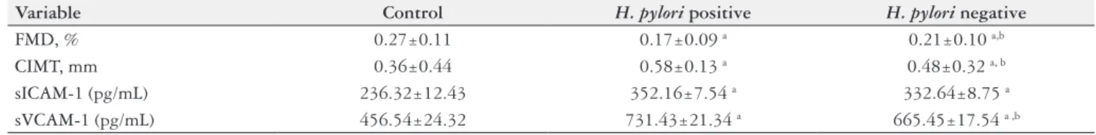
TABLE 2. Compression the level of endothelial dysfunction markers in patients with chronic gastritis associated with H. pylroi infection
Variable Control H. pylori positive H. pylori negative
FMD, % 0.27±0.11 0.17±0.09 a 0.21±0.10 a,b
CIMT, mm 0.36±0.44 0.58±0.13 a 0.48±0.32 a, b
sICAM-1 (pg/mL) 236.32±12.43 352.16±7.54 a 332.64±8.75 a
sVCAM-1 (pg/mL) 456.54±24.32 731.43±21.34 a 665.45±17.54 a ,b
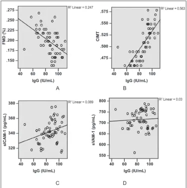
Relationship between IgG level with markers of endothelial dysfunction
Figure 1 shows the correlation between markers of endothelial dysfunction and the level of IgG as the serological marker of H. pylori infection. Based on the results, the level of IgG had a revers correlation with FMD, so that by increasing the serum level of IgG, the level of FMD was reduced, but this correlation was not signiicant (P=0.247). Regardless to FMD, the level of CIMIT increased with increasing the serum level of IgG (P=0.563).
endothelial dysfunction during chronic gastritis disease for the irst time. Another interesting inding in results was the FMD changes in patients with H. pylori infection was signiicantly lower than those in patients without H. pylori infection. Also, early changes in the endothelial dysfunction were higher among chronic gastric patients compared to healthy controls, but these changes in the patients with chronic gastritis associated with H. pylori infection were higher than those in the chronic gastritis without H. pylori in-fection and healthy people. Due to lack of similar studies, we could not compare the results of this study with other indings. Blum and colleagues reported that H. pylori eradication could improve endothelial dysfunction according to attenuated FMD and ankle brachial index after 3 months(3). This study also revealed a reverse
correlation between FMD changes and the serum level of IgG as a serological marker of H. pylori infection, for the irst time. In a study by Ozdogru and colleagues, the relationship between IgG titer and the extent of coronary artery disease was reported among patients with angiography proven coronary artery disease (CAD) as well as those with acute coronary syndrome (ACS)(14), while
Hara and colleagues have rejected this kind of association(6). This
study showed a direct association between CIMT and IgG titer in chronic gastritis associated with H. pylori infection compared to the healthy subjects and furthermore compared to patients with negative H. pylori infection. CIMT is an indicator of structural changes in vascular wall and measuring this marker can predict future risk of cardiovascular events(13). Fortunately, some studies
have limitedly investigated the association between CIMT and
H. pylori infection(10). For example, Akbas and colleagues reported
that the CIMT among positive H. pylori subjects was signiicantly higher than that in those with negative H. pylori infection(1).
Find-ings of this study were in accordance with the results of Akbas et al. Of course, our study was relatively different from Akbas study, so this study compared chronic gastric patients either with or without
H. pylori infection with the healthy subjects. Nevertheless, Altintas and colleagues reported that carotid intima-media thickness was higher among positive H. pylori patients with atrophic gastritis compared to positive H. pylori patients with non-atrophic gastri-tis but their difference was not signiicant(2). In addition, we have
shown a direct linear correlation between the CIMT rate and the serum level of IgG that this inding has not been reported so far. Another important aspect of this study was assessment of blood markers associated with endothelial dysfunction such as sICAM-1 and sVCAM-1 in patients with chronic gastritis associated with
H. pylori infection. We showed that the levels of sICAM-1 and sVCAM-1 in chronic gastritis patients associated with H. pylori
infection were signiicantly higher compared to those without
H. pylori infection. In addition, the level of these two markers in chronic gastritis patients with or without H. pylori infection was signiicantly higher in comparison with healthy individuals. According to Mori’s study, H. pylori infection, directly, induced expression of ICAM-1 during culturing of gastric epithelial cells in a NF-kappa B-dependent manner(11). In another study, indings
showed a signiicant role of ICAM-1 and VCAM-1 in leukocyte-endothelial interaction among patients with chronic gastritis associated H. pylori infection without E-selectin upregulation(7).
This inding was in accordance with our results. We showed that a direct association between serum level of IgG and increasing level of ICAM-1 and VCAM-1 in chronic gastritis patients. This result was not reported so far and this issue was performed by this study for the irst time. As it was previously mentioned, there
FIGURE 1. Association between endothelial dysfunction markers and
H. pylori IgG in patients with chronics gastritis associated with H. pylori
infection. FMD: low mediated dilation; CIMT: carotid intima media; sICAM: soluble inter cellular adhesion molucle-1; sVCAM: soluble vas-cular cell adhesion molucle; FMD: low mediated dilation, CIMT: carotid intima media; sICAM: soluble intera cellular adhesion molucle-1; sVCAM: soluble vascular cell adhesion molucle.
DISCUSSION
The aim of this study was to assess the endothelial dysfunc-tion in patients with chronic gastritis associated with H. pylori
infection. Some blood markers such as sICAM-1 and sVCAM-1 as well as some vascular markers such as FMD and CIMT were measured in this study. Endothelial dysfunction is a key com-ponent of maintaining structure and vascular function through various mechanisms(9). According to the results, changes of FMD
in patients with positive H. pylori infection showed signiicant differences with FMD changes in healthy subjects as well as those with negative H. pylori infection. Although many studies have reported a relationship between H. pylori infection and coronary artery diseases and also vascular events such as atherosclerosis, but these studies were cross sectional or epidemiological reports(5) and
there is not a speciic study to investigate the effects mechanism of H. pylori infection on vascular changes. This study showed the relationship between H. pylori infection and FMD changes and
.275 – .250 – .225 – .200 – .175 – .150 –
I I I I 40 60 80 100
F
MD
(%
)
IgG (IU/mL)
R2 Linear = 0.247
.575 –
.550 –
.525 –
.500 –
.475 –
I I I I 40 60 80 100
C
IMT
R2 Linear = 0.563
IgG (IU/mL)
380 –
360 –
340 –
320 –
I I I I 40 60 80 100
s IC A M-1 ( p g /m L ) IgG (IU/mL)
R2 Linear = 0.089
I I I I 40 60 80 100
IgG (IU/mL) 800 – 750 – 700 – 650 – 600 – 550 – s V A M-1 (p g /m L ) R2 Linear = 0.03
A B
304 • Arq Gastroenterol • 2017. v. 54 nº 4 Out/Nov
are few studies to show the changes in the endothelial dysfunc-tion markers among chronic gastritis patients and we showed a completely elaborated endothelial dysfunction in patients with chronic gastritis. We showed that these dysfunctions were exac-erbated in the presence of H. pylori infection. One of the limita-tions of our study was a small sample size of participants, mostly due to narrow criteria in the selection of patients for inclusion. Considering the role of H. pylori in the endothelial dysfunction, if the effects of Helicobacter pylori eradication on improving of endothelial function to be evaluated by the clinical trials studies, better results could be expected about the pathogenesis of this disease. In addition, new therapeutic strategies for endothelial dysfunction in chronic gastritis disease associated with H. pylori
infection may be achieved.
ACKNOWLEDGEMENTS
We thank colleagues in the Faculty of Medicine, Ilam Uni-versity of Medical Sciences, participants, coordinators and data reviewers who assisted in this study.
Authors’ contributions
Judaki A and Rahmani A contributed to data analysis, inter-pretation of the results and drafted the manuscript. Rahmani A participated in manuscript preparation. Hafezi Ahmadi MR, Mis Ghavam S and Asadollahi KH participated in data collection. Norozi S contributed to the design of the study and participated in the clinical work and manuscript preparation. All authors have read and approved the article for publication.
REFERENCES
1. Akbas HS, Basyigit S, Suleymanlar I, Kemaloglu D, Koc S, Davran F, Demir I, Suleymanlar G.The assessment of carotid intima media thickness and serum paraoxo-nase-1 activity in Helicobacter pylori positive subjects. Lipids Health Dis. 2010;30:92. 2. Altintas E, Ucbilek E, Ulu O, Sezgin O, Uzer C, Tataroglu C, Camdeviren H.
Heli-cobacter pylori-associated atrophic gastritis and carotid intima-media thickness: is there a link? Int J Clin Pract. 2007;61:810-4.
3. Blum A, Tamir S, Mualem K, Ben-Shushan RS, Keinan-Boker L, Paritsky M. Endothelial dysfunction is reversible in Helicobacter pylori-positive subjects. Am J Med. 2011;124:1171-4.
4. Gimbrone MA Jr, García-Cardeña G. Endothelial Cell Dysfunction and the Patho-biology of Atherosclerosis. Circ Res. 2016;118:620-36.
5. Haider AW, Wilson PW, Larson MG, Evans JC, Michelson EL, Wolf PA, O’Donnell CJ, Levy D. The association of seropositivity to Helicobacter pylori, Chlamydia pneumoniae and cytomegalovirus with risk of cardiovascular disease: a prospective study. J Am Coll Cardiol. 2002;40:1408-13.
6. Hara K, Morita Y, Kamihata H, Iwasaka T, Takahashi H. Evidence for infection with Helicobacter pylori in patients with acute myocardial infarction. Clin Chim Acta. 2001;313:87-94.
7. Hatz RA, Rieder G, Stolte M, Bayerdörffer E, Meimarakis G, Schildberg FW, Enders G.Pattern of adhesion molecule expression on vascular endothelium in Helicobacter pylori-associated antralgastritis. Gastroenterology. 1997;112:1908-19.
8. Hu Y, Liu JP, Zhu Y, Lu NH. The Importance of Toll-like Receptors in NF-κB Signaling Pathway Activation by Helicobacter pylori Infection and the Regulators of this Response. Helicobacter. 2016;13(6).
9. Luscher TF, Barton M. Biology of the endothelium. Clin Cardiol. 1997;20:3–10. 10. Markus H, Kapozsta Z, Ditrich R, Wolfe C, Ali N, Powell J, Mendell M, Cullinane
M. Increased common carotid intima-media thickness in UK African Caribbeans and its relation to chronic inlammation and vascular candidate gene polymorphisms. Stroke. 2001;32:2465-71.
Judaki A, Norozi S, Ahmadi MRH, Ghavam SM, Asadollahi K, Rahmani A. Fluxo mediado pela dilatação e espessura da íntima-média da carótida em pacientes com gastrite crônica associada com infecção por Helicobacter pylori. Arq Gastroenterol. 2017;54(4):300-4.
RESUMO – Contexto – A disfunção endotelial é um dos estágios iniciais de doenças vasculares. Objetivo – O objetivo deste estudo foi investigar os mar-cadores de disfunção endotelial em pacientes com gastrite crônica associada com infecção por Helicobacter pylori (H. pylori). Métodos – Através de estudo cruzado seccional, foram analisadas informações básicas e clínicas de 120 participantes (40 pacientes com infecção pelo H. pylori, 40 pacientes sem infecção pelo H. pylori e 40 pessoas saudáveis). A espessura da camada íntima-média da carótida e níveis de dilatação mediada por luxo foram medidos em todos os pacientes e controles. A adesão da molécula-1 solúvel (sVCAM-1) à célula vascular e da molécula de adesão intercelular-1 (ICAM-1) foram medidas pelo método Elisa para todas os indivíduos. O nível de H. pylori IgG foi avaliado em pacientes de gastrite crônica. Resulta-dos – O nível de dilatação mediada por luxo em pacientes com infecção positiva pelo H. pylori foi signiicativamente menor do que em aqueles com infecção negativa (0,17% ±0, 09) X (0,21% ±0,10) P<0,05 e em relação ao grupo controle (0,27% ±0,11) P<0,05). O nível da espessura da íntima-média da carótida em pacientes com infecção positiva pelo H. pylori foi signiicativamente maior (0,58±0,13 mm) do que aqueles com negativa (0,48±0,32 mm)
P<0,05) e em relação ao grupo controle (0,36±0,44 mm) P<0,05). O nível médio de sICAM-1 grupo de infecção H. pylori positiva (352,16±7,54 pg/ mL) foi maior do que o grupo de infecção negativa (332,64±8,75 pg/mL = 0,75) e em relação ao grupo controle (236,32±12,43 pg/mL) P<0,05). Revelou-se uma relação direta entre a dilatação mediada por luxo e alterações da espessura da íntima-média da carótida e sICAM-1 e sVCAM-1, associada com o nível de H. pylori IgG em gastrite crônica. Conclusão – Os níveis de dilatação mediada por luxo, da espessura da íntima-média da carótida e sICAM-1 foram maiores entre os pacientes com infecção positiva pelo H. pylori. Pacientes com gastrite crônica associada a infecção por
H. pylori correm o risco de disfunção endotelial, devido à dilatação mediada por luxo e anormalidades da espessura da íntima-média da carótida e aumento do nível de sICAM-1 e sVCAM-1.
DESCRITORES – Gastrite. Helicobacter pylori. Fatores de crescimento endotelial. Espessura intima-media carotídea.
11. Mori N, Wada A, Hirayama T, Parks TP, Stratowa C, Yamamoto N. Activation of intercellular adhesion molecule 1 expression by Helicobacter pylori is regu-lated by NF-kappa B in gastric epithelial cancer cells. Infect Immun. 2000;68: 1806-14.
12. Muhlestein JB, Anderson JL. Chronic infection and coronary artery disease. Cardiol Clin. 2003;21:333-62.
13. O ’Leary DH, Polak JF, Kronmal RA, et al. Carotid-artery intima and media thickness as a risk factor for myocardial infarction and stroke in older adults. Cardio-vascular Health Study Collaborative Research Group. N Engl J Med. 1999;340:14–22. 14. Ozdogru I, Kalay N, Dogan A, Inanc MT, Kaya MG, Topsakal R, Gul I, Kutukoglu I, Kilic H, Eryol NK. The relationship between Helicobacter pylori IgG titre and coronary atherosclerosis. Acta Cardiol. 2007;62:501-5.
15. Pellicano R. Helicobacter pylori and cardiovascular disease: any missing data? Eur Rev Med Pharmacol Sci. 2015;19:4684-5.
16. Prasad M, McBane R, Reriani M, Lerman LO, Lerman A. Coronary endothelial dysfunction is associated with increased risk of venous thromboembolism. Thromb Res. 2016;139:17-21.
17. Rahmani A, Moradkhani A, Hafezi Ahmadi MR, Jafari Heirdarlo A, Abangah G and et al. Association between serum levels of high sensitive C-reactive protein and inflammation activity in chronic gastritis patients. Scand J Gastroenterol. 2016;51:531-7.
18. Tan HJ, Goh KL. Extragastrointestinal manifestations of Helicobacter pylori infec-tion: facts or myth? A critical review. J Dig Dis. 2012;13:342-9.
19. Taslipinar A, Yaman H, Yilmaz MI, Demirbas S, Saglam M, Taslipi-nar MY, et al. The relationship between inlammation, endothelial dysfunction and proteinuria in patients with diabetic nephropa-thy. Scand J Clin Lab Invest. 2011;71:606-12. 20. Vita JA, Keaney JF Jr. Endothelial function: a barometer for car- diovascular risk?