46
Unilateral idiopathic elevated episcleral
venous pressure in a young woman
Hipertensão venosa episcleral idiopática
unilateral em mulher jovem
Marcelo Mendes Lavezzo¹, Alan Kardec Barreira Junior
1, Ricardo Suzuki
1, Roberto Freire Santiago Malta
1A
BSTRACTThe objective is to report a 33 year old female who came to the emergency room of Ophthalmology complaining of reduced visual acuity on the left eye, in a progressive and insidious way, about two years ago. In the ophthalmological examination, she presented dilated tortuous vessels in her left bulbar conjunctiva, very high intraocular pressure and increased cupping of the optic disc. SITA-SWAP and 24:2 computed perimetry were performed on the right eye, both within normal limits. CT scans of the skull and orbits, and ultrasonography of the eyeball and doppler of the ophthalmic artery and the supra-orbital veins had no abnormalities. Thus, it was suggested the possibility of idiopathic elevated episcleral venous pressure, an exclusion diagnosis, since intra-cranial and intraorbital pathologies were excluded. The patient was treated medically with hypotensive eyedrops, with significant reduction of intraocular pressure on the left eye, but not enough, evolving to trabeculectomy.
Keywords: Glaucoma, Open-angle; Intraocular pressure; Episcleral venous pressure; Ultrasonography, doppler; Case reports
R
ESUMOO objetivo é relatar o caso de uma paciente de 33 anos, que veio ao Pronto Socorro de Oftalmologia apresentando queixa de redução da acuidade visual à esquerda, de caráter insidioso e progressivo, há dois anos. Ao exame oftalmológico, apresentava ingurgitamento dos vasos da conjuntiva bulbar, pressão intraocular muito elevada e nervo óptico com escavação total à esquerda. Foi submetida à campimetria computadorizada 24:2 WW e SITA-SWAP do olho direito, ambas com resultados dentro da normalida-de. As tomografias de crânio e órbitas, bem como ultrassonografiacom doppler do globo ocular, artérias oftálmicas e veias supraorbitárias não apresentavam anormalidades. Diante disso, aventou-se a hipótese diagnóstica de hipertensão venosa episcleral idiopática, um diagnóstico de exclusão, visto que patologias intracranianas e intraorbitárias haviam sido excluídas. Paciente foi tratada clinicamen-te com colírios hipoclinicamen-tensores, com redução importanclinicamen-te da pressão intraocular à esquerda, porém não o suficienclinicamen-te, evoluindo para trabeculectomia.
Descritores: Glaucoma de ângulo aberto; Pressão intraocular; Pressão venosa episcleral; Ultrassonografia, doppler; Relatos de casos
¹ Ophthalmic Clinic, Faculty of Medicine, University Hospital of the São Paulo University (USP), São Paulo (SP), Brazil.
Study conducted in the Ophthalmic Clinic of the University Hospital of the São Paulo University (USP), São Paulo (SP), Brazil.
The authors declare no conflicts of interest
Received for publication: 28/4/2011 - Accepted for publication: 13/2/2012
C
ASER
EPORT47
I
NTRODUCTIONT
he intraocular pressure (IOP) depends on the rate of production of aqueous humour, the ease of its drainage and episcleral venous pressure(1).Most cases of dilated episcleral veins with increased IOP can be attributed to carotid-cavernous fistula, cavernous sinus thrombosis, dural arterio-venous shunt, superior vena cava syndrome, Sturge-Weber syndrome, dysthyroid orbitopathy, obstructive orbital injuries, or orbital varices(2). There are rare
cases of open-angle glaucoma and dilated episcleral veins without an apparent cause. This condition is known as idiopathic episcleral venous hypertension(1,3-7).
The aim of this paper is to report a case of unilateral idiopathic episcleral venous hypertension in a young woman and to describe the diagnostic procedure for this relatively rare condition.
Case report
DISS, 33 year-old mixed-race female housewife born and raised in São Paulo, SP, Brazil. The patient went to the Ophthalmic Emergency Room of the University Hospital of the University of São Paulo (HC-FMUSP) reporting an insidious and progressive reduction of visual acuity in the left eye (LE), starting two years earlier. She reported mild hyperaemia in the left eye starting eight years earlier. She denied pain, history of trauma, use of systemic/topic steroids and changes in the right eye (RE). She was overweight with a history of dyslipidemia and was seeing an endocrinologist. The patient was using no ocular medication and had no relevant ocular or family history.
Ophthalmic examination showed a corrected visual acuity of 20/20 in the RE (refraction: -0,50SD%-0,50CDx145°)
and hand motion vision in the temporal field of the LE. Extrinsic ocular motility was preserved. No proptosis of chemosis were observed, and retropulsion was normal. The patient had a reduced direct photomotor reflex and a prominent relative afferent defect in the LE. Biomicroscopy of the LE showed mild dilation of episcleral vessels (Figure 1), clear cornea, deep and quiet anterior chamber, normal iris and clear lens. Biomicroscopy of the RE was normal. IOP was 16 mmHg (RE) and 56 mmHg (LE). Gonioscopy showed an open angle at 360°, visible to the ciliary band, without visible blood in Schlemm’s canal in both eyes (BE). Fundus examination was normal in the RE, and the LE had a pale optic disc, with total excavation and discrete retinal venous dilation (Figure 1). No carotid or orbital murmurs were found. No relevant neurologic findings.
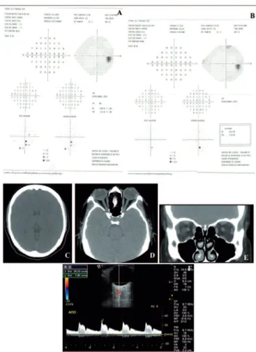
The 24:2 WW and SITA-SWAP (Humphrey Systems, San Leandro, Calif.) automated perimetry of the RE was within nor-mal limits (Figure 2). Perimetry was not performed in the LE due to loss of central fixation. Fluorescein angiography was nor-mal in the RE, and the LE showed a slight delay in venous filling in the region near the superior temporal arcade and dilation of perifoveal capillaries.
Normal chest radiograph. Computed tomography of the brain and orbits was within normal limits, as well as Doppler ultrasound of the eyeballs (Figure 2). Laboratory tests (thyroid hormones, rheumatologic tests, coagulation, serology for infectious diseases) showed no relevant findings.
Given the very high IOP values in the LE, we opted for medical treatment with hypotensive eyedrops (timolol maleate, brimonidine tartrate, dorzolamide hydrochloride and travoprost). The IOP was significantly reduced to 20mmHg, on average, as was the retinal venous dilation (Figure 1). However, due to the risk of glaucomatous damage, we decided to proceed with trabeculectomy.
Figure 1: External examination showed a normal conjunctiva in the right eye (A) and dilated episcleral veins in the left eye (B). Fundoscopy of the right eye (C) was normal, and the left eye (D) showed a pale optic disc with total excavation and discrete retinal venous engorgement. Fundoscopy under hypotensive eye drops showed no changes in the right eye (E) and decreased venous engorgement in the left eye (F)
48
D
ISCUSSIONArterio-venous fistula is the most frequent cause of ocular hyperaemia with elevated IOP, due to episcleral venous hypertension(8,9). The ocular hyperaemia results from dilated and
arterialized conjunctival vessels, which are different from those found in episcleral venous hypertension, which is characterised by dilated episcleral vessels(1).
Idiopathic episcleral venous hypertension is a relatively rare syndrome that progresses with ocular hyperaemia and elevated IOP. Although originally described by Minas and Podos(7), the condition is called the Radius-Maumenee Syndrome
in the German literature(8,10).
Jorgensen and Guthoff, while studying 64 patients with glaucoma due to episcleral venous hypertension, diagnosed this syndrome in 10 patients(8).
In another report of six patients with unilateral dilated episcleral veins and one patient with bilateral involvement, the IOP of affected eyes ranged from 21 to 44 mmHg(4). In the six
patients with unilateral involvement, the IOP was higher in those eyes with dilated conjunctival vessels. The episcleral venous pressure was measured and corresponded to twice the normal value in the affected eyes of these patients(4).
There is some variability in the clinical presentation of episcleral venous hypertension. The onset of signs (dilated episcleral vessels) may vary, usually occurring between the third and fourth decades of life(3). Dilated episcleral veins may occur
unilaterally or bilaterally. Of the 25 cases reviewed in the English literature, 13 involved predominantly the RE, 3 involved predominantly the LE, and 9 had bilateral involvement, although asymmetrical. The ratio of men to women was 13:12, with no apparent relation to age or gender(1,3-7).
Gonioscopy typically shows an open angle, and blood may or may not be visible in Schlemm’s canal. Of the 25 cases reviewed in the English literature, 24 had an open angle, of which 13 had blood in Schlemm’s canal(1,3-7).
The condition should be distinguished from other diseases, the most common of which is arteriovenous fistula, which can cause dilated episcleral vessels and elevated IOP. Although the cause of this disorder remains unclear, a congenital abnormality in the vasculature and familial predisposition have been considered as possible causes(5).
Episcleral venous hypertension is a diagnosis of exclusion that must be considered after intraorbital and intracranial conditions have been discarded. Diagnosis is based on the presence of dilated episcleral vessels, elevated IOP, and open angle on gonioscopy. Blood in Schlemm’s canal is a general sign of episcleral venous hypertension, but it is not always present, as was the case with our patient and other cases in the literature(3). The diagnostic evaluation should include a
com-plete eye examination and imaging. Some reports suggest performing magnetic resonance and even arteriography as part of the diagnostic investigation. However, in this report, we chose to perform a computed tomography of the head and orbits and a Doppler ultrasound; as these tests showed no abnormalities, more invasive and complex diagnostic tests were deemed unnecessary.
These imaging tests are important because they can exclude other conditions. Older reports mention the use of the MRI in a few cases, and although this is a more sensitive test than CT, it yielded no major findings, with most cases showing no abnormalities(3). Doppler ultrasound is a non-invasive method
for examining the orbital and ocular blood flow and, in patients with fistula, it typically shows retrograde blood flow and a dilated superior ophthalmic vein[1].
However, because this is a rare condition, it is difficult to estimate the sensitivity of such tests. The diagnosis of glaucoma secondary to idiopathic episcleral venous hypertension is clinical. Measuring episcleral venous pressure is unpractical, with no commercially-available instruments to this end(3).
The treatment of glaucoma due to idiopathic episcleral venous hypertension with elevated IOP is similar to primary open angle glaucoma(3). When medical treatment is not sufficient,
filtration surgery is the procedure of choice. One report mentions a patient who underwent trabeculectomy with subsequent reduction of the IOP, although the episcleral vessels remained dilated(6). Additionally, there are reports in the literature of
choroidal effusion after filtration surgery, showing that the procedure is not without risks or complications(3).
It is necessary to raise awareness among ophthalmologists about idiopathic episcleral venous hypertension, so that patients with dilated episcleral veins can be managed appropriately given the risks posed by the condition. In addition to medical treatment with eyedrops, such patients may require filtration surgery, and ophthalmologists should exercise caution in performing this procedure because of the risk of postoperative choroidal effusion.
Figure 2: Computer perimetry of the right eye (Humphrey, 24:2, white on white, SITA-FAST (A) and blue-yellow, SITA-SWAP (B)) was within normal limits. Computed tomography of the head (C) and orbits (D and E) was normal, and Doppler ultrasound of the eyes (F) showed symmetric ophthalmic arteries with normal flow velocities and spectral measurements, patent supraorbital veins with symmetrical flow, and no vascular dilation
Rev Bras Oftalmol. 2013; 72 (1): 46-9
49
R
EFERENCES1. Foroozan R, Buono LM, Savino PJ, Sergott RC. Idiopathic dilated epis-cleral veins and increased intraocular pressure. Br J Ophthalmol. 2003;87(5):652-4.
2. Weinreb RN, Karwatowski WS. Glaucoma associated with elevated episcleral venous p ressure. In: Ritch R, Shields MB, Krupin T, editors. The glaucomas. St. Louis: Mosby; c1996. p. 1143-55.
3. Rhee DJ, Gupta M, Moncavage MB, Moster ML, Moster MR. Idio-pathic elevated episcleral venous pressure and open-angle glaucoma. Br J Ophthalmol. 2009;93(2):231-4.
4. Talusan ED, Fishbein SL, Schwartz B. Increased pressure of dilated episcleral veins with open-angle glaucoma without exophthalmos. Ophthalmology. 1983;90(3):257-65.
5. Radius RL, Maumenee AE. Dilated episcleral vessels and open-angle glaucoma. Am J Ophthalmol. 1978;86(1):31-5.
6. Lanzl IM, Welge-Luessen U, Spaeth GL. Unilateral open-angle glau-coma secondary to idiopathic dilated episcleral veins. Am J Ophthalmol. 1996;121(5):587-9.
7. Minas TF, Podos SM. Familial glaucoma associated with elevated epis-cleral venous pressure. Arch Ophthalmol. 1968;80(2):202-8. 8. Jorgensen JS, Guthoff R. [The role of episcleral venous pressure in the
development of secondary glaucomas]. Klin Monbl Augenheilkd.1988;193(5):471-5. German.
9. Henderson JW, Schneider RC. The ocular findings in carotid-cavern-ous fistula in a series of 17 cases. Am J Ophthalmol. 1959;48:585-97. 10. Groh MJ, Küchle M. [Idiopathic episcleral venous stasis with second-ary open-angle glaucoma (Radius-Maumenee syndrome)]. Klin Monbl Augenheilkd. 1997;211(2):131-2. German.