Behavioral and physiological methods
for early quantitative assessment of
spinal cord
injury and prognosis in rats
1Escola de Engenharia de São Carlos, Universidade de São Paulo, São Carlos, SP, Brasil
2Departamento de Morfologia, Estomatologia e Fisiologia, Faculdade de Odontologia de Ribeirão Preto,
3Departamento de Biomecânica, Medicina e Reabilitação do Aparelho
Locomotor, Faculdade de Medicina de Ribeirão Preto, Universidade de São Paulo, Ribeirão Preto, SP, Brasil C.A. Giglio1,
H.L.A. Defino1,3, C.A. da-Silva2, A.S. de-Souza2 and E.A. Del Bel2
Abstract
Methods for reliable evaluation of spinal cord (SC) injury in rats at short periods (2 and 24 h) after lesion were tested to characterize the mechanisms implicated in primary SC damage. We measured the physiological changes occurring after several procedures for produc-ing SC injury, with particular emphasis on sensorimotor functions. Segmental and suprasegmental reflexes were tested in 39 male Wistar rats weighing 250-300 g divided into three control groups that were subjected to a) anesthesia, b) dissection of soft prevertebral tissue, and c) laminectomy of the vertebral segments between T10 and L1. In the
lesion group the SC was completely transected, hemisected or sub-jected to vertebral compression. All animals were evaluated 2 and 24 h after the experimental procedure by the hind limb motility index, Bohlman motor score, open-field, hot-plate, tail flick, and paw com-pression tests. The locomotion scale proved to be less sensitive than the sensorimotor tests. A reduction in exploratory movements was detected in the animals 24 h after the procedures. The hot-plate was the most sensitive test for detecting sensorimotor deficiencies following light, moderate or severe SC injury. The most sensitive and simplest test of reflex function was the hot-plate. The hemisection model promoted reproducible moderate SC injury which allowed us to quantify the resulting behavior and analyze the evolution of the lesion and its consequences during the first 24 h after injury. We conclude that hemisection permitted the quantitation of behavioral responses for evaluation of the development of deficits after lesions. Hind limb evaluation scores and spontaneous exploration events provided a sensitive index of immediate injury effects after SC lesion at 2 and 24 h. Taken together, locomotion scales, open-field, and hot-plate tests represent reproducible, quantitatively sensitive methods for detecting functional deficiencies within short periods of time, indicating their potential for the study of cellular mechanisms of primary injury and repair after traumatic SC injury.
Correspondence
E.A. Del Bel
Departamento de Morfologia, Estomatologia e Fisiologia FORP, USP
Av. do Café, s/n
14049-904 Ribeirão Preto, SP Brasil
Fax: +55-16-3633-0999 E-mail: [email protected]
Part of a Master’s thesis presented by C.A. Giglio to the Faculdade de Medicina de Ribeirao Preto, and Escola de Engenharia de São Carlos, IQSC, USP.
Publication supported by FAPESP, CNPq and CAPES.
Received October 30, 2005 Accepted September 26, 2006
Key words
•Trauma
•Spinal cord injury •Experimental models •Sensorial test
Introduction
Spinal cord (SC) injury causes serious consequences that can persist for an entire life span (1,2). One objective of SC injury research is to understand the post-lesion events so as to minimize injury or perhaps devise strategies for repairing damage. It is known that the immature mammalian cen-tral nervous system can repair itself after injury, but only up to a particular stage of development after which the capacity for repair is lost (2,3). In contrast, in adults peripheral nerves regenerate successfully with recovery of function after injury (4,5). Mechanical force applied to the SC causes primary damage to neural tissue. Over a pe-riod of minutes to hours the affected area spreads through complex cascades that give rise to secondary damage (2,4,5). The acute post-injury phase is critically important for recovery (4,6). The injured nerve cells re-spond with an injury-induced barrage of ac-tion potentials. Accompanying this are signifi-cant electrolytic shifts, principally involving monovalent (Na+, K+) and divalent cations
(Ca2+) (1,2,4). Increases in intracellular Ca2+
to toxic levels contribute to failure of normal neural functions. Spinal shock, which lasts for about 24 h in rats, represents a generalized failure of circuitry in the spinal neural network (2,4,7). Apart from the direct effects on neural tissues, hemorrhage, localized edema, throm-bosis, vasospasm, and loss of vasculature au-toregulation all contribute to the damage, in addition to the compression of the SC that follows vertebral displacement and edema (7-9). In the best circumstances the time to admis-sion after SC injury is usually not less than 3 h (1,2). Hence there is little information about the immediate effects of acute injury. The extent of our understanding is reflected in the limited neuroprotective strategies currently available beyond rapid trauma resuscitation and attentive clinical care (1,2,4).
At present there is no simple or well-de-fined procedure available for complete
char-acterization of the severity of SC injury. One experimental procedure is contusion of the SC (6,7), which mimics the situation in humans. However, with this approach it is not possible to interrupt specific and well-defined spinal tracts in the same way as done by sectioning at specific locations (5). Other less clinically relevant models consist of compression (8,9), transection (10) or hemisection (11). Transec-tion has the distinct advantage of consistent reproducibility. Hemisection permits to com-pare cellular responses ipsi- and contra-lateral to the injury. An extensive review of experi-mental studies postulates that permanent paraplegia induced in rats represents an appro-priate experimental model. Therefore, each lesion mimics a particular aspect of the dam-age to the SC that occur in humans (12,13).
The development of new animal models should provide strategies to help patients with SC injuries to be evaluated in terms of residual motor and sensory function. Ac-cordingly, we have quantitatively investi-gated differences in behavior following SC injury within 2 and 24 h after lesioning in order to characterize the mechanisms impli-cated in primary SC damage.
Material and Methods
Animals
Thirty-nine male Wistar rats weighing 250-300 g were kept in a temperature-con-trolled room (23ºC) with a 12-h light/dark cycle (lights on at 7:00 am) with free access to water and food. The experiments were carried out according to the guidelines of the Brazilian Society of Neuroscience and Be-havior for the care and use of laboratory animals. All efforts were made to minimize animal suffering. Experiments were per-formed between 8:00 and 12:00 am.
Surgical preparation
under aseptic conditions. Animals were anes-thetized by intraperitoneal injection of 250 mg/kg 2,2,2-tribromoethanol (Aldrich Chem-ical Company Inc., Milwaukee, WI, USA). After shaving the skin, animals were kept in a David Kopf stereotaxic apparatus and an incision was made in the skin and underly-ing muscles of the mid-thoracic region (14). Muscles were retracted and a mid-thoracic laminectomy was performed (vertebral seg-ments T10-L1). When required, the dura mater
was exposed. The animals were divided into three control groups and three test groups and then submitted to different types of pro-cedures.
Spinal cord injury
Control groups. Anesthesia. Control un-operated rats (N = 4) were anesthetized for the same periods of time as needed for the surgical procedures.
Removal of soft tissue. A dorsal incision was made in the skin and underlying muscles of anesthetized rats (N = 9). Muscles were retracted (T10-L1) and muscular tissue was
removed to expose the vertebral bone plate which was left intact.
Laminectomy. The vertebra was ap-proached as described above and vertebral bone of the T10-L1 segments (laminectomy)
was removed bilaterally to expose the dura mater, which was left intact (N = 12).
Test groups. Complete SC section. After laminectomy, the dura mater was opened and a sharp tiny blade (0.2 mm) was posi-tioned perpendicularly to the tissue, touch-ing the inferior side of the bone plate. A complete cut of the cord was made with the blade running completely from left to right, twice (N = 6).
Hemisection of the right side of the SC. Anesthetized animals (N = 4) were submit-ted to a procedure similar to that described above. To produce this lesion, the blade (0.2 mm) was positioned perpendicularly to the tissue, touching the inferior side of the bone
plate. A cut of the cord was made with the blade running carefully from the right side to the midline of the cord.
Vertebral compression.Based on the method of Gruner et al. (9), the tips of a hemostat forceps were inserted between the spine and the sur-rounding muscle of each rat (N = 4). The spine was compressed between the T10-L1 segments
by slowly closing the arms of the forceps and holding them closed for 15 s.
The injury sites were irrigated with sa-line and the incisions were closed in layers using silk sutures (4-0 thread, Ethicon, São Jose dos Campos, SP, Brazil). After receiv-ing penicillin subcutaneously, the animals were placed in a warming chamber in indi-vidual boxes and body temperature was main-tained at 37ºC until they were fully awake.
After the end of the behavioral tests (2 and 24 h after surgery), rats were sacrificed with a lethal dose of anesthetic. The lesion site (T10-L1) was confirmed by macroscopic
and microscopic observation.
Behavioral tests of functional deficits
Behavioral data were collected from the rats after dorsal hemisection, complete SC section and vertebral compression or control procedures (anesthesia, removal of tissue and laminectomy groups). We used motor and sensory tasks and quantitative evalua-tion according to defined scaling systems. The animals were evaluated 2 and 24 h after each procedure. The battery of experimental behavioral tests was applied once to each animal in the same order and consisted of: l) hind limb motility index, 2) Bohlman motor evaluation score, 3) open-field, 4) hot-plate, 5) tail flick, and 6) paw compression tests. A varying number of animals per test were analyzed since data were collected from con-trol animals (laminectomy) in several ex-periments.
abil-ity of the hind limbs to move over a period of 2 min (8). Animals were placed on a smooth flat surface and allowed to walk freely. The following scores were applied: 0 = normal moving ability of the hind limbs, 1 = paresis or decreased moving ability of the hind limbs, 2 = plegia or blocked moving ability of the hind limbs.
Bohlman motor evaluation score. A com-monly used instrument is the Tarlov scale (15) which ranks hind limb movements and weight support. The Tarlov score was modi-fied by Bohlman (16). Animals were evalu-ated and assigned to one of the five catego-ries: grade 0, complete paraplegia without movements; grade 1, small articulate move-ments; grade 2, large movemove-ments; grade 3, standing on their feet; grade 4, walking; grade 5, walking on a plane with a 20-degree inclination. Scores 0-4 were attributed after observing the rats on a smooth flat surface for 4 min. Orientation of the body in space was measured by using a 20º inclined board (1 m long, 0.5 m wide and 0.02 m width) covered with rubber. Both the time to orient the body to face upward and the motor pat-terns used to turn the body around were recorded. The rat was placed with all paws on the board, with its body and nose facing down (10). No obvious signs of pain or stress were observed in any group.
Open-field test. To assess exploratory be-havior (17,18), rats were tested in an open-field consisting of a circular wooden box (72 cm in diameter) surrounded by 49-cm high transparent walls with an open top. The floor was divided into twelve equal size fields. Be-havioral activity was measured for periods of 4 min at the beginning of the dark cycle (10:00 am to 11:30 pm) at 2 and 24 h after surgery. Each activity chamber was cleaned with alco-hol between tests to eliminate urine and olfac-tory cues from previous subjects.
Each rat was gently placed in the center of the open-field and hand-operated counters and stopwatches were used to score behaviors. Four different behavioral measures of
sponta-neous activity were determined: locomotion or exploratory movement, measured as the number of floor units entered with the four paws; rearing frequency, as the number of times the animal stood on its hind limbs; groom-ing or stationary movement frequency, and defecation. The four parameters of spontane-ous behaviors were empirically found to be best for measurements of individual and group behaviors. The number of rearing events is important for analyzing the effect of laminec-tomy alone (sham) versus spinal injury. Ex-ploratory measurements are necessary since stationary activity may not reflect the animal’s exploratory behavior in the field in an accurate manner. We evaluated changes in each pa-rameter individually. A white noise generator provided a constant background noise.
Hot-plate test. The animals were placed on a hot-plate maintained at 52 ± 0.5ºC. The thermal nociceptive threshold was defined as the time required to elicit a hind paw lick or a jump (14). The cut-off time was 20 s.
The force at which a rat withdrew its hind paw or struggled was multiplied by 10, as recom-mended by the manufacturer, and recorded as the withdrawal force (g).
Statistical analysis
Well-established scoring methods used for human neurological examinations were used to evaluate and classify symptoms. The non-parametric Kruskal-Wallis test was used for the hind leg motility index and Bohlman motor evaluation score and multivariate anal-ysis of variance (MANOVA) was used for the open-field, hot-plate, tail flick, and paw compression tests. The items analyzed were procedure, time and procedure x time inter-action. In cases of significance of the proce-dure-time interaction, ANOVA was em-ployed, followed by the Duncan post hoc test. The level of significance was set at P < 0.05 for all tests.
Results
Hind limb motility index
The median scores attributed to the ani-mals of each group are shown in Table 1.
The animals belonging to the anesthesia, laminectomy, removed soft tissue, complete section, hemisection, and vertebral compres-sion groups receiveda single score for each group, suggesting homogeneity of the ex-perimental procedure and effects. A score of two for the complete section and hemisec-tion groups denotes paralysis (2 h: χ2 =
66.00, d.f. = 6, P < 0.001; 24 h: χ2 = 61.99,
d.f. = 6, P < 0.001).
Bohlman score
Table 1 also shows the results obtained for the inclined plane and their Bohlman scores. All animals in the anesthesia, re-moved soft tissue and vertebral compression groups could walk 2 or 24 h after treatment. Nine of 12 animals from the laminectomy group were able to walk, but their ability was lower for the 24-h period than for the 2-h period. Animals from the complete section and hemisection groups lost the ability to move down on the inclined plane. There-fore, the results of the inclined plane test differed significantly between the groups anesthesia, removed soft tissue, laminec-tomy, and compression and the groups com-plete section and hemisection (2 h: χ2 =
Table 1. Motility index for fore- and hind legs of rats after spinal cord lesion.
Test Anesthesia Removed soft Laminectomy Complete Hemisection Compression (N = 4) tissue (N = 9) (N = 12) section (N = 6) (N = 4) (N = 4)
Bohlman score
2 h 5 5 5 0 0 5
24 h 5 5 4 0 0 5
Inclined plane
2 h + + + - - +
24 h + + - - - +
Hind limb motility index
2 h 0 0 1 2 2 0
24 h 0 0 1 2 2 0
33.00, d.f. = 6, P = 0.005; 24 h: χ2 = 25.32,
d.f. = 6, P < 0.001).
The animals in the anesthesia, removed soft tissue, and vertebral compression groups scored 5, a normal Bohlman score, whereas the animals in the laminectomy group showed partial motor losses. Those in the complete section and hemisection groups had total loss of motor functions, with the lowest Bohlman score (2 h: χ2 = 21.46, d.f. = 6, P =
0.005; 24 h: χ2 = 22.28, d.f. = 6, P = 0.001).
No signs of pain were seen in any group.
Open-field test
Quantitative march analysis (ambulation) for the evaluation of locomotion ability is shown in Figure 1.
The laminectomy, complete section, he-misection, and vertebral compression groups caused a significantly reduced numbers of floor units to be crossed when compared with the anesthesia and removed soft tissue groups at both 2 and 24 h after surgery (ANOVA; 2 h: F6,26 = 6.68, P < 0.001, 24 h:
F6,26 = 13.21, P < 0.001). In assays
per-formed 2 h after surgery, there was a similar decrease in the number of quadrants crossed by the animals in the laminectomy, complete section, hemisection, and vertebral compres-sion groups compared with the anesthesia and removed soft tissue groups. For the 24-h pe-riod, the anesthesia and removed soft tissue groups crossed a larger number of quadrants compared with other groups. The anesthesia group also crossed a larger number of quad-rants than the removed soft tissue group.
The number of rearing events was de-creased in most experimental groups com-pared with the anesthesia group. There was a significant general effect of lesion (F6,26 =
9.32, P = 0.001) and an interaction between lesion and time (F6,26 = 10.93, P < 0.001).
This decrease was statistically significant by ANOVA (lesion, 2 h: F6,26 = 9.32, P < 0.001;
24 h: F6,26 = 5.66, P = 0.001) for the removed
soft tissue (2 h only), laminectomy, plete section, hemisection, and vertebral com-pression groups compared with the anesthe-sia group.
A decrease in the number of grooming events was observed in most experimental groups compared with the anesthesia and removed soft tissue groups. There was a significant effect of lesion (F6,26 = 5.47, P =
0.001) but not of time (F6,26 = 1.07, P = 0.31).
No interaction between lesion and time (F6,26
Figure 1. Effects of spinal cord lesion on exploratory behavior of rats tested in an open-field. Ambulation, rearing, grooming, and defecation were recorded for 5 min and analyzed 2 h (open bars) and 24 h (hatched bars) after treatment. Data are reported as the means ± SEM. *P < 0.05 compared to anesthesia (control); **P < 0.05 compared to removed soft tissue (RST); +P < 0.05 compared to the anesthesia (control), RST and laminectomy groups
= 1.74, P = 0.15) was found. This decrease was statistically significant by ANOVA (le-sion, 2 h: F6,26 = 3.24, P = 0.005; 24 h: F6,26 =
5.83, P < 0.001) for the laminectomy, plete section, hemisection, and vertebral com-pression groups compared with the anesthe-sia and removed soft tissue groups.
No significant effect on defecation was detected in any experimental group (lesion: F6,26 = 1.30, P = 0.29; time: F6,26 = 0.06, P =
0.80; lesion x time: F6,26 = 1.96, P = 0.11).
Tail flick test
The results of this assay, used to measure thermal sensitivity by medullar reflexes, are shown in Figure 2. One-way ANOVA of the mean values revealed a significant effect of lesion (F6,26 = 9.8, P < 0.001) but not of time
(F6,26 = 3.48, P = 0.073). No interaction
between the lesion and time (F6,26 = 0.23, P =
0.96) was found. Post hoc analysis showed that the animals subjected to anesthesia, re-moved soft tissue, laminectomy, and verte-bral compression had significantly increased tail flick reactions compared to the complete section group at 2 h (F6,26 = 7.32, P < 0.001).
At 24 h, the total section group had a longer lag time (F6,26 = 6.32; P < 0.001), as did the
hemisection group. Thus, the tail flick tests revealed a decreased ability of the total sec-tion and hemisecsec-tion groups to respond to the thermal stimuli.
Hot-plate test
The results of the hot-plate test used to evaluate medullar and brain reflexes are also shown in Figure 2. Statistical analysis indi-cated significant effects of the different pro-cedures (lesion: F6,26 = 7.38, P < 0.001).
There was no time dependence (F6,26 = 1.88,
P = 0.31) and only marginal significance of the interaction between the type of injury and elapsed time (F6,26 = 2.20, P = 0.075).
After 2 h, only the total section and vertebral compression groups differed from the
anes-Figure 2. Effects of spinal cord lesion on sensitivity to pain tested by the tail flick latency and hot-plate tests. Measurements were made 2 h (open bars) and 24 h (hatched bars) after treatment. Results are reported as average tail flick or hind paw lick or jump latency in seconds. Data are reported as the means ± SEM. *P < 0.05 compared to anesthesia; **P < 0.05 compared to removed soft tissue (RST); +P < 0.05 compared to the anesthesia, RST
and laminectomy groups (ANOVA followed by the Duncan test for all comparisons). For number of rats in each group, see Table 1.
thesia and removed soft tissue groups (F6,26 =
2.68, P < 0.005). However, 24 h after sur-gery, all groups differed from the anesthesia and removed soft tissue groups (F6,26 = 23.76,
P < 0.001). Accordingly, the hot-plate tests showed function defects in all groups when compared with controls.
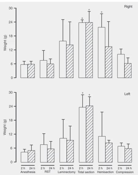
Paw compression test
re-sponses to a stimulus of progressive pres-sure applied to the 3rd interdigital space of the left paw contralateral to the medullar hemisection.
A significant effect was observed on the right paw (F6,26 = 14.49, P < 0.001), with a
significant effect for the factor time (F6,26 =
5.48, P = 0.027). Only marginal significance of time x lesion interaction (F6,26 = 2.11, P =
0.086) was observed. The complete section and hemisection groups (the former at time 2 h) showed a significantly longer response
time to the pain stimulus applied to the right paw ipsilateral to the medullar injury (2 h: F6,26 = 14.43, P < 0.001; 24 h: F6,26 = 9.97, P
< 0.001).
A significant effect was observed on the left paw (F6,26 = 3.45, P = 0.005); no
signifi-cant effect of time (F6,26 = 0.79, P = 0.38) or
time x lesion interaction (F6,26 = 0.64, P =
0.69) was observed. The complete section group showed a longer response time to the pain stimulus applied to the left paw (2 and 24 h: F6,26 = 1.05, P = 0.012).
Discussion
The aim of the present study was to try to identify an SC injury process that could be useful for analyzing mechanisms of initial SC damage. The criteria we sought were reproducibility of changes in well-es-tablished sensory and motor tests 2 and 24 h after lesion. Our results show that motor scores were less sensitive than sensorimotor tests. A decreased ability to explore was detected in the animals 2 and 24 h after the procedures, but the hot-plate was the most sensitive test for detecting sensorimotor de-ficiencies following light, moderate or se-vere SC lesions. Laminectomy seemed to be a more drastic injury than vertebral com-pression. Hemisection induced medium to moderate injury and made it possible to quan-tify behavior and thereby to evaluate the consequences of the lesions. The results show that the locomotor scores and the open-field and hot-plate tests were reproducible, quan-titative and sensitive tests for detecting functional deficiencies 2 and 24 h after SC lesion. The primary mechanisms implicated in SC damage may be investigated quanti-tatively at short times after injury (2 and 24 h).
Considerations on the behavioral tests
The evaluation scales used in the present study detected significant deficiencies in the
Figure 3. Effects of spinal cord lesion on paw compression test on left and right paws. Measurements were made 2 h (open bars) and 24 h (hatched bars) after treatment. Results are reported as weight in g. Data are reported as the means ± SEM. +P < 0.05 compared to
severe SC injury group. The Bohlman test score was 5 for all groups other than the hemisection and complete section groups and the test did not discriminate between lesion effects at 2 or 24 h despite the lami-nectomy procedure. The removed soft tissue and laminectomy groups presented score variations among animals of the same group and, in addition, between the median values corresponding to 2 and 24 h. Hind limb motility scores were consistent for animals belonging to the anesthesia, complete sec-tion, hemisecsec-tion, and vertebral compres-sion groups, suggesting that the experimen-tal procedures caused similar damage. This index indicated that laminectomy was more injurious to the animals than vertebral com-pression, suggesting sensitivity for the de-tection of light or moderate lesions.
The open-field test was sensitive to behav-ioral modifications and provided motor activ-ity indexes for injured animals. This is a rela-tively simple, reproducible and quantitative test with enough sensitivity to detect func-tional deficiencies even in cases of light SC injury. Medium and severe SC injury caused a general reduction of exploratory activity after treatment. After 24 h, the anesthesia and re-moved soft tissue groups presented an in-crease in the number of quadrants crossed. Interestingly, mild lesion induced by remov-ing soft tissue, laminectomy, and vertebral compression, caused a greater decrease of rearing events 24 h after the procedure. There-fore, exploratory activity and rearing and grooming events were sensitive parameters for the detection of sensorimotor deficiencies and weaker responses 2 and 24 h after injury. As a general effect, SC injury reduced spontaneous locomotor activity as measured in an open-field test. These results are in agreement with previous data reported by Basso et al. (21), Mills et al. (17), and Metz et al. (18). However, these investigators evaluated rats between 5 and 10 weeks after lesion. In addition, Metz et al. (18) reported that animals with low locomotion capacity
presented a closer relationship between the 21-point open-field locomotion score (BBB scale) (20,21) and activity in the open-field (18,19,21). In addition, differences in ex-ploratory behavior may reflect pain episodes (18). Mills et al. (17) showed that control animals on postsurgical 14, 28, and 60 days presented a reduction in exploratory activ-ity, grooming and rearing events; in con-trast, rats with SC injury demonstrated a decrease in the same parameters. These in-vestigators suggested that spontaneous be-haviors may be used to evaluate pain imme-diately after SC injury (18-21).
The hot-plate test was the most sensitive test of reflex function for the detection of sensorimotor deficiencies after injury. This test is also simple, reproducible and quanti-tative. The tail flick test showed similar sen-sitivity to the locomotion scales used in the present study (hind limb motility index, Bohlman score). The tail flick test reflects spinally organized reflexive responses, whereas the hot-plate test is thought to re-flect supraspinal nociceptive processing. The paw compression test can be efficiently used in the hemisection model.
Thus, hind limb evaluation scores and spon-taneous exploration events provided a sensi-tive index of the immediate effects of SC injury at 2 and 24 h. The spontaneous locomo-tion, rearing and grooming events were the most sensitive parameters to detect sensorimo-tor deficiencies. The most sensitive test of reflex function was the hot-plate, which is also simple. In addition, the tail flick test had simi-lar sensitivity to the Bohlman score and the paw compression test can be efficiently used in the hemisection lesion.
Experimental spinal cord lesion
behav-ior and analyze the evolution of the lesion and its consequences during the first 24 h after injury. Criticism of the hemisection and complete section models is based on the fact that, in addition to removal of the bone layer, the meninges are also cut (4,12). In injury caused by crushing or contusion, al-though the bone layer is removed, the me-ninges are better preserved (10). Kwon et al. (22) suggested that sharp SC injury in which the cord is completely or partially transected is useful for assessing axonal regeneration anatomically. In contrast, blunt injury mod-els in which the cord is compressed or con-tused more accurately mimic the typical hu-man injury and provide a good setting for the study of secondary pathophysiologic pro-cesses immediately after injury. Our results showed that injury produced by vertebral compression was heterogeneous.
There is evidence that experimental SC injury in rats produces effects similar to those occurring in humans (13). In most patients traumatic injuries do not involve physical transection of the cord, but rather the SC is damaged by contusion, compres-sion or stretch (13,23). The lecompres-sion zone usu-ally presented bleeding and damaged tissue (1,2,11,12). Typically, residual white matter containing portions of the ascending sen-sory and motor tracts remains intact, allow-ing the possibility of recovery. However, during the first minutes and hours following injury, a secondary degenerative process is initiated by the primary mechanical injury that is proportional to the magnitude of the initial injury insult (2,4,11,24). Neverthe-less, the initial anatomical continuity of the
injured SC in most cases, together with the present knowledge of many of the factors involved in the injury process, have led to the notion that treatments which interrupt the secondary cascade, if applied early, could improve SC tissue survival, and thus pre-serve the necessary anatomic substrate for functional recovery to take place. Promising research (17,21,22,25,26) is being carried out to delineate the aspects that may be amenable to pharmacologic intervention. Few agents such as methylprednisolone (1, 12,23) have been subjected to large-scale human trials.
Considering the differences observed between sensitive and motor parameters and section and compression models, structural evaluation and/or biochemical assays could be helpful to explain these results. The ex-tent of our understanding of immediate events after SC lesion may expand the neuroprotec-tive strategies currently available.
The hemisection model was the one that permitted us to quantify the resulting behav-ior and to analyze the evolution of the lesion and its consequences during the first 24 h.
Acknowledgments
The authors are indebted to S. Saltareli and E.C. Zieri for skillful technical assis-tance, and to Dr. M.A. Oliveira, and Profes-sor F.S. Guimarães for the statistical analy-sis. We also thank Professor J.G. Nicholls (Neurobiology, Scuola Internazionale Su-periore di Studi Avanzati, Trieste, Italy) for manuscript discussion and suggestions.
References
1. Dobkin BH, Havton LA. Basic advances and new avenues in thera-py of spinal cord injury. Annu Rev Med 2004; 55: 255-282. 2. Hulsebosch CE. Recent advances in pathophysiology and
treat-ment of spinal cord injury. Adv Physiol Educ 2002; 26: 238-255. 3. Nicholls J, Saunders N. Regeneration of immature mammalian
spi-nal cord after injury. Trends Neurosci 1996; 19: 229-234.
4. Tator CH, Fehlings MG. Review of the secondary injury theory of acute spinal cord trauma with emphasis on vascular mechanisms. J
Neurosurg 1991; 75: 15-26.
morphomet-ric analyses of alterations in the spinal cord. Exp Neurol 1985; 88: 135-149.
7. Beattie MS, Bresnahan JC, Komon J, Tovar CA, Van Meter M, Anderson DK, et al. Endogenous repair after spinal cord contusion injuries in the rat. Exp Neurol 1997; 148: 453-463.
8. Rivlin AS, Tator CH. Effect of duration of acute spinal cord compres-sion in a new acute cord injury model in the rat. Surg Neurol 1978; 10: 38-43.
9. Gruner JA, Yee AK, Blight AR. Histological and functional evaluation of experimental spinal cord injury: evidence of a stepwise response to graded compression. Brain Res 1996; 729: 90-101.
10. Noble LJ, Ellison JA. Effect of transection on the blood-spinal cord barrier of the rat after isolation from descending sources. Brain Res 1989; 487: 299-310.
11. Dusart I, Schwab ME. Secondary cell death and the inflammatory reaction after dorsal hemisection of the rat spinal cord. Eur J
Neurosci 1994; 6: 712-724.
12. Basso DM. Behavioral testing after spinal cord injury: congruities, complexities, and controversies. J Neurotrauma 2004; 21: 395-404. 13. Rosenzweig ES, McDonald JW. Rodent models for treatment of spinal cord injury: research trends and progress toward useful re-pair. Curr Opin Neurol 2004; 17: 121-131.
14. Del-Bel EA, Borges CA, Defino HL, Guimaraes FS. Induction of Fos protein immunoreactivity by spinal cord contusion. Braz J Med Biol Res 2000; 33: 521-528.
15. Tarlov IM, Klinger H, Vitale S. Spinal cord compression studies.
AMA Arch Neurol Psychiatry 1953; 71: 271-290.
16. Bohlman HH, Bahniuk E, Field G, Raskulinecz G. Spinal cord moni-toring of experimental incomplete cervical spinal cord injury: a pre-liminary report. Spine 1981; 6: 428-436.
17. Mills CD, Grady JJ, Hulsebosch CE. Changes in exploratory behav-ior as a measure of chronic central pain following spinal cord injury.
J Neurotrauma 2001; 18: 1091-1105.
18. Metz GA, Merkler D, Dietz V, Schwab ME, Fouad K. Efficient testing of motor function in spinal cord injured rats. Brain Res 2000; 883: 165-177.
19. Randall LO, Selitto JJ. A method for measurement of analgesic activity on inflamed tissue. Arch Int Pharmacodyn Ther 1957; 111: 409-419.
20. Basso DM, Beattie MS, Bresnahan JC. Graded histological and locomotor outcomes after spinal cord contusion using the NYU weight-drop device versus transection. Exp Neurol 1996; 139: 244-256.
21. Basso DM, Beattie MS, Bresnahan JC, Anderson DK, Faden AI, Gruner JA, et al. MASCIS evaluation of open-field locomotor scores: effects of experience and teamwork on reliability. Multicenter Animal Spinal Cord Injury Study. J Neurotrauma 1996; 13: 343-359. 22. Kwon BK, Oxland TR, Tetzlaff W. Animal models used in spinal cord
regeneration research. Spine 2002; 27: 1504-1510.
23. Bracken MB, Shepard MJ, Collins WF, Holford TR, Young W, Baskin DS, et al. A randomized, controlled trial of methylprednisolone or naloxone in the treatment of acute spinal-cord injury. Results of the Second National Acute Spinal Cord Injury Study. N Engl J Med 1990; 322: 1405-1411.
24. Kakulas BA. The clinical neuropathology of spinal cord injury. A guide to the future. Paraplegia 1987; 25: 212-216.
25. Wintzer M, Mladinic M, Lazarevic D, Casseler C, Cattaneo A, Nicholls J. Strategies for identifying genes that play a role in spinal cord regeneration. J Anat 2004; 204: 3-11.