vol. 40, n. 1, jan./mar., 2004
Cyclophosphamide identification in wipe test
by GC-MS and solid phase
extraction
Isarita Martins
1*, Henrique Vicente Della Rosa, Pietro Apostoli
21Department of Clinical and Toxicological Analysis, Faculty of Pharmaceutical Sciences, University of San Paolo, 2Facoltà di Medicina e Chirurgia, Università degli Studi di Brescia
In this study cyclophosphamide was quantified after adapting a prior analytical method using gas chromatography-mass spectrometry after solid phase purification and derivatization with trifluoroacetic anhydride. The analyte was measured by analysis in wipe test from infusion bags, which may be contaminated by contact with the gloves used during preparation of the drugs. Surface of bag contaminated may be an important source of contamination for workers in the others chemoterapy handling areas, such as administration rooms. This drug, in fact, is one of the most frequently used alkylating antineoplastic agents for different types of tumors and it is furthermore classified as a human carcinogen by IARC. Ifosfamide was used as internal standard and the quantification was carried out by reference to calibration curves within a range from 1 to 100 ng/mL. The limit of detection was 0.4 ng/mL. The values of the variation coefficient varied from 0.5 to 10% (intra-assay) and from 0 to 19% (interassay). Frozen reference wipe samples containing cyclophosphamide were analysed over one month and no significant loss was observed. The range obtained for bias assay was 83-116% and the recovery was 98.9%. Cyclophosphamide was measured in 36 of 42 infusion bags collected from different hospitals with values ranging from 90 to 41874 ng (median= 607.5 ng). The results, well related to those reported in the literature, suggest that this method can be used to identify cyclophosphamide from wipe samples and can be considered useful in exposure assessment to this drug.
*Correspondence:
I. Martins
Departamento de Análises Clínicas e Toxicológicas
Faculdade de Ciências Farmacêuticas -USP
Av. Prof. Lineu Prestes, 580 - Bloco 13B - Cidade Universitária 05508-900 - São Paulo - SP - Brasil
Uniterms
• Cyclophosphamide • Wipe test
• Exposure assessment
INTRODUCTION
The possibility of occupational hazards due to exposure to chemotherapeutic and antineoplastic agents
chemotherapeutic agents might also be responsible for teratogenic effects and potentially carcinogenic, for the workers, is quite recent.
The overall evidence, combining the results from a variety of studies and bioassays, of carcinogenic risk to humans has been evaluated to be conclusive for at least ten cytostatic drugs, cyclophosphamide being one of them (Sorsa, Anderson, 1996).
As a genotoxic compound, it is not possible to establish a dose-effect relationship, although exposure is considered inappropriate even in small doses.
At the occupational level, the main contamination route is dermal contact, which may occur by prolonged contact with either contaminated surfaces or instruments (Sessink, Bos, 1999). Glove analysis might be helpful in the evaluation of dermal exposure of workers to antineoplastic drugs. Another possible source of contamination is the inhalation of aerosols dispersed throughout the working place (Alessio et al., 1996; Sorsa, Anderson, 1999; Sessink, Bos, 1999).
The opportunities for exposure are easily identified. During the preparation procedure of some drugs, some steps are particularly hazardous, such as the opening of the glass vial, removal of the needle from the vial (which may produce aerosol formation), bleeding of air from the syringe, transfer of the drug from one vial to another by means of a syringe or a needle, and inappropriate discarding of a partially used vial. In the administration procedure, exposure may occur during the bleeding from the syringe, by aerosol formation or by direct skin contact. In routine hospital practice, the contact with biological material or undergarments from patients treated with these drugs represents another possible source of exposure (Sessink, 1994; Sessink, Bos, 1999).
Some national and international agencies concerned with labor safety have published documents providing precise directives aimed at reducing exposure risks during manipulation of antineoplastic drugs. Commonly, these guidelines ask for periodical exposure evaluation, use of collective and individual protective devices, and establishment of sanitary surveillance and informative programs, in order to guarantee the lowest possible levels of exposure (Minoia, Perbellini, 2000).
Workers at risk are usually exposed to more than one antineoplastic chemotherapeutic agent; for this reason, it is necessary to determine which substances might be considered indicators of such exposure. These pharmacological drugs can be assessed in environmental and biological matrices according to the following hierarchic sequence: a) surface detection, called wipe test; b) detection in biological material and c) environmental
detection (Alessio et al., 1996). For instance, surface analysis of a working space often gives positive results while air samples collected from the same space might not show any contamination (Apostoli et al., 1996).
This work seeks to determine the concentration of cyclophosphamide (CP) in wipe test samples collected from the surfaces of infusion bags, using gas chromatography coupled to mass spectrometry, after solid phase extraction. Infusion bags may become contaminated by contact with gloves donned during the preparation of chemotherapeutic drugs and may constitute an important source of contamination for workers who administer such drugs.
MATERIAL AND METHODS
Study design
CP was chosen as tracing substance, since it is one of the most utilized antineoplastic drugs and also one of the most toxic ones (class 1 of IARC).
Forty-two infusion bags were collected from four different hospital centers. In order to screen for contamination, wipe test were collected from the outer surfaces of the bags. Samples were taken during the first and third weeks of CP usage, providing that at least 6 g of CP were used on the collection days. During the investigation, questionnaires about the characteristics of the work were answered.
For the wipe test sampling procedure, previously delimited surfaces were washed with 20 mL of 0.03 mol
L-1 NaOH and subsequently wiped with two sheets of
absorbent paper. The absorbent paper was then placed in an appropriate recipient and sent to the laboratory packed in dry ice, being kept at -20 °C until analysis.
Chemicals
CP (ACRÓS-USA; purity > 97%) and ifosfamide
(Holoxan®- ASTA- PHARMA- Italy; purity > 97%);
trifluoroacetic anhydride (SIGMA- USA; purity > 99%). The other chemicals were of the highest purity obtainable.
Analytical conditions
The analyses were performed with a 6890 GC coupled to a 5972-A MS (gas chromatography-mass
spectrometry-Hewlett Packard®), both controlled by a
Vectra XM series 3 5/9- Hewlett Packard® personal
thickness-Hewlett Packard® connected with a deactivated fused
silica retention gap (HP Retention Gap 5 m, 0.53 mm internal diameter- Hewlett Packard®). 1 µL of the cleaned
sample was injected in the on-column mode; the initial injector temperature was 80 °C. After 1 min, the injector temperature was increased at 75 °C/min to 300 °C, remaining constant. The initial oven temperature was 70 °C. After 1 min, the temperature was increased at 15 °C/min to 250 °C, where it remaining constant for 3 min and, after this, was increased at 30 °C/min to 300 °C, and held constant for 5 min. Helium (He) was used as carrier gas (column inlet pressure 82 kPa). The interface temperature was 310 °C.
Initially, CP and the internal standard identification was carried out in the SCAN mode, with full spectra (50 to 350 m/z) from the tenth to the fifteenth minute of the chromatographic run. From the full spectra, ions fragments 307 and 309 were selected for analysis in the SIM mode. Finally, the analyte was identified by the presence of ions 307 and 309 and by the relative retention time of N-trifluoroacetylated CP/ifosfamide.
Quantification was performed on the selected ion fragment 307 and the peak area ratio of N-trifluoroacetylated derivatives CP/ifosfamide was calculated. By reference was carried out a calibration cur-ve, ranging from 1 to 100 ng/mL of CP, treated as samples and injected before each set of samples.
Sample treatment
The following procedure is a modification of Sessink et al. (1992). The original technique utilized liquid-liquid extraction, whereas the present work used solid phase extraction with a modified form of a diatomaceous earth packed into polypropylene cartridge.
Extraction procedure
Thirty milliliters of 0.03 mol L-1 NaOH were added
to the sample flask, together with 20 mL of aqueous NaOH solution previously collected. After sonication for 90 min and shaking for 10 min, the sample was centrifuged at 3000 rpm for 10 min. To the supernatant (1 mL), 1 mL of TRIS buffer (0.2 mol/L; pH 4.5) and 20 µL of internal standard (ifosfamide 5 mg/mL) were added. One milliliter of this mixture was placed on the extraction column. After 5 min, it was eluted twice with 5 mL of ethyl acetate and the extract was dried in an evaporator centrifuge under vacuum. Ethyl acetate (50 µL) and trifluoroacetic anhydride (50 µL) were added and mixed. The vial was closed for derivatization at 70 ºC for 20 min. After cooling at room temperature, the sample was dried
with the evaporator centrifuge under vacuum, and 50 µL of toluene were then added. The sample was then sonicated for 3 min and stored at -20 ºC until analysis.
Validation of the method
The validation of the method was performed by establishing limits of detection and quantification, linearity, accuracy, recovery, precision and stability, acording to FDA, 1996 and FDA, 1998.
Statistical analysis
A Kruskall-Wallis test was performed to assess
significant differences between the levels of CP in
different hospital centers as mentioned above. For the statistical calculations the InStat software package was used.
RESULTS AND DISCUSSION
The analytical conditions were selected for measurement of CP surface contamination because they provided a suitable method. CP determination in environmental matrices, such as a wipe test collected from the surface of the infusion bags, was performed according to the method proposed by Sessink et al., in 1992, that employed gas chromatography coupled to mass spectrometry to identificate the analyte.
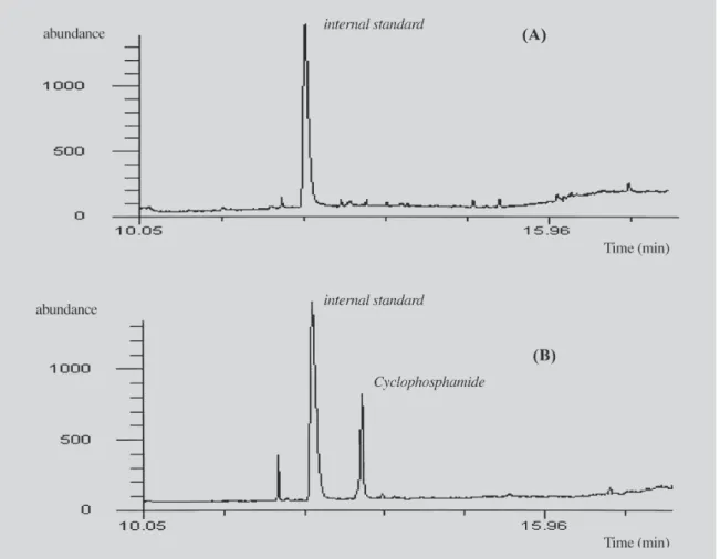
Most of the several methods for CP determination in wipe samples employ extraction procedures using liquid-liquid extraction with ethyl acetate (Sessink et al., 1992; Minoia et al., 1998; Connor et al., 1999; Schmaus et al., 2002). In the present work, the sample was extracted with a solid phase, using a diatomaceous earth column. The use of this column provided a clean extract (Figure 1). It furthermore reduces the occupational exposure of the analyst to the solvent ethyl acetate, an important characteristic when a method is in routine use. Evelo et al. (1986) utilized an amberlite XAD-2 column to substitute the liquid-liquid extraction of CP from complete 24 h uri-ne, concentrating on XAD-2 of only 10% of the urine. In this case, the method is more suitable as an exposure test for routine purposes.
and Schmaus et al. (2002). In the present study, the ion 309 had a response three times lower than ion 307. The use of both ions led to some loss of instrumental sensitivity, but it conferred increased certainty in identification.
The relative retention time obtained by this procedure (1.06) was another parameter used to identify the substance. Peaks of N-trifluoroacetylated CP and ifosfamide were completely separated, without interference within their respective retention times, as shown in Figure 1. Our study is characterized by a relatively simple and fast chromatographic run, permitting the analysis of a considerable number of samples within a work day.
The validation results (Table I) were considered satisfactory because they are comparable to previous studies. However, the interassay precision coefficient exceed 15%, when analysed at the limit of quatífication. Connor et al. (1999), using gas chromatography coupled
to a tandem mass spectrometer, obtained lower coefficients.
TABLE I - Confidence parameters of the proposed method for cyclophosphamide determination in wipe test by GC-MS and previous solid phase extraction
Parameter Result
limit of detection (LOD) 0.4 ng/ml
limit of quantification (LOQ) 1.0 ng/ml
dynamic range 1-100 ng/ml
correlation coefficient 0.9993
accuracy (bias) 83-116%
recovery 98.9%
precision (range of coefficient intra-assay: 0.5-10%
of variation) interassay: 0-19%
stability 30 days at -20 °C
The method accuracy varied from 116% for wipe test with a nominal concentration of 30 ng/mL of CP, to 83% for 100 ng/mL samples. Minoia et al. (1998) obtained an accuracy ranging from 98 to 109% for CP analysis in pads, analyzed by liquid chromatography coupled to a tandem mass spectrometer.
Our method resulted in a recovery of 98.9%, which is comparable to other work. Pyy et al. (1988) obtained 97% recovery in CP analysis in air samples. Minoia et al. (1998) obtained mean values above 85%, McDevitt et al. (1993) reported a similar recovery and Schmaus et al. (2002) obtained a recovery between 60 and 100%. In these studies a liquid-liquid extraction was used.
The described method produced a linear curve in the concentration range of 1-100 ng/mL, with a correlation coefficient of 0.9982. We were able to detect CP within this range in the majority of the samples (85.7%). A simi-lar correlation coefficient was found by Sessink et al. (1993), in urinary CP analysis.
The analyte remains stable for one month, if stored at -20 °C. The CP concentration decreases up to two standard deviation values during this period. Pyy et al. (1988) observed that in 8 days there is a decrease of 15% of the CP concentration on filters, for air sampling, and of 25% in the water extracts. Sessink et al. (1992) did not observe significant CP losses during a three months period for urine analysis, with concentrations of 11.7 ng/mL. The disagreement between our results and those obtained by Sessink et al. (1992) could be explained by analysis of different types of samples.
Table II shows median concentrations of CP and the respective intervals found in the analyzed infusion bags. CP levels ranged from <1.0 ng/mL to 41874 ng/ infusion
bag. Sessink et al. (1992) found 2100 ng of CP in this kind of analysis.
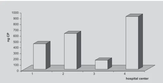
Of the 42 analyzed samples, 6 presented CP concentrations below the quantification level (1.0 ng/mL). The other 36 samples revealed CP contamination of infusion bags in some level from all of the four hospital centers. Although center 4 showed the highest individual levels of contamination (Figure 2), it also presented the highest number of samples with no detectable contami-nation. As the p value obtained was 0.1516 (Kruskall-Wallis test), the statistical difference was considered not significant, between the four hospital centers.
The results obtained in the present study reinforce those reported in the specialized literature and show CP contamination on several surfaces of infusion bags in the chemotherapy handling areas. Such contamination could result in ingestion and/or dermal absorption of CP. These positive samples suggest that the drug handling procedures are not adequate. Infusion bags may be
TABLE II - Average concentrations of cyclophosphamide (ng) in infusion bags samples from four different hospital centers
Hospital Average concentration Range of center of cyclophosphamide concentration
(ng/ infusion bag) (ng/infusion bag)
1 441 115.0-1077.2
2 616 320.0-21975
3 155 NQ(1)-850.0
4 911 NQ(1)- 41874
Note: (1) NQ: non quantified- below 1.0 ng/mL
FIGURE 2- Average concentration of cyclophosphamide (CP), in ng, from infusion bags samples collected in four
contaminated by gloves utilized by hospital workers. In addition, contamination of surfaces also increases the risk of exposure to others in the areas.
Sessink et al. (1997) suggest that cyclophosphamide excretion can be lower when we know the exact relationship between sources of exposure, distribution of sources and absorption of this agent.
The present methodology can be used to identify CP in wipe test samples. Furthermore, our results suggest that infusion bags could be valuable monitoring targets for assessment of dermal exposure during preparation and administration of antineoplastic drugs.
ACKNOWLEDGEMENTS
This work was conducted with doctoral fellowship sponsored by CAPES-Brazil.
RESUMO
Identificação de ciclofosfamida em wipe teste por CG-EM com prévia extração em fase sólida
A ciclofosfamida é uma agente alquilante freqüentemente utilizado na prática clínica para diferentes tipos de tumo-res e, é classificado como carcinógeno para humanos pelo IARC. Neste estudo, o fármaco foi quantificado, após adaptação de um método analítico, utilizando a croma-tografia gasosa acoplada à espectrometria de massa com prévia extração em fase sólida e derivação com anidrido trifluoroacético. Foram utilizadas como amostras, bolsas de infusão as quais podem estar contaminadas pelo con-tato com luvas usadas durante o preparo dos medicamen-tos, constituindo importante fonte de contaminação para trabalhadores em outras áreas da quimioterapia, tais como a sala de administração do medicamento ao pacien-te. A ifosfamida foi utilizada como padrão interno e a quantificação foi realizada com auxílio de curva de calibração, em um intervalo dinâmico de 1 a 100 ng/mL. O limite de detecção do método foi 0,4 ng/mL. Os coeficien-tes de variação obtidos, no ensaio de precisão, foram de 0,5 a 10% (intra-ensaio) e de 0 a 19% (interensaio). O analito permaneceu estável na matriz após um mês de congelamento das amostras, sem perdas significativas. No teste de exatidão os resultados variaram de 83 a 116% e a recuperação do método foi de 98,9%. A ciclofosfamida foi determinada em 36 das 42 bolsas coletadas em quatro diferentes hospitais, com valores que variaram de 90 a 41874 ng (mediana= 607,5 ng). Os resultados, bem correlacionados com os anteriormente relatados na lite-ratura, sugerem que este método pode ser usado na
iden-tificação da ciclofosfamida e pode ser considerado impor-tante ferramenta na monitorização da exposição aos fármacos antineoplásicos.
Unitermos: ciclofosfamida, wipe teste; avaliação da expo-sição
REFERENCES
ALESSIO, L.; APOSTOLI, P.; DRAICCHIO, F.; FORNI, A.; LUCCHINI, R.; MERLER, E.; PALAZZO, S.; SCARSELLI, R.; SOSSAI, D. Prevenzione dei rischi da esposizione professionale a chemioterapici antiblastici. Documento de consenso. Med. Lav., Milano, v.87, n.3, p.194-200, 1996.
APOSTOLI, P.; CLONFERO, E.; COTTICA, D.; BERGAMASCHI, A.; MOCCALDI, R.; DRAICCHIO, F.; TRANFO, G.; SANNOLO, N.; SOSSAI, D. Criteri e metodi per lo studio dell’esposizione occupzionale a chemioterapici antiblastici. Med. Lav., Milano, v.87, n.3, p.230-254, 1996.
CONNOR, T. H.; ANDERSON, R. W.; SESSINK, P. J.; BROADFIELD, L.; POWER, L. A. Surface contamination with antineoplastic agents in six cancer treatment centers in Canada and United States. Am. J. Health Syst. Pharm., Wisconsin, v.56, p.1427-1432, 1999.
EVELO, C. T. A.; BOS, R. P.; PETERS, J. G. P.; HENDERSON, P. TH. Urinary cyclophosphamide assay as a method for biological monitoring of occupational exposure to cyclophosphamide. Int. Arch. Occup. Environ. Health, Berlin, v.58, p.151-155, 1986.
FOOD AND DRUG ADMINISTRATION. Center for Drug Evaluation and Research. Q2B validation of analytical procedures: methodology: guidance for industry. Rockville: FDA, 1996. p. 1-10.
FOOD AND DRUG ADMINISTRATION. Center for Drug Evaluation and Research. Bioanalytical methods validation for human studies: guidance for industry. Rockville: FDA, 1998. p. 1-13.
MINOIA, C.; TURCI, R.; SOTTANI, C.; SCHIAVI, A.; PERBELLINI, L.; ANGELERI, S.; DRAICCHIO, F.; APOSTOLI, P. Application of high-performance liquid chromatography/ tandem mass spectrometry in the environmental and biological monitoring of health care personnel occupationally exposed to cyclophosphamide and ifosfamide. Rapid Commun. Mass Spectrom., London, v.12, p.1485-1493, 1998.
MINOIA, C.; PERBELLINI, L. Monitoraggio ambientale e biologico dell´esposizione professionale a xenobiotici: chemoterapici antiblastici. Milano: Morgan, 2000. v.3, 265p.
PYY, L.; SORSA, M.; HAKALA, E. Ambient monitoring of cyclophosphamide in manufacture and hospitals. Am. Ind. Hyg. Assoc. J., Guilford, v.49, n.6, p.314-317, 1988.
SCHMAUS. G.; SCHIERL, R.; FUNCK, S. Monitoring surface contamination by antineoplastic drugs using gas chromatography-mass spectrometry and voltammetry. Am. Soc. Health Syst. Pharm., Bethesda, v.59, n.10, p.956-961, 2002.
SESSINK, P. J. M.; BOER, K. A.; SCHEEFHALS, A. P. H.; ANZION, R. B. M.; BOS, R. P. Occupational exposure to antineoplastic agents at several departments in a hospital. Int. Arch. Occup. Environ. Health, Berlin, v.64, p.105-112, 1992.
SESSINK, P. J. M.; SCHOLTES, M. M.; ANZION, R. B. M.; BOS, R. P. Determination of cyclophosphamide in urine by gas chromatography-mass spectrometry. J. Chromatogr., Amsterdam, v.616, p.333-337, 1993.
SESSINK, P. J. M.; Van de KERKHOLF, M. C. A. Environmental contamination and assessment of exposure to antineoplastic agents by determination of cyclophosphamide in urine of exposed pharmacy technicians: is skin absorption an important exposure route? Arch. Environ. Health, Chicago, v.49, p.165-169, 1994.
SESSINK, P. J. M.; WITTENHORST, B. C. J.; ANZION, R. B. M.; BOS, R. P. Exposure of pharmacy technicians to antineoplastic agents: reevaluation after additional protective measures. Arch. Environ. Health, Chicago, v.52, n.3, p. 240-244, 1997.
SESSINK, P. J. M.; BOS, R. P. Drugs hazardous to healthcare workers. Evaluation of methods for monitoring occupational exposure to cytostatic drugs. Drug Safety, Auckland, v.20, n.4, p.347-359, 1999.
SORSA, M.; ANDERSON, D. Monitoring of occupational exposure to cytostatic anticancer agents. Mutat. Res., Amsterdam, v.355, p.253-261, 1996.