Breast fat grafting: experimental or established
procedure?
Lipoenxertia nas mamas: procedimento consagrado ou experimental?
ABSTRACT
There has recently been an increase in the use of fat grafting for the correction of congenital or acquired breast deformities and for aesthetic breast treatment. This increase is due to the emergence of novel fat grafting techniques that produce lasting and reliable results despite some authors’ beliefs that the procedure might hinder breast cancer screening. This literature review aims to identify the most frequently used breast fat grafting techniques, evaluate
their eficacy and safety, and report their major complications. The review was performed
after a search in the PubMed and LILACS databases, and only English-language articles published over the past 5 years were assessed. There have been an increased number of
studies on the topic in recent years, and a speciic fat grafting technique has been primarily
reported. Most studies in this area have shown that breast fat grafting does not affect radio-logical breast cancer screening, whereas some studies have suggested that the use of grafted fat may enhance the development of breast cancer. Despite the increased number of published
studies, few have a good level of scientiic evidence. This review concludes that fat grafting
is an alternative technique for the correction of breast deformities and moderate aesthetic breast enlargement and should be performed by properly trained surgeons accompanied by a radiology team that is experienced in breast imaging. Further studies with appropriate
scientiic methodologies are needed to evaluate breast fat grafting.
Keywords: Breast fat grafting. Fat graft
RESUMO
Atualmente vem ocorrendo aumento da incidência de utilização de enxertia de gordura para correção de defeitos mamários congênitos ou adquiridos e para tratamento mamário estético. Esse aumento é decorrente do surgimento de novas técnicas de lipoenxertia, que
produzem resultados mais duradouros e coniáveis, apesar da crença de alguns autores de que o procedimento possa diicultar o rastreamento do câncer mamário. Esta revisão de literatura tem como objetivo identiicar as técnicas de lipoenxertia mamárias mais utilizadas, avaliá-las quanto à eicácia e à segurança, e relatar as principais complicações associadas. A
revisão foi realizada nas bases de dados PubMed e LILACS, e os fatores de inclusão foram: artigos em idioma inglês, publicados nos últimos cinco anos. Como resultado observou-se aumento do número de trabalhos abordando o tema nos últimos anos, e predomínio de uma
técnica de lipoenxertia especíica. A maioria dos trabalhos demonstra que a lipoenxertia mamária não prejudica o rastreamento radiológico para câncer mamário, e alguns sugerem que a gordura enxertada potencializa o desenvolvimento de câncer nas mamas. Apesar
do aumento do número de trabalhos, existem ainda poucos com bom nível de evidência
cientíica. Esta revisão permitiu concluir que a lipoenxertia é uma técnica alternativa para
This study was performed at the Faculdade de Medicina da Universidade Federal de Goiás (Faculty of Medicine of the Federal University of Goiás),
Goiânia, GO, Brazil.
Submitted to SGP (Sistema de
Gestão de Publicações/Manager
Publications System) of RBCP (Revista Brasileira de Cirurgia
Plástica/Brazilian Journal of
Plastic Surgery).
Article received: February 8, 2012
Article accepted: June 7, 2012
AlexAndre roriz
Blumenschein1
ruffo freitAs-Junior2
AndreA thomAzine
tuffAnin3
dAnielle isAdorA
Blumenschein4
1. Volunteer Physician, Mastology Service, Hospital of the Federal University of Goiás (UFG); Plastic Surgeon at the Instituto de Mastologia e Oncologia, Goiânia, GO, Brazil.
2. PhD, Professor, Department of Mastology UFG, Mastology Program Coordinator, Faculty of Medicine UFG, Goiânia, GO, Brazil. 3. MSc, Volunteer Physiotherapist, Mastology Program, Faculty of Medicine at UFG, Goiânia, GO, Brazil.
reparação de defeitos mamários e discreto aumento estético das mamas, devendo ser
uti-lizada por cirurgiões com treinamento adequado e acompanhada por equipe de radiologia experiente em imagenologia mamária. Mais trabalhos com metodologia cientíica adequada
são necessários para avaliar a lipoenxertia mamária.
Descritores: Lipoenxertia mamária. Enxerto de gordura.
INTRODUCTION
Autologous fat grafts have long been used as a illing material to correct soft tissue defects. One of the irst reports
on fat grafting described a case of breast defect correction1.
Fat is considered an ideal illing material since it is auto -logous, easily accessed, widely abundant in the body, and has low immunogenic and allergic reactivity2. However, fat
grafts often have high rates of resorption and replacement
with ibrous scar tissue or calciied oil cysts, which can generate aftereffects and result in dificulties in interpreting
radiological breast images3.
Publications on this subject are increasing due to the deve -lopment of new techniques for fat harvest, preparation, and grafting, despite the belief of some authors that the procedu re may hinder breast cancer screening3. Novel techniques have
been developed to improve the viability and aesthetic results of fat grafts2,4,5. Fat grafting, which has primarily been used
to correct breast deformities, has also been used for aesthetic purposes and to increase breast volume in an attempt to re -place the use of breast implants6-8.
Several studies have shown that fat grafting did not effecti-vely hinder radiological breast screening2,9-16. However, Wang
et al.17 challenge these indings. In cases in which fat grafting is used for breast reconstruction after mastectomy or breast conservation therapy, new concerns have arisen regarding the possibility that the transplanted cells stimulate carcinogenesis in breasts with a predisposition to developing cancer18,19.
In this context, this literature review aims to identify the main techniques used for breast fat grafting, evaluate their
eficacy and safety, and report their results and associated
complications.
METHODS
Article Selection Process
An article search was performed in the PubMed and LILACS databases using the following key words: “breast fat graft”, “breast fat transplantation”, “breast adipose tissue
injection”, “breast lipomodelling”, and “breast lipoilling”. The number of articles retrieved per keyword was 37, 35,
151, 4, and 11, respectively.
Articles published in the last 5 years (between 2006 and 2011) in English were selected. We included all original
articles indexed between January 2006 and November 2011,
including experimental (clinical trials, randomized or not) and observational studies (casecontrol, cohort studies) per -formed in humans. Duplicate references were excluded, as were articles “ahead of print,” reviews, letters to the editor, and editorials.
A total of 24 studies were assessed for study design, num ber of patients, indication for surgery, surgical technique, compli-cations, follow-up imaging, and oncological follow-up.
RESULTS
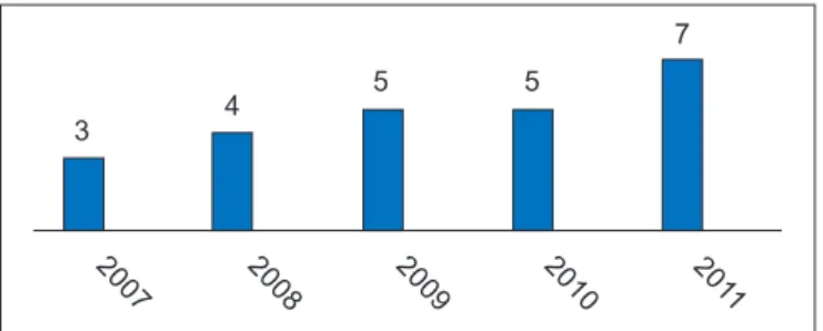
After the selected articles were analyzed, we noted that the number of publications had grown in recent years (Figure 1). The studies were mostly retrospective (n = 13) or prospec-tive and descripprospec-tive (n = 10), and there were no randomized controlled studies.
Studies involving large numbers of patients involved re trospective analyses of medical records or prospective eva
-luations of patients undergoing a speciic technique and did
not allow for an overall assessment of fat grafting8,13-16,19-21.
The publications mentioned various techniques for obtaining, treating, and grafting fat. The main differences bet -ween the techniques were present in the treatment of the obtained fat (Figure 2). The studies generally reported that the grafts must be obtained by liposuction using the tumes-cent technique from body areas with an abundance of fat tissue (abdomen, flank, inner thigh, lateral thigh, medial knees, and back, as the commonlyused donor areas, in des -cending order)2,13-16,20,21.
Most of these studies emphasized that liposuction should be gentle, involve the use of syringes or a low-pressure appa-ratus, and avoid exposure of the fat to ambient air (closed technique) to ensure that the fatty material is handled as little as possible2-8,13-16,19-24. The obtained fat should then be puriied
using low-speed centrifugation (Figure 2).
Fat grafting is performed through small cannulas in small
quantities, and retroinjection occurs after tunnels are establi -shed using the cannulas. More fat grafting procedures empha-size that the grafting of fat should be performed using small quantities to ensure that the grafted tissue is in full contact with the vascularized tissue of the receptor region2,16,21.
procedures were required per patient (Table 1). However, some papers presented techniques for performing larger grafts and fewer procedures5,6,8,25,26. All studies that evalua ted
breast reconstruction after radiotherapy noted that additio nal fat grafting procedures are required23-25,27.
No conclusive studies have examined the percentage of fat remaining after the procedure. Most studies estimate
that 30%-40% of the volume is lost after the irst procedure,
thus requiring subsequent procedures5,21,22,25,26 or graft
over-correction14,16,25.
Several studies have shown high rates of patient satisfac-tion after fat grafting as well as good aesthetic results that
are subjectively evaluated using photographs2,5-8,14,16,20-24.
These studies considered breast fat grafting as a safe te
-ch nique with a low number of major complications (Table
1). The main complications of most studies were liponecro sis and oil cyst formation (Table 1), whereas local infection of the grafted area2,13-15,21 occurred in a few cases. Most studies
reported no dificulty with radiological breast screening
after fat grafting procedures. Screening was conducted primarily using mammography and breast ultrasono-graphy4,7,8,10-13,15,16,21,22,25,26 by radiology teams experienced in
discerning images of benign and malignant breast calciica -tion4,7,13,15,16,21. All tissues with suspected abnormal
radiolo-gical indings were biopsied, which revealed a low number of malignant disease conirmations7-12,16,17,22. Rietjens et al.13
concluded that the only case of recurrence detected in their study was likely due to underdiagnosis of the initial cancer. Petit et al.15 and Ilouz and Sterodimas21 stated that the number
of cancer cases detected after fat grafting was similar to the cancer incidence in the general population, although they did not present data on this statement in their studies. Rigotti et al.19 found a similar incidence of tumor recurrence in patients
treated with fat grafting compared to patients who did not receive fat grafting treatment. However, their comparison of the same group of patients at different time windows has been criticized by other authors15.
After a review of studies in vitro and in animals conirmed
their hypothesis, Lohsiriwat et al.18 hypothesized that fat
grafting in predisposed breasts could lead to carcinogenesis
but concluded that there is insuficient evidence for this as
-sociation in humans.
DISCUSSION
Fat grafting is a widely used and established technique that is used to correct soft tissue deformities1. Nevertheless,
it is no longer used by some surgeons as a result of the large resorption of graft material, the loss of results, and
cal ciication and oil cyst formation within the graft. This
could possibly interfere with radiological follow-up of the breasts, thereby affecting the diagnosis of new breast tumors or recurrences3. With the development of new techniques
for obtaining, processing, and grafting fat material2, more
durable grafts could be obtained initially, thus presenting good results in the correction of soft tissue defects of the face and subsequently in other locations such as the breast. Coleman & Saboeiro2 questioned the restriction on the
use of fat grafting by the American Society of Plastic and
Reconstructive Surgery in 19873 and argued that the same
re gions of calciications and liponecrosis that are observed after fat grafting procedures are also observed after other breast procedures such as breast reduction and mastopexy. According to this study, the number of published studies on breast fat grafting have increased in recent years. Despite
this increase, few questions have been answered scientii -cally, primarily because most studies were related to case series or reports of new techniques and had small numbers of patients. Moreover, few observational, case-control26, or
large studies have been performed.
Fat grafting was used mainly in cases of breast recons-truction, particularly to correct deformities after primary reconstructions13-15,16,20,22 or adjuvant treatments such as
radiotherapy19,23,27 (Table 1). Some articles mention its use to
Figure 1 – Number of publications on fat grafting by publication year.
3 4
5 5
7
2007 2008 2009 2010 201
1
1
6
17
Centrifugation Decantation *Lipivage®
Figure 2 – Distribution of published studies according to fat preparation technique. *Closed system for collecting,
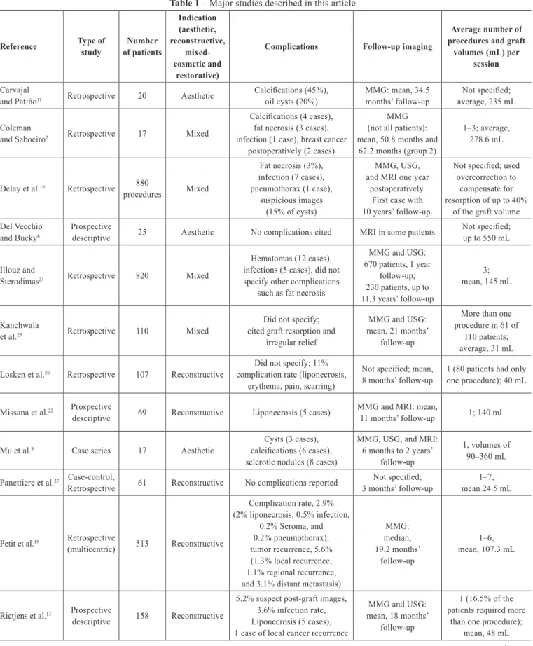
Table 1 – Major studies described in this article.
Reference Type of
study
Number of patients
Indication (aesthetic, reconstructive,
mixed-cosmetic and
restorative)
Complications Follow-up imaging
Average number of procedures and graft
volumes (mL) per session
Carvajal
and Patiño11 Retrospective 20 Aesthetic
Calciications (45%),
oil cysts (20%)
MMG: mean, 34.5 months’ follow-up
Not speciied;
average, 235 mL
Coleman
and Saboeiro2 Retrospective 17 Mixed
Calciications (4 cases),
fat necrosis (3 cases), infection (1 case), breast cancer
postoperatively (2 cases)
MMG (not all patients): mean, 50.8 months and
62.2 months (group 2)
1–3; average,
278.6 mL
Delay et al.16 Retrospective 880
procedures Mixed
Fat necrosis (3%),
infection (7 cases),
pneumothorax (1 case), suspicious images
(15% of cysts)
MMG, USG, and MRI one year
postoperatively. First case with 10 years’ follow-up.
Not speciied; used
overcorrection to compensate for resorption of up to 40%
of the graft volume
Del Vecchio and Bucky6
Prospective
descriptive 25 Aesthetic No complications cited MRI in some patients
Not speciied;
up to 550 mL
Illouz and
Sterodimas21 Retrospective 820 Mixed
Hematomas (12 cases), infections (5 cases), did not specify other complications
such as fat necrosis
MMG and USG:
670 patients, 1 year
follow-up; 230 patients, up to 11.3 years’ follow-up
3; mean, 145 mL
Kanchwala
et al.25 Retrospective 110 Mixed
Did not specify; cited graft resorption and
irregular relief
MMG and USG: mean, 21 months’
follow-up
More than one procedure in 61 of
110 patients; average, 31 mL
Losken et al.20 Retrospective 107 Reconstructive
Did not specify; 11% complication rate (liponecrosis,
erythema, pain, scarring)
Not speciied; mean,
8 months’ follow-up
1 (80 patients had only one procedure); 40 mL
Missana et al.22 Prospective
descriptive 69 Reconstructive Liponecrosis (5 cases)
MMG and MRI: mean,
11 months’ follow-up 1; 140 mL
Mu et al.9 Case series 17 Aesthetic
Cysts (3 cases),
calciications (6 cases),
sclerotic nodules (8 cases)
MMG, USG, and MRI: 6 months to 2 years’
follow-up
1, volumes of 90–360 mL
Panettiere et al.27 Case-control,
Retrospective 61 Reconstructive No complications reported
Not speciied;
3 months’ follow-up
1–7,
mean 24.5 mL
Petit et al.15 Retrospective
(multicentric) 513 Reconstructive
Complication rate, 2.9% (2% liponecrosis, 0.5% infection,
0.2% Seroma, and 0.2% pneumothorax); tumor recurrence, 5.6% (1.3% local recurrence, 1.1% regional recurrence, and 3.1% distant metastasis)
MMG: median, 19.2 months’
follow-up
1–6,
mean, 107.3 mL
Rietjens et al.13 Prospective
descriptive 158 Reconstructive
5.2% suspect post-graft images, 3.6% infection rate, Liponecrosis (5 cases), 1 case of local cancer recurrence
MMG and USG: mean, 18 months’
follow-up
1 (16.5% of the patients required more
than one procedure); mean, 48 mL
correct deformities after aesthetic surgeries, such as breast augmentation and mammoplasty5,21,26, and others describe
the use of fat grafting for aesthetic breast augmentation, as an alternative to prostheses use6-9.
Several studies have emphasized that the fat must be
collected and puriied through low-pressure liposuction,
which is usually performed using syringes, to minimize the resultant trauma to the autologous material being collected. Moreover, prolonged contact between the fat and the air must
be avoided, and the samples should be puriied by low-speed centrifugation or decantation. Puriied fat grafts are then
constructed through the use of smaller cannulas in small Continuation of the Table 1 – Major studies described in this article.
Reference Type of
study
Number of patients
Indication (aesthetic, reconstructive,
mixed-cosmetic and
restorative)
Complications Follow-up imaging
Average number of procedures and graft
volumes (mL) per session
Rigotti et al.23 Prospective
descriptive 20 Reconstructive No complications reported
Not speciied;
31 months’ follow-up 3; mean, 70 mL
Rigotti et al.19 Prospective
descriptive 137 Reconstructive
Evaluated recurrence after fat grafting only,
11% (16 cases)
Not speciied; mean, 7.6 years’
follow-up
3; volume not speciied
Salgarello et al.4 Retrospective 42 Mixed
Not speciied;
average 0.4% liponecrosis and oil cysts
MMG and USG:
9 months’ follow-up 2; 117 mL
Serra-Renom et al.24
Prospective
descriptive 65 Reconstructive No complications reported
Not speciied;
12 months’ follow-up 1–3; average 150 mL
Sinna et al.14 Retrospective 200 Reconstructive
Liponecrosis (5 cases), infection (2 cases), pneumothorax (1 case)
Not speciied;
median, 14.5 months’ follow-up
Most with 1;
2 (37 patients), 3 (7 patients); mean, 176 mL
Veber et al.10 Retrospective 76 Mixed
16%, benign microcalciications; 9%, macrocalciications;
25%, cystic lesions
MMG: median, 16.2 months’
follow-up
Most with 1; mean, 100 mL
Wang et al.12 Prospective
descriptive 41 Aesthetic
Nodules of fat necrosis in 82.9% of patients postoperatively. There was a correlation between graft volume
and the amount of nodules
USG: mean, 16 months’ follow-up
Most with 1; mean, 55 mL
Wang et al.17 Retrospective 48 Aesthetic 16.7% macrocalciications MMG: mean,
45 months’ follow-up 2; mean, 110 mL
Yoshimura et al.26
Prospective
descriptive 15 Aesthetic No complications reported
MMG and MRI: minimum 12 months’
and maximum 18 months’ follow-up
Not speciied;
mean, 250 mL
Yoshimura et al.5 Prospective
descriptive 40 Aesthetic
Cysts (2 cases),
microcalciications (2 cases) 6-42 months’ follow-upNot speciied:
Not speciied; mean, 270 mL
Zheng et al.7 Retrospective 66 Aesthetic 16.7% necrosis images
USG, MMG and MRI:
mean, 37 months’
follow-up
1 (28 patients), 2 (21 patients),
and 3 (17 patients); mean 17 mL
Zocchi and
Zuliani8 Retrospective 181 Aesthetic
1.2% liponecrosis, 1.8% oil cysts,
3.9% microcalciications
USG and MMG: 12 months’ follow-up
Not speciied; mean, 375 mL
portions or amounts such that the graft material maintains maximum contact with the receptor tissue and its blood vessels. This protocol ensures appropriate nutrition in the early days after grafting. The same reasoning is used for fat grafts in the tissue after radiotherapy, when vascularization is scarce but the graft still shows good results19,23.
Although several studies have shown good results through photographs and high levels of satisfaction by patients and
surgeons, few have quantiied the percentage of grafted fat
that did not undergo resorption, degeneration, or necrosis. Some authors recommended overcorrection to allow for re -sorption14,16,25. Del Vecchio & Bucky6 evaluated the amount
of graft absorbed by comparing magnetic resonance imaging (MRI) of the breast before and after the procedure, indicating that this can be a good tool to evaluate fat graft absorption. However, in their study, tests were not performed on all pa -tients since they were asked to finance them, thus affecting
the inal assessment of this variable. Most surgeons subjec -tively estimate the amount of fat that is needed to correct the defect and state that the fat grafting procedure should be performed in more than one step, particularly in cases of reconstruction after radiotherapy.
Most of these studies cited a low number of compli-cations. Liponecrosis and oil cyst formation comprise the most common complications, followed by local infection of the graft material. Despite a considerable number of li -ponecrosis events and oil cysts after the breast fat grafting process, most studies concluded that these images appear benign when evaluated by a radiologist who is experienced in mammography or breast ultrasonography2,7,9-16,21. Results that generate doubts should be better evaluated using MRI or guided breast biopsy7,9,15,16,22,25. Some studies report that the number of liponecrosis events was higher at the begin ning of the series, and following the development of more suitable fat grafting techniques, the number of liponecrosis events
un derwent a signiicant reduction that approached zero16.
Coleman and Saboeiro2 mentioned that the number of
suspect images after breast fat grafting is similar to that of post-operative breast surgeries such as mastopexy and breast reduction. Wang et al.17 challenged the published stu dies and
concluded that the calciications observed after fat grafting
in the breast can be a confounding factor in the diagnosis of future breast cancer. In their study, the fat grafting techni que is not very detailed, but it is inferred that, although the authors used a moderate amount of fat in each procedure (mean, 110 mL), the grafts were introduced in large incre-ments and required a “massage” after grafting to accommo-date the graft, which would result in a higher rate of necrosis in the central portion of the grafts12.
Although some experimental studies in vitro and in ani -mals suggested that fat grafting could lead to cancer18, no
studies have properly evaluated a cause and effect rela-tionship between breast fat grafting and cancer in humans.
Some studies have reported on the incidence or recurrence of breast cancer events after fat grafting but did not draw conclu-sions about the causal effect. Most of the cases observed were attributed to “underdiagnosis” of the initial cancer, which would have occurred regardless of the fat grafting13,15,16.
CONCLUSIONS
Although none of the analyzed studies have indicated a
high level of scientiic evidence, fat grafting seems to be an
adequate and safe technique to repair breast deformities and is a good alternative to moderate aesthetic breast en -large ment. Fat grafting presents a low number of compli-cations when performed by experienced professionals and yields good results and high levels of patient satisfaction. It should be performed by well-trained breast grafting teams, and patients should be monitored by an experienced breast imaging ra diology team.
Issues regarding the effective evaluation of the integra -tion of the graft with the breast tissue, the percentage of graft resorption according to the technique used, and the
long-term changes in graft material as well as their inluence
on the grafted area have yet to be addressed and require randomized studies with larger numbers of patients and a
better scientiic design.
REFERENCES
1. Czerny V. Plastischer ersatz der brustdruse durch ein lipom. Zentralbl
Chir.1895;27-72.
2. Coleman SR, Saboeiro AP. Fat grafting to the breast revisited: safety
and eficacy. Plast Reconstr Surg. 2007;119(3):775-85.
3. ASPRS Ad-Hoc Committee on New Procedures. Report on autologous
fat transplantation. Plast Surg Nurs. 1987;7(4):140-1.
4. Salgarello M, Visconti G, Rusciani A. Breast fat grafting with plate let-rich plasma: a comparative clinical study and current state of the art.
Plast Reconstr Surg. 2011;127(6):2176-85.
5. Yoshimura K, Sato K, Aoi N, Kurita M, Hirohi T, Harii K. Cell-assisted lipotransfer for cosmetic breast augmentation: supportive use of
adipose-derived stem/stromal cells. Aesthetic Plast Surg. 2008;32(1):
48-55.
6. Del Vecchio DA, Bucky LP. Breast augmentation using preexpansion and autologous fat transplantation: a clinical radiographic study. Plast
Reconstr Surg. 2011;127(6):2441-50.
7. Zheng DN, Li QF, Lei H, Zheng SW, Xie YZ, Xu QH, et al. Autolo -gous fat grafting to the breast for cosmetic enhancement: experience
in 66 patients with long-term follow up. J Plast Reconstr Aesthet Surg. 2008;61(7):792-8.
8. Zocchi ML, Zuliani F. Bicompartmental breast lipostructuring. Aesthe-tic Plast Surg. 2008;32(2):313-28.
9. Mu DL, Luan J, Mu L, Xin MQ. Breast augmentation by autologous fat injection grafting: management and clinical analysis of complications. Ann Plast Surg. 2009;63(2):124-7.
10. Veber M, Tourasse C, Toussoun G, Moutran M, Mojallal A, Delay E. Radiographic indings after breast augmentation by autologous fat transfer. Plast Reconstr Surg. 2011;127(3):1289-99.
-cation of compli-cations of cosmetic augmentation with autologous fat obtained by liposuction. Ann Plast Surg. 2010;64(4):385-9.
13. Rietjens M, De Lorenzi F, Rossetto F, Brenelli F, Manconi A, Martella
S, et al. Safety of fat grafting in secondary breast reconstruction after
cancer. J Plast Reconstr Aesthet Surg. 2011;64(4):477-83.
14. Sinna R, Delay E, Garson S, Delaporte T, Toussoun G. Breast fat grafting (lipomodelling) after extended latissimus dorsi lap breast reconstruc
-tion: a preliminary report of 200 consecutive cases. J Plast Reconstr Aesthet Surg. 2010;63(11):1769-77.
15. Petit JY, Lohsiriwat V, Clough KB, Sarfati I, Ihrai T, Rietjens M, et al. The oncologic outcome and immediate surgical complications of lipoilling in
breast cancer patients: a multicenter study: Milan-Paris-Lyon experience
of 646 lipoilling procedures. Plast Reconstr Surg. 2011;128(2):341-6. 16. Delay E, Garson S, Tousson G, Sinna R. Fat injection to the breast: te
-chnique, results, and indications based on 880 procedures over 10 years.
Aesthet Surg J. 2009;29(5):360-76.
17. Wang CF, Zhou Z, Yan YJ, Zhao DM, Chen F, Qiao Q. Clinical analyses of clustered microcalciications after autologous fat injection for breast augmentation. Plast Reconstr Surg. 2011;127(4):1669-73.
18. Lohsiriwat V, Curigliano G, Rietjens M, Goldhirsch A, Petit JY. Auto -logous fat transplantation in patients with breast cancer: “silencing” or
“fueling” cancer recurrence? Breast. 2011;20(4):351-7.
19. Rigotti G, Marchi A, Stringhini P, Baroni G, Galiè M, Molino AM, et al. Determining the oncological risk of autologous lipoaspirate grafting for post-mastectomy breast reconstruction. Aesthetic Plast Surg. 2010; 34(4):475-80.
20. Losken A, Pinell XA, Sikoro K, Yezhelyev MV, Anderson E, Carlson GW. Autologous fat grafting in secondary breast reconstruction. Ann Plast Surg. 2011;66(5):518-22.
21. Illouz YG, Sterodimas A. Autologous fat transplantation to the breast: a personal technique with 25 years of experience. Aesthetic Plast Surg.
2009;33(5):706-15.
22. Missana MC, Laurent I, Barreau L, Balleyguier C. Autologous fat transfer
in reconstructive breast surgery: indications, technique and results. Eur J Surg Oncol. 2007;33(6):685-90.
23. Rigotti G, Marchi A, Galiè M, Baroni G, Benati D, Krampera M, et al. Clinical treatment of radiotherapy tissue damage by lipoaspirate transplant: a healing process mediated by adipose-derived adult stem cells. Plast
Reconstr Surg. 2007;119(5):1409-22.
24. Serra-Renom JM, Muñoz-Olmo JL, Serra-Mestre JM. Fat grafting in
postmastectomy breast reconstruction with expanders and prostheses in patients who have received radiotherapy: formation of new subcutaneous tissue. Plast Reconstr Surg. 2010;125(1):12-8.
25. Kanchwala SK, Glatt BS, Conant EF, Bucky LP Autologous fat grafting to the reconstructed breast: the management of acquired contour deformities. Plast Reconstr Surg. 2009;124(2):409-18.
26. Yoshimura K, Asano Y, Aoi N, Kurita M, Oshima Y, Sato K, et al. Proge -nitor-enriched adipose tissue transplantation as rescue for breast implant
complications. Breast J. 2010;16(2):169-75.
27. Panettiere P, Marchetti L, Accorsi D. The serial free fat transfer in irra -diated prosthetic breast reconstructions. Aesthetic Plast Surg. 2009;
33(5):695-700.
Correspondence to: Alexandre Roriz Blumenschein Instituto de Mastologia e Oncologia