Rev Bras Cir Plást. 2012;27(4):523-6 523 Detection of venous thrombosis in free laps by measurement of capillary blood glucose
Detection of venous thrombosis in free laps by
measurement of capillary blood glucose
Detecção de trombose venosa em retalhos livres por medidas
de glicemia capilar
ABSTRACT
Background: Monitoring of free laps after surgery is vitally important, especially in the irst few hours because the timing of reoperation can determine lap salvage or loss. To date, no study has examined the decision to reoperate on a lap based on the objective measure of glycemia or a comparison between laps that showed good outcomes and those that showed vascular damage. The objective of this study was to evaluate the validity of blood glucose measurements within the lap as a method for monitoring free laps and to compare the efi
-cacy of this method with that of clinical assessments. Methods: The study was prospective, included 16 patients with free laps, and was conducted from May 2012 to July 2012. A team of professionals not involved in the surgery evaluated capillary glycemia. Flaps were clinically evaluated during the immediate postoperative period, on ICU admission, at every 3 hours, and as needed. Results: Of the 16 patients, 5 (31.3%) had venous thrombosis in the irst 24 hours. Statistically signiicant differences were noted in capillary glycemia in patients with or without venous thrombosis in measurements obtained 6, 9, and 12 hours after surgery (P < 0.05). Conclusions: The measurement of capillary glycemia was not superior to clinical evaluation by an experienced professional for the detection of venous thrombosis within free laps.
Keywords: Microsurgery. Surgical laps. Venous thrombosis.
RESUMO
Introdução: A monitorização do retalho livre após a cirurgia é de vital importância, espe
-cialmente nas primeiras horas de pós-operatório, pois o momento de reabordagem pode ser o deinidor entre o salvamento ou a perda do retalho. Até o momento, não existe trabalho na literatura estudando a decisão de abordagem do retalho baseada em medidas objetivas ou a comparação da glicemia entre retalhos que evoluíram bem com os que sofreram sofri
-mento vascular. O objetivo deste estudo é avaliar a validade da medida da glicemia capilar do retalho como método de monitorização de retalhos microcirúrgicos comparando com a avaliação clínica. Método: Foram estudados prospectivamente 16 pacientes portadores de retalhos livres, realizados de maio de 2012 a julho de 2012. A glicemia capilar foi avaliada
This study was performed in the Division of Plastic Surgery and Burns, Hospital das Clínicas da Faculdade de Medicina da Universidade de São Paulo (Hospital das Clinicas, Faculty of Medicine, University of São Paulo), São Paulo, SP, Brazil.
Submitted to SGP (Sistema de Gestão de Publicações/Manager Publications System) of RBCP (Revista Brasileira de Cirurgia Plástica/Brazilian Journal of Plastic Surgery). Article received: September 26, 2012 Article accepted: November 11, 2012 LincoLn Saito MiLLan1
Luiz carLoS iShida2
ESthEr Mihwa oh choi3
Enio cESar GiacchEtto
Junior4
tEnG hSianG wEi5
raMES Mattar Júnior6
MarcuS caStro FErrEira7
Franco T et al. Vendramin FS et al.
ORIGINAL ARTICLE
1. Preceptor Physician of the Hospital das Clínicas da Faculdade de Medicina da Universidade de São Paulo (Hospital of the Faculty of Medicine, Uni
-versity of São Paulo – HCFMUSP), Assistant Physician Plastic Surgery Service at Hospital do Servidor Público Estadual de São Paulo (Hospital for State Civil Servants of St. Paul), Associate Member of the Sociedade Brasileira de Cirurgia Plástica (Brazilian Society of Plastic Surgery – SBCP), São Paulo, SP, Brazil.
2. Doctor, full member of the SBCP, Physician, Division of Plastic Surgery HCFMUSP, São Paulo, SP, Brazil. 3. Resident in General Surgery HCFMUSP, São Paulo, SP, Brazil.
4. Resident of Plastic Surgery HCFMUSP, São Paulo, SP, Brazil.
5. Physician Group of Hand and Reconstructive Microsurgery, Institute of Orthopaedics and Traumatology HCFMUSP, São Paulo, SP, Brazil.
6. Livre docente, Associate Professor of the University of São Paulo, Head of Group of Hand and Reconstructive Microsurgery, Institute of Orthopaedics and Traumatology HCFMUSP, São Paulo, SP, Brazil.
Rev Bras Cir Plást. 2012;27(4):523-6
524
Millan LS et al.
por equipe formada por proissionais não envolvidos com a cirurgia realizada. A avaliação clínica do retalho foi realizada no pós-operatório imediato, na chegada à UTI, a cada 3 horas e sempre que necessário. Resultados: Dos 16 pacientes, 5 (31,3%) apresentaram complicações nas primeiras 24 horas. Todas as complicações observadas foram trombose venosa. Foi observada diferença estatisticamente signiicante na glicemia capilar de por
-tadores de retalhos que apresentaram trombose venosa em comparação àqueles que não tiveram a complicação, nas medidas realizadas 6 horas, 9 horas e 12 horas após a operação (P < 0,05). Conclusões: A medida da glicemia capilar não foi superior à avaliação clínica por proissional experiente na detecção de trombose venosa de retalhos livres.
Descritores: Microcirurgia. Retalhos cirúrgicos. Trombose venosa.
INTRODUCTION
Monitoring of free laps after surgery is of vital impor
-tance, especially during the irst few hours, because the ti ming of reoperation can deine lap salvage or loss1.
However, monitoring parameters and reoperation deci
-sions are challenging for surgeons. Classically, lap viability is evaluated based on clinical parameters such as temperature, color, turgor, capillary reill, and bleeding after scariication. Wilson et al.2 recommend lap evaluation by trained nurses every 30 minutes in the irst 24 postoperative hours and every 4 hours thereafter.
Although physical examination is essential in a reopera -tion decision, indings of physical examina-tion may be faul ty if the surgical team lacks experience or if other non-ideal factors such as inadequate lighting or visualization dificulties are present. This limitation led to the study and development of other monitoring methods such as assessment of tempe
-rature differences between the lap and the ad jacent skin2, Doppler ultrasonography of the vascular pe dicle3, measure -ment of tissue oxygenation4, analysis by microdialysis5, use of intra-arterial or intravenous catheters6, and quantitative spectral imaging (which measures distance, oxyhemoglobin, deoxyhemoglobin, total hemoglobin, and tissue saturation)7. Using objective measures, health professionals who are inex
-perienced in microsurgery can monitor the lap.
The use of capillary glycemia can be useful because it provides objective data without requiring complex apparatus. Glucometers are routinely used in all intensive care units and cost less than other monitoring systems.
The objective of this study was to evaluate the validity of the measurement of capillary glycemia of the lap as a method for monitoring free laps and compare its eficacy with that of clinical evaluation.
METHODS
This study was prospective and included 16 patients who had undergone free lap surgery performed by the Division
of Plastic Surgery and Burns, Hospital das Clínicas da Facul
-dade de Medicina da Universi-dade de São Paulo (Hospital das Clinicas, Faculty of Medicine, University of São Paulo), between May 2012 and July 2012.
The institutional research ethics committee approved this project. All patients were informed about the project’s ob -jectives and procedures and participated voluntarily after signing an informed consent form.
A team of professionals not involved in the surgery evaluated capillary glycemia and temperature. A physician performed clinical evaluation of the lap during the imme
-diate postoperative period, on ICU admission, at every 3 hours, and when necessary.
Measurements of capillary glycemia were always ob -tai ned using the same device (One Touch® Ultra, LifeScan, Inc., Milpitas, CA, USA). The evaluator collected a drop of blood from each lap for each measurement. The team performing clinical monitoring of the lap had no access to the blood glucose monitoring results to prevent inluencing the medical caregivers’ decisions and maintain a standard treatment for all patients.
RESULTS
Of the 16 patients, 5 (31.3%) had complications (venous thrombosis) in the irst 24 hours (Table 1). Figures 1 and 2 illustrate the lap capillary glycemia of patients with and without complications, respectively. Figure 3 shows the average lap capillary glycemia of patients with and without complications. A nonparametric Mann-Whitney U-test showed that the differences were statistically signiicant (P < 0.05) at 6, 9, and 12 hours (Table 2).
Because of the limited number of patients with complica
-tions, it was not possible to determine a lap glycemia value that was indicative of venous thrombosis. Moreover, it was not possible to determine whether measurement of capillary glycemia before clinical diagnosis of thrombo sis would iden
Rev Bras Cir Plást. 2012;27(4):523-6 525 Detection of venous thrombosis in free laps by measurement of capillary blood glucose
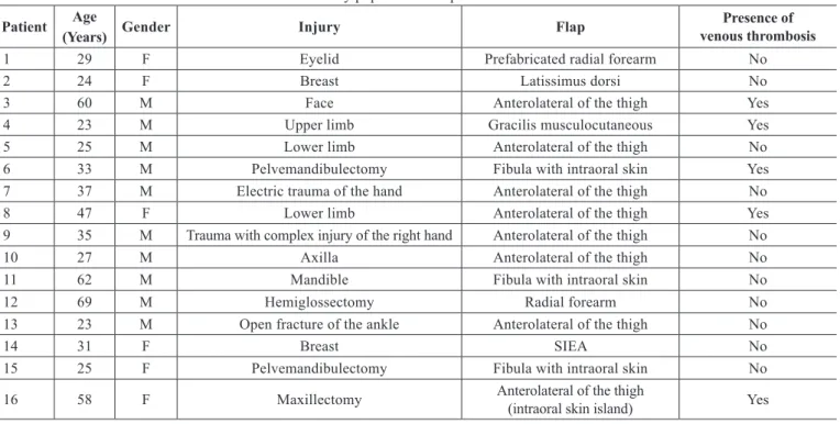
Table 1 – Characteristics of the study population and presence or absence of venous thrombosis.
Patient Age
(Years) Gender Injury Flap
Presence of venous thrombosis
1 29 F Eyelid Prefabricated radial forearm No
2 24 F Breast Latissimus dorsi No
3 60 M Face Anterolateral of the thigh Yes
4 23 M Upper limb Gracilis musculocutaneous Yes
5 25 M Lower limb Anterolateral of the thigh No
6 33 M Pelvemandibulectomy Fibula with intraoral skin Yes
7 37 M Electric trauma of the hand Anterolateral of the thigh No
8 47 F Lower limb Anterolateral of the thigh Yes
9 35 M Trauma with complex injury of the right hand Anterolateral of the thigh No
10 27 M Axilla Anterolateral of the thigh No
11 62 M Mandible Fibula with intraoral skin No
12 69 M Hemiglossectomy Radial forearm No
13 23 M Open fracture of the ankle Anterolateral of the thigh No
14 31 F Breast SIEA No
15 25 F Pelvemandibulectomy Fibula with intraoral skin No
16 58 F Maxillectomy Anterolateral of the thigh (intraoral skin island) Yes
F = female. M = male. SIEA = lap of the supericial inferior epigastric artery. DISCUSSION
Although free laps enable transfer of various tissues and rebuilding of complex architectures, surgeons face the challenge of microvascular anastomosis. Anastomosis of small-diameter structures, some measuring < 1 mm, requires reinement of suture techniques. Because handling and
introduction of the needle into the vessel wall can be throm
-bogenic, venous thrombosis is the most feared complica -tion8. Therefore, monitoring the lap after surgery is of vital importance, especially during the irst few hours, because the timing of reoperation may determine lap salvage or loss1.
Surgeons experienced in microsurgery usually monitor these laps. However, these highly specialized professionals
Figure 1 – Flap capillary blood glucose (mg/dL) of the patients without venous thrombosis.
0 3 6 9 12 15 18 21 24 250
200
150
100
50
0
1 2 5 7 9 10 11 12 13 14 15 Mean mg/dl
Hours
Figure 2 – Flap capillary blood glucose (mg/dL) of the patients with venous thrombosis.
0 3 6 9 12 15 18 21 24 140
120
100
80
60
40
20
0
3
4
6
8
16
Mean mg/dl
Rev Bras Cir Plást. 2012;27(4):523-6
526
Millan LS et al.
Table 2 – Mann-Whitney U-test comparison of the
mean blood glucose levels in laps with and without complications (venous thrombosis).
Statistical test 0 3
hours 6 hours
9 hours
12 hours
Mann-Whitney U 23.000 16.000 4.000 2.000 __
Wilcoxon W 89.000 26.000 14.000 8.000 6.000
Z -0.511 -0.785 -2.352 -2.260 -2.572
P 0.661 0.489 0.018 0.022 0.005
may not be available in all hospitals. Therefore, the use of an objective measurement technique can reduce the need for human resources, thereby reducing cost.
Flap glycemia has been studied as a diagnostic method for venous thrombosis by other authors, but none of the studies used a blinded system. In 2012, Hara et al.9 reported that a blood glucose level of 62 mg/dL had a sensitivity of 88% and speciicity of 82% for the detection of venous thrombosis. In their study, 3 false positives and 1 false negative were con -sidered indicative of venous thrombosis. More studies with larger data series are needed to determine the exact value that is indicative of thrombosis.
The present study conirms that there is a decrease in capillary glycemia in laps with venous thrombosis; however,
evaluation of this parameter did not enable earlier detection of complications. Therefore, if experienced microsurgery professionals conduct postoperative monitoring of laps, such routine measurements are unnecessary.
Finally, in this study, only 5 complications were observed in the irst 24 hours after surgery, most within the irst 12 hours. Moreover, all complications were venous thrombosis. Other complications such as hematomas did not occur, and possibly cannot be detected using capillary blood glucose measurements. Therefore, evaluation by a professional expe
-rienced in postoperative changes in lap condition is still highly recommended.
CONCLUSIONS
The measurement of free lap capillary glycemia was not superior to clinical evaluation by an experienced professio nal for the detection of venous thrombosis.
REFERENCES
1. Novakovic D, Patel RS, Goldstein DP, Gullane PJ. Salvage of failed free laps used in head and neck reconstruction. Head Neck Oncol. 2009;1:33.
2. Wilson JL, Morritt AN, Morrison WA. Avoiding complications. In: Wei FC, Mardini S, eds. Flaps and reconstructive surgery. Philadelphia: Saunders; 2009. p.117-24.
3. Wise JB, Talmor M, Hoffman LA, Gayle LB. Postoperative monitoring of microvascular tissue transplants with an implantable Doppler probe. Plast Reconstr Surg. 2000;105(6):2279-80.
4. Kamolz LP, Giovanoli P, Haslik W, Koller R, Frey M. Continuous free-lap monitoring with tissue-oxygen measurements: three-year experience. J Reconstr Microsurg. 2002;18(6):487-91.
5. Jyränki J, Suominen S, Vuola J, Bäck L. Microdialysis in clinical practice: monitoring intraoral free laps. Ann Plast Surg. 2006;56(4): 387-93.
6. Sakurai H, Nozaki M, Takeuchi M, Soejima K, Yamaki T, Kono T, et al. Monitoring the changes in intraparenchymatous venous pressure to ascertain lap viability. Plast Reconstr Surg. 2007;119(7):2111-7. 7. Pharaon MR, Scholz T, Bogdanoff S, Cuccia D, Durkin AJ, Hoyt DB,
et al. Early detection of complete vascular occlusion in a pedicle lap model using quantitative [corrected] spectral imaging. Plast Reconstr Surg. 2010;126(6):1924-35.
8. Evans BC, Evans GR. Microvascular surgery. Plast Reconstr Surg. 2007;119(2):18e-30e.
9. Hara H, Mihara M, Iida T, Narushima M, Todokoro T, Yamamoto T, et al. Blood glucose measurement for lap monitoring to salvage laps from venous thrombosis. J Plast Reconstr Aesthet Surg. 2012;65(5):616-9.
Correspondence to: Lincoln Saito Millan
Av. Doutor Enéas Carvalho de Aguiar, 255 – 8o andar – sala 8128 – São Paulo, SP, Brazil – CEP 05403-900
E-mail: [email protected] Figure 3 – Mean lap capillary blood glucose (mg/dL)
in patients with and without venous thrombosis.
Venous thrombosis Without thrombosis
0 3 6 9 12 15 18 21 24 140
120
100
80
60
40
20
0 mg/dl