Rev. Bras. Cir. Plást. 2011; 26(3): 390-3
390
Figueiredo JCA et al.
Inluence of sildenail and bulomedil on survival of
randomized laps in rats: an experimental study
Inluência do bulomedil e do sildenail na sobrevivência de retalhos
randomizados em ratos: estudo experimental
ABSTRACT
Background: Microcirculation dysfunction, as a consequence of localized vascular insufi -ciency, is considered to be one of the dominant causes of surgical lap necrosis. Several vasoactive drugs have been tested for the pharmacological treatment of tissue ischemia, with varying degrees of success. This study aimed to assess the impact of bulomedil and sildenail on the viability of random skin laps in rats. Methods: Caudally pedicled skin laps (10 x 3 cm) were created on the
backs of rats. The animals were randomly assigned, in groups of 10, to three treatment groups: one group served as the vehicle control group, one group received bulomedil (10 mg/kg/d, orally), and a third group received the same dosage of sildenail. Following seven days of dosing, the animals were sacriiced, and the viable lap area was determined. Results: The average viable
lap area for each group was: 16.2 + 3.56 cm² (control group), 17.69 + 2.54 cm² (bulomedil group), and 18.28 + 3.74 cm² (sildenail group). Data analysis by the Kruskal-Wallis test failed to show a statistically signiicant difference between the three groups. Conclusions: Neither
bulomedil nor sildenail showed a reduction in the necrotic area of random skin laps in rats.
Keywords: Surgical laps. Piperazines. Pyrrolidines. Necrosis/prevention & control. Rats.
RESUMO
Introdução: A insuiciência no aporte sanguíneo e a consequente disfunção gerada no luxo
da microcirculação são consideradas causas dominantes de sofrimento de um retalho cirúrgi -co. Várias drogas vasoativas têm sido testadas para o tratamento farmacológico da isquemia tecidual, porém com graus variáveis de sucesso. Este estudo teve como objetivo avaliar a inluência do bulomedil e do sildenail na viabilidade de retalhos cutâneos ao acaso, em ratos.
Método: Foram confeccionados retalhos cutâneos no dorso de ratos, com dimensões de 10
x 3 cm e base caudal. Foram utilizados 30 ratos, divididos em três grupos de 10 ratos cada: um grupo que recebeu apenas o veículo da solução (grupo controle); um grupo que recebeu bulomedil (grupo bulomedil); e um terceiro grupo que recebeu sildenail (grupo sildenail). A via de administração foi a oral e a dose foi de 10 mg/kg/dia para cada droga, durante sete dias. Ao inal desse período, os animais foram sacriicados, sendo realizada a determinação das áreas viáveis dos retalhos. Resultados: A média das áreas viáveis dos retalhos foi de 16,2
+ 3,56 cm² para o grupo controle, de 17,69 + 2,54 cm² para o grupo bulomedil, e de 18,28 + 3,74 cm² para o grupo sildenail. A análise dos dados pelo teste de Kruskal-Wallis não de -monstrou signiicância estatística entre os três grupos. Conclusões: A utilização do bulomedil
e do sildenail demonstrou não diminuir a área de necrose de retalhos randomizados em ratos.
Descritores: Retalhos cirúrgicos. Piperazinas. Pirrolidinas. Necrose/prevenção & controle.
Ratos. Study conducted at Instituto de
Cirurgia Plástica Santa Cruz, São Paulo, SP, Brazil. Submitted to SGP (Sistema de Gestão de Publicações/Manager Publications System) of RBCP (Revista Brasileira de Cirurgia Plástica/Brazilian Journal of Plastic Surgery). Received: April 5, 2011 Accepted: August 18, 2011
1. Ph.D. at Universidade de São Paulo (USP), full member of the Brazilian Society of Plastic Surgery (SBCP), assistant physician at Instituto de Cirurgia Plástica Santa Cruz, São Paulo, SP, Brazil.
2. Associate member of SBCP, fellow assistant physician at Instituto de Cirurgia Plástica Santa Cruz, São Paulo, SP, Brazil. 3. Associate member of SBCP, plastic surgeon, former resident at Instituto de Cirurgia Plástica Santa Cruz, São Paulo, SP, Brazil. 4. Ph.D. at USP, pathologist at Hospital Santa Cruz, São Paulo, SP, Brazil.
5. Pharmacist at Hospital Santa Cruz, São Paulo, SP, Brazil.
6. Postgraduate studies at USP-Ribeirão Preto, resident physician at Instituto de Cirurgia Plástica Santa Cruz, São Paulo, SP, Brazil. 7. Full member of SBCP, head of the Plastic Surgery Service of Instituto de Cirurgia Plástica Santa Cruz, São Paulo, SP, Brazil.
JASON C. ABRANTES
FIGUEIREDO1
ANTONIO GUSTAVO ZAMPAR2
CRISTINA DESTRO3
VICTOR EDUARDO A. ARIAS4
REBECA MORRO5
ADIVÂNIA DE SOUZA
PINHEIRO6
JOSÉ MARCOSDE ANDRADE
MÉLEGA7
Franco T et al.
Vendramin FS et al.
Rev. Bras. Cir. Plást. 2011; 26(3): 390-3 391 Inluence of sildenail and bulomedil on survival of randomized laps in rats
INTRODUCTION
Despite improvements in plastic surgery, necrosis remains a major complication in the cosmetic use of skin laps. Its occurrence can have a devastating impact on the envisioned result. Insuficient blood supply to the skin lap and the consequent microcirculation dysfunction are considered to be the dominant causes of necrosis in surgical laps1,2. Diffe
-rent vasoactive drugs have been tested, with varying degrees of success, on improving localized ischemia, and a standard reference drug for the pharmacological treatment of tissue ischemia has not been identiied to date3-5.
Bulomedil has been clinically used to treat peripheral circulatory deicits. Previously published studies have indi -cated that this drug decreases peripheral vascular resistance resulting in increased blood perfusion6-8.
Phosphodiesterase (PDE) inhibitors have also been studied for their vasodilating effects. PDEs are a group of related enzymes, comprised of 11 subtypes, that cleave c-AMP (cyclic adenosine monophosphate) and/or c-GMP (cyclic guanosine monophosphate), inactivating it. Sildenail is a type 5 PDE inhibitor, which acts speciically on c-GMP molecules abundant in smooth muscle cells such as those found in vessel walls5,9. When endothelial cells release nitric
oxide, it diffuses into the smooth muscle cells of the micro -vasculature and binds guanylate cyclase, activating it and leading to the formation of c-GMP10. c-GMP, in turn, acts to
relax myosin (a protein in the vessel walls), resulting in vessel dilation. Thus, sildenail’s inhibition of the enzyme that cleaves c-GMP would be expected to result in an increased concentration of c-GMP, with consequent vasodilation5,9.
This study aimed to assess the impact of bulomedil and sildenail on tissue viability in randomized skin laps.
METHODS
Thirty adult, male, Wistar rats weighing 250–280 g were used in this study. The animals were kept in the climate controlled animal laboratory of Instituto de Cirurgia Plástica Santa Cruz (São Paulo, SP, Brazil), with water and feed ad
libitum, for one week prior to the study. Animals were treated
in accordance to the resolutions of the Brazilian Association of Animal Experimentation (COBEA).
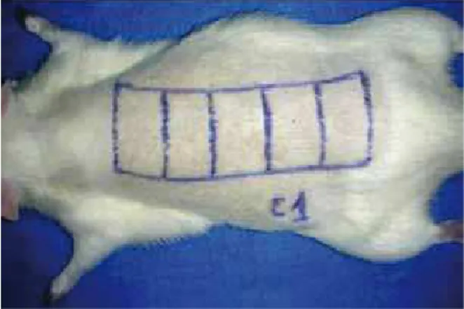
Anesthesia was induced by intraperitoneal administration of a mixture of ketamine (100 mg/ml) and xylazine (20 mg/ ml) at a total dose of 0.2 ml per 100 mg of body weight. The backs of the animals were shaved and cleaned with an anti -septic. Thereafter, a skin lap with a caudal pedicle (10 x 3 cm) was made on the back of each animal with sterile instruments; the iliac crests served as anatomical reference points for the beginning of each lap (Figure 1). The lap was elevated above the panniculus carnosus and then continuously resutured to
the wound bed using 4-0 monoilament nylon. The laps were sutured in place on the wound bed with markings placed every 2 cm along the lap length in order to facilitate observation of the necrotic area (Figures 1 and 2).
The animals were randomly divided into three groups, with 10 animals in each group: control group, to which only the vehicle solution was administered; sildenail group, to which a solution containing sildenail was administered; and bulomedil group, to which a solution containing bulomedil was administered.
The suspensions were previously prepared in a laboratory and the drug vehicle consisted of a 1:1 mixture of 1% carbo -xymethylcellulose and a simple syrup, prepared according to the Brazilian Pharmacopoeia IV-4th ed. (1988). Seven bottles
containing 300 mL of the vehicle were prepared. Four bottles were used in the preparation of the suspensions, as described below, and three were reserved for the control group.
The sildenail suspension was prepared by grinding 30 Viagra® tablets (50 mg) into a ine powder, which was suspended in the above-mentioned vehicle to form a paste. The paste was subsequently mixed with 1% carboxymethyl -cellulose and simple syrup in a test tube to obtain a inal
Figure 1 – Flap (10 x 3 cm) created on the back of a rat with transverse marks every 2 cm to observe the necrotic area.
Rev. Bras. Cir. Plást. 2011; 26(3): 390-3
392
Figueiredo JCA et al.
volume of 600 mL (2.5 mg/mL, final concentration) and stored in two opaque plastic bottles (300 mL/bottle).
The buflomedil suspension was prepared by mixing a fine powder of 5 tablets of Bufedil® 300 mg with the base vehicle of a 1:1 mixture of 1% carboxymethylcellulose and a simple syrup to form a paste. This paste was transferred to a test tube, and the volume was made up to 600 mL to obtain a concentration of 2.5 mg/mL. The suspension was then transferred to two opaque plastic bottles of 300 mL each.
The solutions were administered by oral gavage. The first dose was administered in the immediate postoperative period and the remaining doses were administered once a day, for 7 days. The daily dose of sildenafil and buflomedil was 10 mg/kg, with an equivalent volume of vehicle admi -nistered to the control group animals.
Post-surgery, the animals were kept in individual cages and the flaps were observed and photographed for seven days. At the end of this period, the rats were sacrificed. The viable area of the flap was designed and transposed to a paper carton (Figure 2), which was scanned and analyzed using Image Tool 2.0®, a specific software designed for
area calculation.
The flaps were cleaved and fixed in 10% formalin and a tissue slice, representing the viable area, was processed in a gradient of alcohol and xylene solutions. The dehydrated tissue was embedded in paraffin, sectioned (4 µm thick) and stained with hematoxylin and eosin. The images were scanned and the vessel diameters were calculated using Image Tool 2.0®.
The results were statistically analyzed using the Kruskal-Wallis test.
RESULTS
The average viable areas of the laps from each group were 16.2 + 3.56 cm² (control group), 17.69 + 2.54 cm² (bulomedil group), and 18.28 + 3.74 cm² (sildenail group) (Tables 1 and 2, Figure 3). Data analysis showed no statistically signiicant difference among the three groups (H = 1.6475; degrees of freedom = 2; P = 0.4388).
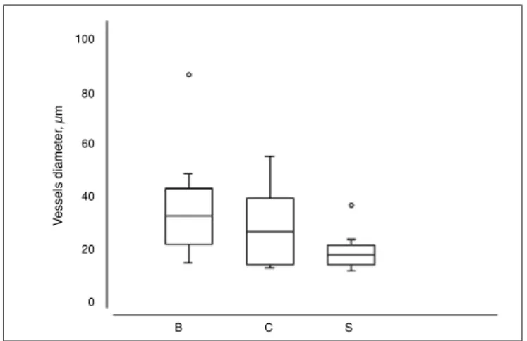
The average vessel diameter observed in flaps from the buflomedil group was 38.35 µm (median = 34.25 µm; standard deviation = 19.85); from the control group, 27.1 µm (median = 24.75 µm; standard deviation = 12.8); and from the sildenafil group, 19.9 µm (median = 18.5 µm; standard deviation = 6.99). Statistical analysis indicated a statistical difference in at least one of the groups (P = 0.02); the post-test, using the method of Dunn’s multiple comparisons, revealed significant differences between the buflomedil and sildenafil groups, without a significant difference between either treatment group and the control groups (Figure 4).
DISCUSSION
The data demonstrated that bulomedil’s reported bene -icial effect on microcirculation was not correlated with a reduction in ischemia in tissue laps. However, some studies
Table 1 – Viable lap areas from individual animals (cm²) Rats (n = 10) Control
group
Sildenail group
Bulomedil group
1 12.34 14.84 18.85 2 20.87 22.54 18.79
3 14.4 18.53 17.9
4 12.04 13.55 18.48 5 11.39 15.75 13.13 6 15.75 14.69 20.23 7 20.58 21.66 17.47 8 19.37 24.18 21.61 9 17.72 16.53 14.95 10 17.54 20.53 15.53
n = total number of rats in each group.
Table 2 –Average viable areas of laps after seven days (cm²).
Group Average + standard
deviation
Control 16.2 + 3.56
Bulomedil 17.69 + 2.54 Sildenail 18.28 + 3.74
V
ia
b
le
a
re
a
,
c
m
2
C S B
Figure 3 – Analysis of the viable areas, with no signiicant statistical difference observed among the three groups
(H = 1.6475, degrees of freedom = 2; P = 0.4388). B = bulomedil; C = control; S = sildenail.
35
30
25
20
15
Rev. Bras. Cir. Plást. 2011; 26(3): 390-3 393 Inluence of sildenail and bulomedil on survival of randomized laps in rats
Correspondence to: Adivânia de Souza Pinheiro
Rua Santa Cruz, 398 – Vila Mariana – São Paulo, SP, Brazil – CEP 04122-000 E-mail: [email protected]
Figure 4 – Microvessel diameter calculations from representative histological sections of the viable areas of laps from animals in the bulomedil, control, and sildenail groups. The Kruskal-Wallis
(P = 0.02) test indicated statistically different results between the bulomedil and sildenail groups (P < 0.05). The results of the bulomedil and sildenail groups did not differ signiicantly
from the results observed in the control group. B = bulomedil; C = control; S = sildenail.
100
80
60
40
20
0
V
essels diameter
,
µ
m
B C S
have shown conlicting results3,6,7,11. The studies that reported
reduced tissue ischemia tested bulomedil at a dose of 3 mg/ kg, intraperitoneally6,7 or intravenously3,8. As the oral route
was used in this study, a higher dose (10 mg/kg) of drug was administered to account for potentially poor absorption from the gastrointestinal tract. In humans, it is known that only approximately 30% of the drug is absorbed through the gastrointestinal tract. Therefore, although doses of 50–200 mg/ day are recommended when dosed intravenously, those doses are increased to 300–600 mg/day when administered orally12.
The ineffectiveness of bulomedil in this study was not unprecedented. A study conducted by Quirinia et al.4
indi-cated the failure of this drug to increase lap viability in rats at doses of 10 mg/kg using the intraperitoneal route. The eficacy of this drug for other applications (e.g., the treatment of intermittent claudication) is currently being questioned due to conlicting published results13.
Sildenail, however, is a drug with vasodilating effects that could potentially beneit randomized laps. Although sildenail still has been infrequently evaluated for this indi -cation, two relatively recent studies have suggested some degree of randomized lap viability improvement in rats5,9.
However, in the study of Hart et al.5, the action of the drug
throughout the postoperative period was followed and the observed improvement in tissue viability on the third posto -perative day did not remain statistically different, relative to the control group, through the ifth and seventh days. Although the administration route and dosages were similar to these studies, a signiicant improvement in the area of lap necrosis was not evidenced, compared to the control and bulomedil groups.
CONCLUSION
The use of bulomedil or sildenail failed to show a reduc -tion in the necrotic area of randomized laps in rats.
REFERENCES
1. Kerrigan CL. Skin lap failure: pathophysiology. Plast Reconstr Surg. 1983;72(6):766-77.
2. Myers MB, Cherry G. Causes of necrosis in pedicle laps. Plast Reconstr Surg. 1968;42(1):43-50.
3. Galla TJ, Saetzler RK, Hammersen F, Messmer K. Increase in skin-lap survival by the vasoactive drug bulomedil. Plast Reconstr Surg. 1991;87(1):130-6.
4. Quirinia A, Gottrup F, Viidik A. Failure of bulomedil to improve wound healing in ischaemic skin laps. Scand J Plast Surg Hand Surg. 1996;30(2):81-7.
5. Hart K, Baur D, Hodam J, Lesoon-Wood L, Parham M, Keith K, et al. Short- and long-term effects of sildenail on skin lap survival in rats. Laryngoscope. 2006;116(4):522-8.
6. Dias LC, Foustanos A, Carreirão S, Souza Filho S, Pitanguy I. Inlu
-ência do bulomedil na viabilidade de retalhos cutâneos. (Bulomedil inluence in skin laps viability) Rev Bras Cir. 1990;80(1):49-55. 7. Mauad RJ Jr, Shimizu MH, Mauad T, Tolosa EM. Bulomedil and
pentoxifylline in the viability of dorsal cutaneous laps of rats treated with nicotine. J Plast Reconstr Aesthet Surg. 2006;59(4):387-92. 8. Saetzler RK, Lerh HA, Barker JH, Kamler M, Galla TJ, Messmer K.
Visualization of nutritive perfusion following tourniquet ischemia in ar
-terial pattern skin laps: effect of vasoactive medication. Plast Reconstr Surg. 1994;94(5):652-60.
9. Sarifakioglu N, Gokrem S, Ates L, Akbuga UB, Aslan G. The inluence of sildenail on random skin lap survival in rats: an experimental study. Br J Plast Surg. 2004;57(8):769-72.
10. Um SC, Suzuki S, Toyokuni S, Kim BM, Tanaka T, Hiai H, et al. In
-volvement of nitric oxide in survival of random pattern skin lap. Plast Reconstr Surg. 1998;101(3):785-92.
11. Uhl E, Rösken F, Curri SB, Menger MD, Messmer K. Reduction of skin lap necrosis by transdermal application of bulomedil bound to liposomes. Plast Reconstr Surg. 1998;102(5):1598-604.
12. Fredj GM, Clenet M, Rousselet F. Dosage du bulomédil dans les milieux biologiques: détermination des différents paramètres pharmacociné
-tiques. Therapie. 1978;33(3):321-32.