Received: January 31, 2012 Accepted: April 17, 2012
Conflict of Interests: The authors state that there are no financial and personal conflicts of interest that could have inappropriately influenced their work.
Copyright: © 2012 Damman et al.; licensee EDIPUCRS. This is an Open Access article distributed under the terms of the Creative Commons Attribution-Noncommercial-No Derivative Works 3.0 Unported License.
Coronal microleakage of restorations with or
without cervical barrier in root-filled teeth
Microinfiltração coronária de restaurações com ou sem barreira
cervical em dentes endodonticamente tratados
Daniela Damman a
Renata Grazziotin-Soares a
Ana Paula Farina a
Doglas Cecchin a
a Department of Restorative Dentistry, School of
Dentistry of Passo Fundo, University of Passo Fundo (UPF), Passo Fundo, RS, Brazil
Correspondence:
Doglas Cecchin
Universidade de Passo Fundo – Campus I – Faculdade de Odontologia
Bairro São José, 285 Passo Fundo, RS – Brazil 99052-900
E-mail: [email protected]
Abstract
Purpose: To evaluate the ability sealing of glass ionomer and composite resin with or without 1-mm thickness of Coltosol on the root-canal filling material.
Methods: Root-canal treatment was completed on 50-extracted human mandibular unirradicular. The teeth were divided into six groups: G1, positive control; G2, negative control; G3, glass ionomer (Vidrion R); G4, Coltosol + Vidrion R; G5, composite resin; and, G6, Coltosol + composite resin. For G1 and G2 five teeth each were used and for the other groups, ten teeth each. The teeth were thermocycled and evaluated for microleakage using methylene blue dye. Specimens were sectioned and measurements made to the maximum point of dye penetration. The mean dye penetration (%) for each group was compared by ANOVA and Tukey’s test for post-hoc comparisons (α=0.05).
Results: The means and standard deviations of microleakage were: G1, 96.56 (±2.31); G2, 0.00 (±0.00); G3, 68.76 (±24.63); G4, 24.42 (±8.33); G5, 20.06 (±9.35); and, G6, 12.86 (±6.08).
Conclusion: It was concluded that none of the materials were able to prevent microleakage. Composite resin alone or combined with coltosol and glass ionomer associated with coltosol resulted in less microleakage than the glass ionomer used alone.
Key words: Coronal microleakage; adhesive system; composite resin; temporary fillings
Resumo
Objetivos: Avaliar a capacidade de selamento do ionomero de vidro e resina composta, com ou sem 1 mm de coltosol, sobre o material obturador endodôntico.
Métodos: O tratamento endodôntico foi realizado em 50 dentes humanos mandibulares unirradiculares. Os dentes foram divididos em 6 grupos: G1, controle positivo; G2, controle negativo; G3, ionômero de vidro (Vidrion R); G4, Coltosol + Vidrion R; G5, resina composta; e, G6, Coltosol + resina composta. Os dentes foram submetidos a termociclagem e a avaliação da microinfiltração usando azul de metileno. Os especimes foram seccionados e a quantidade maxima de penetração de corante avaliado. As medias e desvios-padrão de penetração do corante (em %) para cada grupo foram analisadas pelo teste ANOVA e pelo teste de multiplas comparações de Tukey’s (α=0.05).
Resultados: As medias e desvios-padrão de midroinfiltração foram: G1: 96,56 (±2,31); G2: 0,00 (±0,00); G3: 68,76 (±24,63); G4: 24,42 (±8,33); G5: 20,06 (±9,35); and, G6: 12,86 (±6,08).
Conclusão: Pode-se concluir que nenhum material obturador preveniu completamente a microinfiltração. Resina composta utilizada isoladamente ou associada ao coltosol e o ionômero de vidro associado ao coltosol apresentaram menor microinfiltração do que o ionomero de vidro utilizado sozinho.
Introduction
The sealing in the access cavity during endodontic
treatment is important in order to prevent the entrance of
saliva and microorganisms into the root-canal system (1).
However, studies showed that coronal microleakage can
occur around temporary restorations (2). If the coronal
restoration becomes defective or is lost, the coronal leakage
can compromise the success of root canal therapy (3,4). Ray
and Trope (5) found that the quality of the coronal seal is just
as important as the technical quality of the root canal illing
for periapical health after root canal therapy.
Coronal microleakage introduces the oral microlora
into the root canal system, which can eventually lead to
the failure of the endodontic treatment (3,4). To reduce
microleakage Roghanizad and Jones (6) suggested placing a
coronal seal in the oriice of the root canal immediately after
root canal illing. According to Schwartz and Fransman (7)
oriice barriers provide a second line of defence against the
leakage of bacteria. In an
in vivo
study, Yamauchi et al. (8)
reported a substantial reduction in apical periodontitis when
coronal plugs were used.
In relation to the temporary illing coronary studies have
shown that Coltosol was signiicantly better in preventing
microleakage other temporary materials (2,9). However, the
hygroscopic expansion of Coltosol in a cavity may lead
to cusp delection, infraction development and fracture.
Furthermore,
in vivo
masticatory forces will aggravate
this unfavourable condition. Therefore this material is not
recommended for temporary illing in root-illed teeth (10).
Adhesive dentistry concepts have increasingly been
applied to endodontics to prevent coronal leakage. Some
characteristics as ease and speed of placement, sealing
eficacy, and high bond strength qualify the ideal restorative
material to barrier (11), and the use of dentin bonding agents
has been advocated to help provide a better intracoronal
seal (12). Therefore the aim of this
in vitro
study was to
evaluate the ability sealing of glass ionomer and composite
resin with or without 1-mm thickness of Coltosol on the
root-canal illing material.
Methodology
A total of 50 extracted single-rooted human maxillary
anterior teeth were collected under a protocol reviewed
and approved by the Ethics Committee for Human Studies,
Passo Fundo School of Dentistry, FO-UPF, Brazil. After
extraction, teeth were stored in 0.1% thymol solution for
no more than 1 month. Organic debris and calculus were
detached with scalers (Hu-Friedy Co; Chicago, IL, USA),
and standard access to the pulp chamber was performed and
pulp tissue was removed with a barbed broach (Dentsply
Maillefer, Ballaigues, Switzerland). A crown-down root
canal preparation was performed using Gates-Glidden
drills sizes 5, 4, 3 and 2 (Dentsply Maillefer) placed to a
length where resistance was met in the coronal and middle
thirds of the root canal. This was followed by step-back
instrumentation of the apical third to create a size 45 apical
stop. Root canals were irrigated during instrumentation using
5 mL of 2.5% sodium hypochlorite (NaOCl) (Natufarma
Pharmacy; Passo Fundo, RS, Brazil) solution and rinsed
with 3 mL of 17% ethylenediaminetetraacetic acid (EDTA)
(Natufarma Pharmacy) for 5 min to remove the smear layer.
Subsequently, a inal lush with 10 mL of distilled water
(Natufarma Pharmacy) was performed to wash out the EDTA
solution, and root canals were then dried with paper points
(Dentsply Maillefer).
The teeth were obturated with gutta-percha and Endo-Fill
(Dentsply, Petrópolis, Rio de Janeiro, Brazil) using lateral
compaction. After illing the root canal, excess material was
removed with a heated instrument Dulex number 2 (SS
White; Rio de Janeiro, RJ, Brazil) 1 mm below root canal
oriices. The root canal sealer was removed from the pulp
chamber with cotton pellets soaked in 70% isopropyl alcohol
(Natufarma Pharmacy).
Forty teeth were randomly assigned to 4 experimental
groups (n=10/experimental group), and 10 teeth were
randomly assigned to 2 control groups (n=5/control group)
as follows: group 1, positive control; group 2, negative
control; group 3, glass ionomer chemically cured (Vidrion R,
S. S. White Artigos Dentários Ltda., Rio de Janeiro, Brazil);
group 4, a 1-mm intracanal barrier of Coltosol (Colten,
Langenau, Germany) + Vidrion R; group 5, adhesive system
(Adper Scotchbond Multi-Purpose, 3M ESPE, St Paul, MN)
associated with composite resin (Z250, 3M ESPE); group
6, a 1-mm intracanal barrier of Coltosol + adhesive system
and composite resin.
The glass ionomer was placed into the pulp chamber by a
syringe (Centrix Incorporated, Shelton, USA). Coltosol was
placed with a number 1 spatula Dulex (SS White). In the
Scotch Bond group, the specimens were etched with 37%
phosphoric acid for 15 s, washed for 10 s and gently dried with
cotton pellets; a thin layer of Primer (3M ESPE) and Bond
(3M ESPE) was applied and light cured for 20 s (Ultraled,
Dabi Atlante, Ribeirao Preto, SP, Brazil – power density:
500 mW cm
-2). Composite resin was inserted by incremental
technique and light cured for 20 s each increment.
The teeth received 3 layers of nail polish (Dote Belulla
Cosméticos Ltda; Diadema, SP, Brazil), leaving only the
area of canal’s oriice exposed to provide uniform control of
any lateral or accessory canals. The cavit access was restored
with resin composite and all surfaces of the negative control
group were completely sealed with 3 layers of nail polish.
The positive control did not receive material in access cavity.
Then they were longitudinally sectioned in a buccolingual
direction using a low-speed diamond saw under constant
water-cooling. Under a D.F. Vasconcellos microscope (São
Paulo, SP, Brazil) at ×20 magniication, methylene blue dye
penetration was measured in millimetres at both sides of the
specimen as an indicator of coronal microleakage.
The total length of the illing within the root-canal
obturation, and the greatest depth of dye penetration along
each canal were recorded in millimetres. These measurements
were converted to percentages of microleakage related to the
total length of the root illing for each root canal. The means
and standard deviations of microleakage were calculated,
and the data were analysed using ANOVA and Tukey’s test
for post-hoc comparisons (
α
=0.05).
Results
The degree of dye penetration for each group is presented
in Table 1. There was no dye penetration for teeth in the
negative control group, whereas the positive control group
showed dye penetration in all specimens.
Table 1. Mean percentages of microleakage for each group
Material Mean microleakage (%)* Positive control 96.56 (2.31) a
Negative control 0.00 (0.00) d
Vidrion R 68.76 (24.63) b
Coltosol + Vidrion R 24.42 (8.33) c Composite resin 20.06 (9.35) c,d Coltosol + Composite resin 12.86 (6.08) c,d
* Different letters in superscript mean statistically significant differences (P<0.05).
Coltosol + composite resin, composite resin and
Coltosol + Vidrion R sealed signiicantly better than the
other groups (
P
<0.05). Vidrion R exhibited the highest
microleakage of the experimental groups (
P
<0.05). All
experimental groups showed a statistically signiicant better
seal (
P
<0.05) than the positive control group, independent
of the sealing material used.
Discussion
Studies have shown that gutta-percha and root canal
sealers cannot prevent the passage of saliva and bacteria
to the periapical tissues (4,5,13). The results of the current
study show that all samples obturated with gutta-percha and
sealer without restoration (positive control group) exhibited
extensive dye penetration. Therefore, after obturation of
the root canal system, the occlusal access cavity should be
properly sealed to improve the prognosis of endodontically
treated teeth.
Restorative dentistry concepts using adhesive materials
have increasingly been applied to endodontics to prevent
coronal leakage (2,11), such as use of ibre posts in the
restoration of endodontically treated teeth (14), and even for
the illing of the root canal (15). The use of these materials is
based on the premise that because of the intimate contact with
dentine, these materials could remain micromechanically
retained, reinforcing tooth structure and preventing root
contamination (16). Therefore, the present study focused
on the sealing ability of composite resin and glass ionomer
materials associated with a thickness of 1.0 mm of Coltosol
in the entrance root canal.
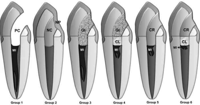
Fig. 1. Schematic illustration of the groups under study. PC: positive control; NC: negative control; NP: nail polish; GI: glass ionomer;
The results of this
in vitro
study indicated composite
resin and Coltosol + composite resin provided a better seal
than glass ionomer, and no difference was found with the
negative control. This fact is consistent with some other
leakage studies that used adhesives materials for sealing (2,
17-20). Uranga et al. (18) reported that composite resin did
not demonstrate any leakage, whereas other experimental
materials leaked signiicantly more. Leonard et al. (19)
showed that dentin-bonding agents and resin seal more
completely than glass ionomer. Bonded restorations are also
recommended by Howdle et al. (20) to decrease leakage.
The possibility of direct restoration of non-vital teeth
with resin-based composites has increased owing to
the development of adhesive systems (21). These have
increased bond strength of composite resins to dentin
(22), and produced less microleakage (23). It is assumed
that these bondings improve the adhesive capability
and bonding strength of resins to the tooth structure by
promoting penetration, impregnation, and entanglement
of the coupling agents into dentinal substrates where they
polymerise in situ and create zones of resin-reinforced dentin
layers (24).
The glass ionomer used without Coltosol, whose setting
is an acid-base reaction, showed poor sealing ability, as
reported by Zaia et al. (2). This is probably due to shrinkage
of the material upon setting, resulting in a potential avenue
for microleakage and its use may be more technique
sensitive (2). Moreover, shrinkage during setting as well
as the presence of a smear layer can adversely affect the
coronal seal ability of glass ionomer (18). The use of EDTA
is recommended to remove the smear layer.
In this study, the use of a 1-mm thickness of Coltosol
to seal the pulp chamber after root-canal treatment reduced
coronary microleakage. The excellent results obtained by
an root canal barrier Coltosol sealing material agree with
previous indings (2,9). Coltosol is a pre-manipulated
material composed of a single paste free of eugenol, which
hardens when exposed to moisture. It is widely used
as a temporary restorative material in endodontics and
has outstanding sealing properties when compared with
other materials for the same purpose (2,9). Coltosol is a
hygroscopic cement that expands when it comes into contact
with moisture. This expansion provides good adaptation
between the restorative material and cavity walls (2,10,25).
According Laustsen et al. (10) the expansion of Coltosol
might cause stress in the material as well as the surrounding
walls. The stress might partly dissipate because of expansion
of material out of the cavity, by a deformation of the walls
and by creep or other stress-releasing mechanism. When the
stress-induced deformation reaches a certain limit, cracks
will develop both in the inner part of the dentine walls, as
well as between enamel and dentine, which may lead to
fracture of the tooth. Due to the good sealing capability of
the Coltosol, it is recommend the use of 1-mm thickness
of Coltosol to seal the pulp chamber, and the rest of the
cavity must be sealed with composite resin because this
material has the ability to reduce microleakage and also has
satisfactory mechanical properties.
It was concluded that none of the materials were able
to prevent microleakage in all specimens. Composite
resin alone or combined with coltosol and glass ionomer
associated with coltosol resulted in less microleakage than
the glass ionomer used alone.
Acknowledgements
The authors deny any inancial afiliations related to this
study or its sponsors.
1. Swanson K, Madison S. An evaluation of coronal microleakage in endodontically treated teeth: part I – time periods. J Endod 1987;13:56-9.
2. Zaia AA, Nakagawa R, De Quadros I, Gomes BP, Ferraz CC, Teixeira FB, et al. An in vitro evaluation of four materials as barriers to coronal microleakage in root-filled teeth. Int Endod J 2002;35:729-34.
3. Heling I, Gorfil C, Slutzky H, Kopolovic K, Zalkind M, Slutzky-Goldberg I. Endodontic failure caused by inadequate restorative procedures: review and treatment recommendations. J Prosthet Dent 2002;87:674-8.
4. Saunders WP, Saunders EM. Coronal leakage as a cause of failure in root canal therapy: a review. Endod Dent Traumatol 1994;10:105-8.
5. Ray HA, Trope M. Periapical status of endodontically treated teeth in relation to the technical quality of the root filling and the coronal restoration. Int Endod J 1995;28:12-8. 6. Roghanizad N, Jones JJ. Evaluation of coronal microleakage after endodontic treatment.
J Endod 1996;22:471-3.
7. Schwartz RS, Fransman R. Adhesive dentistry and endodontics: materials, clinical strategies and procedures for restoration of access cavities: a review. J Endod 2005;31:151-65. 8. Yamauchi S, Shipper G, Buttke T, Yamauchi M, Trope M. Effect of orifice plugs on periapical
inflammation in dogs. J Endod 2006;32:524-6.
9. Madarati A, Rekab MS, Watts DC, Qualtrough A. Time-dependence of coronal seal of temporary materials used in endodontics. Aust Endod J 2008;34:89-93.
10. Laustsen MH, Munksgaard EC, Reit C, Bjørndal L. A temporary filling material may cause cusp deflection, infractions and fractures in endodontically treated teeth. Int Endod J 2005;38:653-7.
11. Santos J, Tjäderhane L, Ferraz C, Zaia A, Alves M, De Goes M, et al. Long-term sealing ability of resin-based root canal fillings. Int Endod J 2010;43:455-60.
12. Fathi B, Bahcall J, Maki JS. An in vitro comparison of bacterial leakage of three common restorative materials used as an intracoronal barrier. J Endod 2007;33:872-4.
13. Shipper G, Teixeira FB, Arnold RR, Trope M. Periapical inflammation after coronal microbial inoculation of dog roots filled with gutta-percha or Resilon. J Endod 2005;31:91-6. 14. Cecchin D, Almeida JF, Gomes BP, Zaia AA, Ferraz CC. Effect of chlorhexidine and
ethanol on the durability of the adhesion of the fiber post relined with resin. J Endod 2011;37:678-83.
15. Cecchin D, Souza M, Carlini-Junior B, Barbizam JVB. Bond strength of Resilon/Epiphany compared with Gutta-percha and sealers Sealer 26 and Endo Fill. Aust Endod J 2012;38:21-5.
16. Teixeira FB, Teixeira ECN, Thompson JY, Trope M. Fracture resistance of endodontically treated roots using a new type of resin filling material. J Am Dent Assoc 2005;135:646-52. 17. Jenkins S, Kulild J, Williams K, Lyons W, Lee C. Sealing ability of three materials in the orifice
of root canal systems obturated with gutta-percha. J Endod 2005;32:225-7.
18. Uranga A, Blum JY, Esber S, Parahy E, Prado C. A comparative study of four coronal obturation materials in endodontic treatment. J Endod 1999;25:178-80.
19. Leonard JE, Gutmann JL, Guo IY. Apical and coronal seal of roots obturated with a dentine bonding agent and resin. Int Endod J 1996;29:76-83.
20. Howdle MD, Fox K, Youngson CC. An in vitro study of coronal microleakage around bonded amalgam coronal-radicular cores in endodontically treated molar teeth. Quintessence Int 2002;33:22-9.
21. Hernandez R, Bader S, Boston D, Trope M. Resistance to fracture of endodontically treated premolars restored with new generation dentine bonding systems. Int Endod J 1994;27:281-4.
22. Pashley DH, Tay FR, Imazato S. How to increase the durability of resin-dentin bonds. Compend Contin Educ Dent 2011;32:60-4.
23. Deliperi S, Bardwell DN, Wegley C. Restoration interface microleakage using one total-etch and three self-etch adhesives. Oper Dent 2007;32:179-84.
24. Goerig AC, Mueninghoff LA. Management of the endodontically treated tooth. Part II: Technique. J Prost Dent 1983;49:491-7.