ORIGIN
AL RESEAR
CH
Mailing address: Luana Godinho Maynard – Department of Physiotherapy, Tiradentes University (UNIT) – Av. Murilo Dantas, 300, Farolândia – Aracaju (SE), Brazil – CEP 49030-270 – E-mail: [email protected] – Funding source: Nothing to declare – Conlict of interest: Nothing to declare – Presentation: Mar. 2015– Accepted for publication: Aug. 2016– Approved by the Ethics Committee CEP/UNIT: 070510; ACTRN12610000786099.
Study carried out at Tiradentes University (UNIT) and presented in I Congresso Nordestino (I CONEFIR) & 4º Congresso
Pernambucano de Fisioterapia Cardiorrespiratória e Fisioterapia em Terapia Intensiva (IV COPEFIR), Pôster. Nov. 3, 2012, Jaboatão dos Guararapes, Pernambuco, Brazil. Published in ASSOBRAFIR Ciência. 2012 Dec;3(Suppl):39-87 – Aracaju (SE), Brazil.
1Department of Physiotherapy, Tiradentes University – UNIT, Aracaju (SE), Brazil.
2Department of Education and Health, Federal University of Sergipe – UFS, Lagarto (SE), Brazil. 3Department of Physiotherapy, Federal University of Sergipe – UFS, São Cristóvão (SE), Brazil.
4Department of Physiology, Ribeirão Preto Medical School, University of São Paulo – USP, Ribeirão Preto (SP), Brazil.
ABSTRACT | The transcutaneous electrical diaphragmatic stimulation (TEDS) is a technique of respiratory muscle activation that afects breathing pattern and rhythm. In an attempt to evaluate changes in cardiac autonomic balance in response to TEDS in healthy individuals, we used a well-established TEDS model. Twenty-two volunteers aged between 22 and 35 years old, with no cardiac pathology history, were randomized into two groups (control, n = 8; TEDS, n = 14). The individuals were allowed to rest in supine position and were then subjected to the electrical stimulation protocol. The control group was subjected to electrical stimulation at perceptive level, whereas for the TEDS group the electric stimulus generated diaphragm contraction. Cardiac intervals (CI) were sampled by a Polar RS800CX monitor. Cardiac interval variability was studied in the time and frequency domains. In the control group, electrical stimulation did not change cardiac interval length and variability (CI: 761±44 vs. 807±39 ms; RMSSD: 37±9 vs. 42±13 ms ; LF: 69±6 vs. 67±5 nu; HF: 31±6 vs. 33±5 nu; all comparisons versus baseline). Nevertheless, as compared to baseline, TEDS group showed decreased sympathetic cardiac modulation (LF: 43±3 vs. 63±4 nu) and increased parasympathetic cardiac modulation (RMSSD: 109±10 vs. 41±6 ms; HF: 57±3 vs. 37±4 nu) during diaphragmatic stimulation. However, cardiac interval length was not changed by electrical stimulation (CI:
248
686±59 vs. 780±31 ms). It can be suggested that the use of TEDS stimulus leads to pronounced changes in the cardiac sympathovagal balance, with higher parasympathetic cardiac modulation, possibly induced by increased diaphragmatic excursion.
Keywords | Transcutaneous Electrical Nerve Stimulation; Respiratory Mechanics; Autonomic Nervous System; Cardiovascular Physiological Phenomena; Physical Therapy Specialty.
RESUMO | A estimulação diafragmática elétrica transcutânea (EDET) é uma técnica de mobilização da musculatura respiratória que interfere no padrão e no ritmo respiratório. Na tentativa de avaliar as alterações no balanço autonômico cardíaco à EDET em indivíduos saudáveis, foi utilizado um modelo já estabelecido de eletroestimulação diafragmática. 22 voluntários com idades entre 22 e 35 anos, sem histórico cardíaco, foram randomizados em dois grupos (controle, n=8; EDET, n=14). O protocolo de eletroestimulação foi aplicado nos indivíduos em repouso (posição supina). O grupo controle foi submetido a estimulação elétrica em nível perceptivo, enquanto no grupo EDET o estímulo gerava contração diafragmática. Os intervalos cardíacos (CI) foram registrados por cardiofrequencímetro Polar (RS800CX). A variabilidade do intervalo cardíaco foi estudada nos domínios de tempo e frequência. No grupo
Efects of transcutaneous electrical diaphragmatic
stimulation on the cardiac autonomic balance in
healthy individuals: a randomized clinical trial
Efeitos da estimulação diafragmática elétrica transcutânea sobre o balanço autonômico
cardíaco de indivíduos hígidos: ensaio clínico randomizado
Los efectos de la estimulación eléctrica transcutánea del diafragma en el balance autónomo
cardiaco en individuos sanos: estudio clínico aleatorio
Luana Godinho Maynard
1, André Sales Barreto
2, Valter Joviniano Santana-Filho
3,
controle, a estimulação elétrica não alterou a duração do intervalo cardíaco e sua variabilidade (CI: 761±44 vs. 807±39ms; RMSSD: 37±9 vs. 42±13ms; LF: 69±6 vs. 67±5nu; HF: 31±6 vs. 33±5nu), em comparação às condições basais). No entanto, o grupo EDET apresentou diminuição na modulação simpática cardíaca (LF: 43 ±3 vs. 63±4nu) e aumento da modulação parassimpática cardíaca (RMSSD: 109±10 vs. 41±6ms; HF: 57±3 vs. 37±4nu) durante a eletroestimulação diafragmática. No entanto, a duração do intervalo cardíaco não foi alterada por estimulação elétrica (CI: 686±59 vs. 780±31ms). Pode-se sugerir que o uso da EDET promove mudanças acentuadas no balanço simpatovagal, resultando em maior modulação parassimpática cardíaca, possivelmente induzida pelo aumento da mobilidade diafragmática.
Descritores | Estimulação Elétrica Nervosa Transcutânea; Mecânica Respiratória; Sistema Nervoso Autônomo; Fenômenos Fisiológicos Cardiovasculares; Fisioterapia.
RESUMEN | La estimulación eléctrica transcutánea del diafragma (EETD) es un método que moviliza la musculatura respiratoria que interiere en el patrón y en la frecuencia respiratoria. Para evaluar las alteraciones en el balance autónomo cardiaco por la EETD en individuos sanos, se empleó un modelo prestablecido de estimulación eléctrica muscular del diagrama. Han participado 22 voluntarios con edad entre 22 y 35 años, sin enfermedades cardiacas, puestos en grupos aleatorios: Grupo control (n=8) y
EETD (n=14). Se aplicó el protocolo de estimulación eléctrica en individuos en posición decúbito supino. Al grupo control se lo sometió a la estimulación eléctrica a nivel perceptual, mientras que en el EETD la estimulación le generaba contracción del diafragma. Los intervalos cardiacos (CI) se registraron a través del monitor de ritmo cardiaco polar (RS800CX). Se estudió la variabilidad del intervalo cardiaco en los dominios tiempo y frecuencia. En el grupo control, la estimulación eléctrica no presentó alteraciones en la duración del intervalo cardiaco y su variabilidad (CI: 761±44 vs. 807±39ms; RMSSD: 37±9 vs. 42±13ms; LF: 69±6 vs. 67±5nu; HF: 31±6 vs. 33±5nu; comparado con las condiciones de base). Pero si comparado las condiciones de base en el grupo EETD presentó una disminución en la modulación simpática cardiaca (LF: 43±3 vs. 63±4nu) y un aumento en la modulación parasimpática cardiaca (RMSSD: 109±10 vs. 41±6ms; HF: 57±3 vs. 37±4nu) durante la realización de este método. Sin embargo, la duración del intervalo cardiaco no presentó alteraciones por la estimulación eléctrica del diafragma (CI: 686±59 vs. 780±31ms). Se puede concluir que el empleo de la EETD promueve cambios signiicativos en el balance simpático, resultando en una modulación parasimpática cardiaca más grande, posiblemente inducida del aumento de la movilidad del diafragma.
Palabras clave| Estimulación Eléctrica Transcutánea del Nervio; Mecánica Respiratoria; Sistema Nervioso Autónomo; Fenómenos Fisiológicos Cardiovasculares; Fisioterapia.
INTRODUCTION
Under physiological conditions, the proper
functioning of the respiratory system depends on
appropriate strength and endurance of respiratory
muscles
1. Any dysfunction that compromise the
functional excursion of the diaphragm, the main
inspiratory muscle, will reduce the operational lung
volume and lead to changes in ventilation/perfusion
ratio, which is crucial for appropriate gas exchange
1,2.Inspiratory muscle weakness reduces lungs’ inlation
capacity, leading to decreased overall lung capacity.
However, the literature shows that endurance and
strength training
3of inspiratory muscles could be
helpful in reversing the dyspnea and exercise intolerance
arising from respiratory muscle weakness
4,5.
he transcutaneous electrical diaphragmatic
stimulation (TEDS) is one of the techniques used for
respiratory muscle training, aiming an increase in strength
and endurance of respiratory muscles
6-8. In brief, the
TEDS consists on applying rhythmic electrical stimuli
with a short duration biphasic waveform pulse through
electrodes placed on the surface of the skin, i.e., diaphragm
area, causing an inspiratory contraction of the diaphragm
muscle
9,10.It is noteworthy to mention that this is a
noninvasive technique that allows normal diaphragmatic
breathing patterns, with no damage to muscle ibers
11.
he inluence of the breathing luctuation on the
heart rate variability (HRV) is described in clinical trials
8.
Studies show that luctuations in the respiratory frequency
have major efect on heart rate (HR) and its variability,
a physiological phenomenon known as respiratory sinus
arrhythmia (RSA)
12-14. he increase in the intrathoracic
aferents
15,16. hen, eferent impulses to the heart modulate
cardiac rhythm during the respiratory cycle, producing a
tachycardic response during the inspiratory phase and a
bradycardic response during the expiratory phase
12. hese
coupled luctuations are known as RSA
14.
he RSA relects the synchronism between HRV
and respiration by the cardiac vagal outlow shortened
during inspiration and prolonged during expiration
14.
his phenomenon seems to be commanded by two
major mechanisms involving respiratory and circulatory
centers in the brainstem. First, the inhibition of cardiac
vagal eferent activity by lung inlation, which evokes
tachycardia by stimulating the pulmonary C-iber
aferents, i.e., pulmonary stretch receptors
14,15,17; second,
the acetylcholine-mediated inhibitory postsynaptic
potential in the cardiac vagal preganglionic neurons
by central respiratory drive, which makes neurons less
responsive to excitatory inputs during inspiration
18-20.
Over the last years, the analysis of HRV has been
extensively used to evaluate the autonomic modulation
of the cardiovascular system
21-23. he information
regarding the cardiac autonomic modulation obtained
from HRV analysis has been proven to be accurate
and feasible
24. HRV can be examined in the time and
frequency domains. Among the various existing time
domain measures of HRV, we can mention the RMSSD
(root mean square of successive diferences), which is
correlated with rapid changes in the HR and relects the
parasympathetic modulation of the heart
25. Furthermore,
HRV can be evaluated in the frequency domain by
spectral analysis. HR oscillations at low frequency (i.e.,
when expressed in normalized units) are accepted as an
index of cardiac sympathetic modulation, whereas high
frequency oscillations of HR are considered to relect
parasympathetic modulation of the heart
22. he LF/HF
ratio has been used to assess sympathovagal balance
22.
herefore, this study was carried out to evaluate
the efects of TEDS, a technique used for respiratory
muscle training on the cardiac autonomic modulation
of healthy individuals.
METHODOLOGY
Individuals and experimental design
A convenience sample (n=30) was invited to
participate and was tested for the inclusion criteria.
Eight subjects were excluded and the remaining 22 were
assigned into the study. Experimental protocols were
conducted at the Rehabilitation Center of Tiradentes
University (UNIT), Aracaju, Sergipe, Brazil. Inclusion
criteria were as follows: (1) aged from 22 to 35 years old;
(2) enrolled in a moderate physical exercise program (no
more than once a week); (3) absence of cardiovascular
diseases. We excluded individuals who were taking drugs
known to inluence the cardiac autonomic modulation,
as well as individuals taking tobacco and alcohol.
Eligibility standards were not changed throughout the
whole study.
hose who met the eligibility criteria were randomly
assigned into two groups as follows: Control group
(subjected to electrical stimulation at perceptive level;
n=8; 2 men, 6 women) and TEDS group (electric
stimulus generated diaphragm contraction; n=14; 4 men,
10 women). he unbalanced randomization between
groups (1:1.75) was preferred, since individuals assigned
to the TEDS group could present low acceptability to
the electrical stimulation protocol, leading to leave the
study. he lowchart of participants through the study is
shown in Figure 1.
Intervention and data collection
One day before the beginning of the experimental
protocol, the individuals underwent a session of
habituation to the procedures (i.e., synchronizing the
breathing rate to the electrical stimulation frequency
and resting in supine position during the whole
experimental protocol) to reduce stress. Participants
were asked to avoid drinking cafeinated beverages or
taking strenuous exercise in the 12 hours prior to the
test. Research trials were conducted in the morning (8
a.m. to 12 p.m.), in the months of May and June of
2010. Prior to investigation, the research protocol was
verbally explained and volunteers were given written
information. All participants signed the informed
consent form. All experimental protocols performed
in this study were approved by the Research Ethics
Committee of Tiradentes University – UNIT (Protocol
#070510; CEP/UNIT).
Group randomization was performed by the
sealed envelope technique
26,27. In order to keep group
experimental trial, the researcher opened an envelope
revealing the allocation to each group. We asked the
participants’ age, measured their height and weight to
calculate their body mass index, and kept them blind to
the group assignment. hen, they were instrumented
with a wrist HR monitor (RS800CX, Polar Electro
Inc., Lake Success, New York, USA), equipped with
a transmitter (Polar Electro Inc., Lake Success, New
York, USA) that was tied around their chests. For
implementation of TEDS, skin electrodes (5×5 cm)
were bilaterally placed (Figure 2A) in the sixth to
eighth intercostal space, between the anterior axillary
line and the midaxillary line
10. Using an electrical
stimulator (FesVif® 995 Dual, Quark, Piracicaba,
São Paulo, Brazil), TEDS was performed with the
following parameters: pulse frequency of 30 Hz, pulse
width of 2 ms, ON time 2 s, OFF time 3 s. We have
chosen the minimum current capable of eliciting
palpable contraction of the diaphragm muscle, and
participants were instructed to keep their BR constant
at 12 cpm during the TEDS protocol (Figure 2B) by
following a metronome. Individuals in the Control
group had their BR monitored and counted by the
researcher.
Data analysis
We evaluated cardiac autonomic balance by cardiac
interval variability (CIV) analysis. For that purpose,
cardiac intervals were sampled through a HR monitor
(RS800CX, Polar Electro Oy, Kempele, Finland) during
the 5 minutes prior to TEDS therapy, in the course of
TEDS (12 minutes), and during the next 5 minutes
following TEDS.
Following data acquisition, we exported
beat-to-beat series of cardiac intervals and HR from the HR
monitor to a computer
software (Polar Pro Trainer 5,
Kempele, Finland
) by an infrared interface.
he cardiac
interval variability was assessed in the time-frequency
domain by a software (
HRV Kubios, Department of
Physics, University of Kuopio, Kuopio, Finland
),
which allowed the precise adjustment of parameters
related to the time-frequency domain analysis (e.g. time
period for analysis, interpolation rate, segment length,
and boundaries of frequency bands). Non-interpolated
beat-by-beat time series of cardiac intervals were used
for RMSSD evaluation. However, for the assessment of
cardiac interval variability in the frequency domain, the
time series were converted to data points every 250 ms
using cubic spline interpolation (4 Hz). he interpolated
series were divided into half-overlapping sequential sets
of 256 data points (64 s). A Hanning window was used to
attenuate side efects and all segments had the spectrum
calculated using a direct Fast Fourier Transform (FFT)
algorithm for discrete time series. Finally, the spectra
were integrated in low-frequency (LF; 0.04
–
0.15
Hz) and high-frequency (HF; 0.15
–
0.4 Hz) bands,
and results were obtained in normalized units (nu). To
assess the sympathovagal balance, the LF/HF ratio was
calculated
21.
Statistical analysis
he results are shown as mean±SEM (standard
error of the mean). Demographic and hemodynamic
characteristics were compared between groups using
Student’s t test. he efects of TEDS on cardiac
autonomic modulation were assessed by one-way analysis
of covariance (ANCOVA). When appropriate,
post-hoc comparisons were performed by Bonferroni’s test.
Diferences were considered signiicant when p < 0.05. All
statistical tests were performed with SigmaPlot software
v13.0 (Systat Software Inc., San Jose, California, USA).
RESULTS
he studied groups showed no diferences in the
demographic and hemodynamic characteristics (Table
1). Table 2 shows the HR and breathing rate (BR) values
obtained before, during, and after electrical stimulation on
control (n=8) and TEDS (n=14) groups. No diferences
were observed in HR and BR between groups.
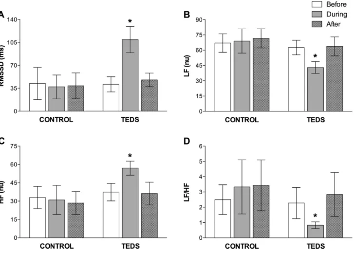
Figure 3 shows the RMSSD; the power of the LF and
HF bands of the cardiac interval spectrum; and the LF/
HF ratio before, during, and after electrical stimulation
in control (n
=
8) and TEDS (n
=
14) groups. Before the
electrical stimulation, both groups showed equivalent
values for all parameters. We observed no diferences
in the RMSSD, LF, HF, and LF/HF ratio following
electrical stimulation in the control group. Conversely,
we observed an increase in RMSSD and HF power,
and a decrease in LF power and LF/HF ratio in TEDS
group after electrical stimulation. After ceasing electrical
stimulation, all parameters were restored to baseline levels
in TEDS group.
Table 1. Demographic and hemodynamic characteristics
Variables Control (n=8) TEDS (n=14) p
Gender (M/F) 2/6 4/10 ---Age (y) 27±1.7 (23–30) 27±1.3 (24–29) 0.908 BMI (Kg/m2) 22±0.9 (20–24) 23±0.7 (22–24) 0.527
SBP (mmHg) 111±4.0 (103–119) 107±2.9
(102–113) 0.416 DBP (mmHg) 70±3.3 (64–76) 69±2.7 (63–74) 0.742
M – male; F – female; BMI – body mass index; SBP – systolic blood pressure; DBP – diastolic blood pressure. Values are expressed as mean±standard error of mean (95% conidence interval)
Table 2. Heart rate and breathing rate values obtained before, during, and after electrical stimulation in Control and TEDS groups
Before During After
Control HR (bpm; n=8) 76±3.5
(69–82) 81±5.0 (71–91)
74±2.5 (69–79)
TEDS HR (bpm; n=14) 78±2.6 (73–83)
78±2.5 (72–83)
75±2.2 (71–80)
Control BR (cpm; n=8) 13±0.8
(11–14) 12±0.0
12±0.8 (11–14)
TEDS BR (cpm; n=14) 12±0.4
(12–13) 12±0.0 12±0.3 (11–12)
HR – heart rate; BR – breathing rate; bpm – beats per minute; cpm – cycles per minute. Values are expressed as mean±standard error of mean (95% conidence interval)
Figure 3. Cardiac interval variability. Root mean square of successive diferences (RMSSD; Panel A); power of the low frequency (LF; Panel B) and high frequency (HF; Panel C) bands of the cardiac interval spectrum; and LF/HF ratio (Panel D) before, during, and after electrical stimulation in Control and TEDS groups. Values are expressed as mean and 95% conidence intervals. * p<0.05 vs. TEDS before
DISCUSSION
he results of this study show that TEDS elicited
diferent changes in the CIV parameters in both
groups studied. control groups presented no changes
of electrical stimulation, all CIV parameters were
restored to baseline.
he studied groups showed no diferences in the
demographic and hemodynamic characteristics (Table
1). We observed no diferences in HR and BR before,
during, and after electrical stimulation in both groups
(Table 2).
In our study, TEDS was able to reduce cardiac
sympathetic modulation (43
±
3 vs. 63
±
4
nu) and
increase cardiac parasympathetic modulation (57
±
3
vs. 37
±
4
nu). he results suggest that transcutaneous
electric stimulation of the diaphragm at perceptive level
(control group) does not change cardiac autonomic
balance. In addition, we observed a decrease in LF/
HF ratio (0.82
±
0.11 vs. 2.28
±
0.52) during electrical
stimulation in TEDS group, as compared to data
obtained in baseline conditions, suggesting a shift
of sympathovagal balance towards parasympathetic
predominance.
he large variety of HR monitors has aroused the
curiosity of the comparison of its reproducibility with
the conventional electrocardiogram (ECG).
Wrist-worn models are worldwide employed because of their
practical use and low-cost, in addition to the advantage
of being used during free movement or dynamic
physical activity in exercise and sports practice. On the
clinical setting, data acquisition should be performed
using a controlled, reliable, and practical method. Riscili
et al.
28have shown that HR data acquisition by Polar
heart monitors is a safe and feasible method that can be
used without any interference in cardiac functionality.
he literature show that R-R interval (period between
two successive R waves in the ECG) data samples with
5 minutes of duration, obtained with HR monitors,
can be used for HRV analysis
29,30. Some studies have
already shown that HRV analysis, i.e., frequency
domain, performed on data sampled with HR monitors
over a short period (short-term) or with ECG devices
over a long period (long-term), produces comparable
and accurate results
31-34. In addition, Pimentel et al.
34showed that HR monitors can be used for R-R interval
sampling during exercise, a condition marked by
pronounced changes in the sympathovagal balance.
Our indings clearly showed that TEDS produces
changes in the cardiac autonomic modulation with
a shift towards parasympathetic predominance, i.e.,
decrease in the LF/HF ratio.
his suggests that the perception of electric current
applied is not responsible for changes in CIV, but the
negative pressure of inlation triggered by diaphragm
contraction after the application of the stimulus on
motor points
35. he cardiac rhythmicity is continually
modulated by the autonomic nervous system, leading
to oscillations in the cardiac interval length that can be
evaluated on a beat-by-beat basis
21,22.Since the power of the HF band of the cardiac
interval spectrum and the HF peak location at the
spectrum are associated with respiration, we can say
that they are inluenced by changes in RSA. From our
results, we can speculate that TEDS stimulus possibly
increased inspiratory volume without noticeable
changes in BR, contributing to changes in RSA.
Studies in the literature
36,37show that changes
in respiratory parameters can inluence markedly
the RSA amplitude. In addition, increases in RSA
contribute to an extended action of acetylcholine on
muscarinic receptors in the sinoatrial node, slowing
down the nerve conduction during expiration
38. In this study,BR remained unchanged during and after TEDS, as
compared to baseline values. Hence, it is suggested that
the changes in the CIV observed in this study may be
due to an increase in depth of breathing generated by
diaphragmatic electrical stimulation.
Corroborating our indings, studies conducted
on chronic obstructive pulmonary disease patients
revealed that, when BR is acutely reduced, changes in
the cardiac sympathovagal balance are observed, i.e.,
reduced sympathetic activity and a trend to an increased
barorelex sensitivity
39.Study limitations and implications
he current trial had a small sample size and the
methodology for BR assessment could be improved by
using a respiration monitor belt around the individuals’
chest. However, our indings contribute to a better
understanding of the autonomic cardiac modulation
during TEDS in healthy adults.
CONCLUSION
TEDS. hus, it can be suggested that TEDS stimulus
can be efectively used not only for respiratory muscles
training, but also as a technique to improve the cardiac
sympathovagal balance in patients.
REFERENCES
1. Hassoun PM, Celli BR. Bilateral diaphragm paralysis secondary to central von Recklinghausen’s disease. Chest. 2000 [acesso em 27 out. 2016];117(4):1196-200. Disponível em: http://bit.ly/2dPZnEb
2. Moreno MA, Catai AM, Teodori RM, Borges BLA, Cesar MC, Silva E. Efect of a muscle stretching program using the Global Postural Reeducation method on respiratory muscle strength and thoracoabdominal mobility of sedentary young males. J Bras Pneumol. 2007 [acesso em 27 out. 2016];33(6):679-86. Disponível em: http://bit.ly/2fk5GVU 3. Reid WD, Dechman G. Considerations when testing and
training the respiratory muscles. Phys Ther. 1995 [acesso em 27 out. 2016];75(11):971-82. Disponível em: http://bit. ly/2eKVXXO
4. Shoemaker MJ, Donker S, Lapoe A. Inspiratory muscle training in patients with chronic obstructive pulmonary disease: the state of the evidence. Cardiopulm Phys Ther J. 2009 [acesso em 27 out. 2016];20(3):5-15. Disponível em: http://bit.ly/2eKitx2
5. Barbalho-Moulim MC, Miguel GPS, Forti EMP, Campos FA, Costa D. Efects of preoperative inspiratory muscle training in obese women undergoing open bariatric surgery: respiratory muscle strength, lung volumes, and diaphragmatic excursion. Clinics (São Paulo). 2011 [acesso em 27 out. 2016];66(10):1721-7. Disponível em: http://bit.ly/2fbDRQf
6. Robinson AJ, Snyder-Mackler L. Eletroisiologia clínica: eletroterapia e teste eletroisiológico. Artmed; 2002.
7. Forti EMP, Pachani GP, Montebelo MIL, Costa D. Transcutaneous diaphragmatic electrostimulation in healthy individuals. Fisioter Bras. 2005 [acesso em 27 out. 2016];6(4):261-4. Disponível em: http://bit.ly/2eKdcpp 8. Costa D, Forti EMP, Barbalho-Moulim MC, Rasera-Junior
I. Study on pulmonary volumes and thoracoabdominal mobility in morbidly obese women undergoing bariatric surgery, treated with two diferent physical therapy methods. Braz J Phys Ther. 2009 [acesso em 27 out. 2016];13(4):294-301. Disponível em: http://bit.ly/2e1sDey
9. Geddes LA, Voorhees WD, Babbs CF, Deford JA. Electroventilation. Am J Emerg Med. 1985 [acesso em 27 out. 2016];3(4):337-9. Disponível em: http://bit.ly/2eAdWj7 10. Cuello A, Masciantonio L, Mendoza S. Estimulación
diafragmática eléctrica transcutânea. Med Intensiva. 1991 [acesso em 27 out. 2016];8(4):194-202. Disponível em: http:// bit.ly/2eRihNj
11. Pavlovic D, Wendt M. Diaphragm pacing during prolonged mechanical ventilation of the lungs could prevent from respiratory muscle fatigue. Med Hypotheses. 2003 [acesso em 27 out. 2016];60(3):398-403. Disponível em: http://bit. ly/2eRkI2F
12. Bernardi L, Spadacini G, Bellwon J, Hajric R, Roskamm H, Frey AW. Efect of breathing rate on oxygen saturation and exercise performance in chronic heart failure. Lancet. 1998 [acesso em 27 out. 2016];351(9112):1308-11. Disponível em: http://bit.ly/2efJ2tl
13. Stauss HM. Heart rate variability. Am J Physiol Regul Integr Comp Physiol. 2003 [acesso em 27 out. 2016];285(5):R927-31. Disponível em: http://bit.ly/2fbBmNY
14. Yasuma F, Hayano J-I. Respiratory sinus arrhythmia: why does the heartbeat synchronize with respiratory rhythm? Chest. 2004 [acesso em 27 out. 2016];125(2):683-90. Disponível em: http://bit.ly/2eAhfHa
15. Guz A, Innes JA, Murphy K. Respiratory modulation of left ventricular stroke volume in man measured using pulsed Doppler ultrasound. J Physiol (Lond). 1987 [acesso em 27 out. 2016];393:499-512. Disponível em: http://bit.ly/2eV105y 16. Taha BH, Simon PM, Dempsey JA, Skatrud JB, Iber C.
Respiratory sinus arrhythmia in humans: an obligatory role for vagal feedback from the lungs. J Appl Physiol. 1995 [acesso em 27 out. 2016];78(2):638-45. Disponível em: http://bit.ly/2eRlRqW
17. Daly MD, Kirkman E. Cardiovascular responses to stimulation of pulmonary C ibres in the cat: their modulation by changes in respiration. J Physiol (Lond). 1988 [acesso em 27 out. 2016];402:43-63. Disponível em: http://bit.ly/2eWdqsC 18. Cohen MA, Taylor JA. Short-term cardiovascular oscillations
in man: measuring and modelling the physiologies. J Physiol (Lond). 2002 [acesso em 27 out. 2016];542(Pt 3):669-83. Disponível em: http://bit.ly/2fbGIbE
19. Eckberg DL. The human respiratory gate. J Physiol (Lond). 2003 [acesso em 27 out. 2016];548(Pt 2):339-52. Disponível em: http://bit.ly/2e1v2pq
20. Denver JW, Reed SF, Porges SW. Methodological issues in the quantiication of respiratory sinus arrhythmia. Biol Psychol. 2007 [acesso em 27 out. 2016];74(2):286-94. Disponível em: http://bit.ly/2eUU5JA
21. Montano N, Ruscone TG, Porta A, Lombardi F, Pagani M, Malliani A. Power spectrum analysis of heart rate variability to assess the changes in sympathovagal balance during graded orthostatic tilt. Circulation. 1994 [acesso em 27 out. 2016];90(4):1826-31. Disponível em: http://bit.ly/2eKPvQH 22. Task Force. Heart rate variability: standards of measurement,
physiological interpretation and clinical use. Task Force of the European Society of Cardiology and the North American Society of Pacing and Electrophysiology. Circulation. 1996 [acesso em 27 out. 2016];93(5):1043-65. Disponível em: http://bit.ly/2eKeOPT
23. Lanfranchi PA, Somers VK. Arterial barorelex function and cardiovascular variability: interactions and implications. Am J Physiol Regul Integr Comp Physiol. 2002 [acesso em 27 out. 2016];283(4):R815-26. Disponível em: http://bit.ly/2eRkMiF 24. Lombardi F, Malliani A, Pagani M, Cerutti S. Heart rate
variability and its sympatho-vagal modulation. Cardiovasc Res. 1996 [acesso em 27 out. 2016];32(2):208-16. Disponível em: http://bit.ly/2eUXAjm
SUDEP-7 Inventory. Epilepsy Behav. 2010 [acesso em 27 out. 2016];19(1):78-81. Disponível em: http://bit.ly/2eUWDHy 26. Viera AJ, Bangdiwala SI. Eliminating bias in randomized
controlled trials: importance of allocation concealment and masking. Fam Med. 2007 [acesso em 27 out. 2016];39(2):132-7. Disponível em: http://bit.ly/2dPTXc8
27. Torgerson DJ, Roberts C. Randomisation methods: concealment. BMJ. 1999 [acesso em 27 out. 2016];319(7206):375-6. Disponível em: http://bit.ly/2e1vLXF 28. Riscili CE, Hinds M, Voorhees WD, Bourland JD, Geddes
LA. The safety factor for electroventilation measured by production of cardiac ectopy in the anesthetized dog. Chest. 1989 [acesso em 27 out. 2016];95(1):214-7. Disponível em: http://bit.ly/2eKdU68
29. Sinnreich R, Kark JD, Friedlander Y, Sapoznikov D, Luria MH. Five minute recordings of heart rate variability for population studies: repeatability and age-sex characteristics. Heart. 1998;80 [acesso em 27 out. 2016] (2):156-62. Disponível em: http://bit.ly/2eRfbS
30. Min KB, Min J-Y, Paek D, Cho S-I, Son M. Is 5-minute heart rate variability a useful measure for monitoring the autonomic nervous system of workers? Int Heart J. 2008 [acesso em 27 out. 2016];49(2):175-81. Disponível em: http:// bit.ly/2dPYQCc
31. Gamelin FX, Berthoin S, Bosquet L. Validity of the polar S810 heart rate monitor to measure R-R intervals at rest. Med Sci Sports Exerc. 2006 [acesso em 27 out. 2016];38(5):887-93. Disponível em: http://bit.ly/2dPUyuo
32. Vanderlei LCM, Silva RA, Pastre CM, Azevedo FM, Godoy MF. Comparison of the Polar S810i monitor and the ECG for the analysis of heart rate variability in the time and frequency domains. Braz J Med Biol Res. 2008 [acesso em 27 out. 2016];41(10):854-9. Disponível em: http://bit.ly/2dPUWsV
33. Porto LGG, Junqueira LF Jr. Comparison of time-domain short-term heart interval variability analysis using a wrist-worn heart rate monitor and the conventional electrocardiogram. Pacing Clin Electrophysiol. 2009 [acesso em 27 out. 2016];32(1):43-51. Disponível em: http://bit. ly/2eRhdco
34. Pimentel AS, Alves ES, Alvim RO, Nunes RT, Costa CMA, Lovisi JCM, et al. Polar S810 as an alternative resource to the use of the electrocardiogram in the 4-second exercise test. Arq Bras Cardiol. 2010 [acesso em 27 out. 2016];94(5):580-4. Disponível em: http://bit.ly/2dPZjUY
35. Fernandes G. A Eicácia de um protocolo utilizando a estimulação diafragmática elétrica transcutânea (Edet) sobre a força muscular do diafragma, avaliada através da Pimax, e sobre a expansibilidade torácica, veriicada através da cirtometria [Course Completion Assignment]. [Cascavel, PR, Brazil]: Universidade Estadual do Oeste do Paraná; 2004 [acesso em 27 out. 2016]. Disponível em: http://bit.ly/2dMtt0i 36. Brown TE, Beightol LA, Koh J, Eckberg DL. Important
inluence of respiration on human R-R interval power spectra is largely ignored. J Appl Physiol. 1993 [acesso em 27 out. 2016];75(5):2310-7. Disponível em: http://bit.ly/2eUX7xv 37. Kobayashi H. Normalization of respiratory sinus arrhythmia
by factoring in tidal volume. Appl Human Sci. 1998 [acesso em 27 out. 2016];17(5):207-13. Disponível em: http://bit. ly/2eKh35R
38. Giardino ND, Glenny RW, Borson S, Chan L. Respiratory sinus arrhythmia is associated with eiciency of pulmonary gas exchange in healthy humans. Am J Physiol Heart Circ Physiol. 2003 [acesso em 27 out. 2016];284(5):H1585-91. Disponível em: http://bit.ly/2dPZEqz