MICROLEAKAGE ASSOCIATED WITH
RETRO-GRADE FILLING AFTER ROOT END RESECTION
(in vitro study)
Elka Radeva1, Tsonko Uzunov1, Dimitar Kosturkov2
1) Department of Conservative Dentistry, Faculty of Dental Medicine, Medical University - Sofia
2) Student 6th course, Faculty of Dental Medicine - Sofia, Medical University – Sofia, Bulgaria.
Journal of IMAB - Annual Proceeding (Scientific Papers)2014, vol. 20, issue 3 Journal of IMAB
ISSN: 1312-773X
http://www.journal-imab-bg.org
SUMMARY:
The purpose of the study is to compare microleakage after root end resection of the two materials (MTA and Biodentine) for two different apical cavity preparation using the method of penetration of dye - 0, 2 % Rodamine B. Ma-terials and Methods: Forty-eight extracted single-rooted human teeth were used in this study. The resection was made at 3 mm from the root tip with a high speed diamond bur at an angle of 90 degree to the long axis of the tooth. For the retrofilling, ProRoot MTA and Biodentine were used. The teeth were divided into 5 groups: 1st group (10 teeth) – the apical cavity was prepared with stainless steel fissure bur #10 at 3 mm depth in the root canal parallel to the long axis of the tooth and is filled retrograde with MTA. 3rd group (10 teeth) - retrofilling with Biodentine. 2 nd group (10 teeth) -with a round bur apical cavity was prepared -with a concave shape and cavity along the root canal with a depth of 3 mm and retrograde obturation with MTA. 4th group (10 teeth) -retrofilling with Biodentine. 5th group (8 teeth) - control group - with preparation of the cavity after resection with-out retrofilling. The with-outer surface of the root is covered with two layers of varnish, with the exception of the apical 3 mm then immersed in 0.2% Rodamine B for 72 h. The degree of penetration of the dye is measured in millimeters. Results: Relative highest median value of penetration of the dye in mm is in the control group. MTA group has a higher value in mm versus the Biodentine. The apical preparation with a concave shape and cavity along the root canal with a depth of 3 mm after apicoectomy is important to reduce apical microleakage. Conclusion: Different apical cavity prepara-tions in both types of material have led to the microleakage dye, but to varying degrees.
Key words: apical microleakage, bevel angle, Biodentine, endodontic surgery, root-end filling materials, MTA
INTRODUCTION
In cases, when healing process in peri-radicular tis-sues does not occur after conventional endodontic treatment or it is impossible to carry out re-treatment, apicoectomy is needed to implement in order to eliminate the root apex and
removal of the pathological tissues of the periapical area. 2. Resection of the root tip 3. Apical root canal preparation. 4. Retrograde filling of the root canal [5].
Kim and Kratchman [8] reported that resection of 3 mm root tip reduced the apical ramification to 98% and lat-eral canals to 93%. In conventional techniques the resection is at an angle of 45 or 30 degrees. In modern techniques 0 -10 degrees resection is recommended, which reduces the number of exposed dentinal tubules [2, 6, 7, 8].
Root-end cavity preparation is an important procedure in periapical surgery. In conventional techniques, the apical preparation was performed with a round bur [1, 6, 7]. In our country no comparative studies for microleakage in differ-ent from traditional apical preparation after root tip resection have been carried out so far.
The primary goal in apical resection is to perform a hermetic sealing between the apical portion of the root ca-nal and periapical tissue by retrograde root end filling. Many materials have been used for root-end fillings in endodontic surgery - amalgam, glass ionomer cements (Vitremer), zinc oxide-eugenol based materials (SuperEBA, IRM, Rickert), mineral trioxide aggregate - MTA, zinc - phosphate cements, calcium hydroxide cements (Sealapex, Sealer 26) sealer based on epoxy resins (AH 26, AH Plus) [6, 9, 10, 11, 12].
MTA is well known and studied material [4, 13, 14]. It has good biological compatibility as well as good sealing properties. Its drawbacks associated with its long setting time (170 min) and difficulties in its application are also familiar. Some new calcium-silicate materials that appear in recent years are trying to compensate for MTA disadvantages. One of these materials is Biodentine (Septodont, France). It has a reduced curing time (12-15 min) and better handling prop-erties. The main difference between Biodentine and MTA is the absence of calcium aluminate and calcium sulphate in Biodentin formation. It is known that these compounds tend to reduce the mechanical strength and determine longer cur-ing time, typically for MTA [13, 15, 16].
Biodentine is a relatively new material (since January 2011) and there have not been many studies and observations of its use in endodontic surgery up to now, which is why it was included in recent study.
MATERIALS AND METHODS
Forty-eight extracted single- rooted human teeth were used in this study. The resection was made at 3 mm from the root tip with a high speed diamond bur at an an-gle of 90 degree to the long axis of the tooth. For the retrofilling, ProRoot MTA and Biodentine were used. The
teeth were divided into 5 groups:
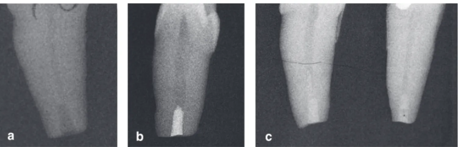
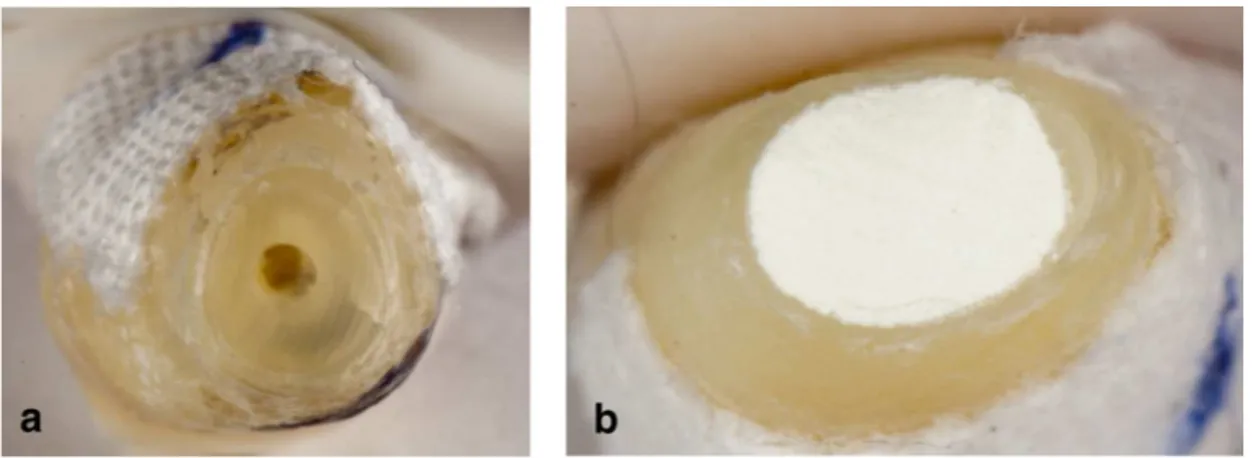
First group (10 teeth) – the apical cavity was prepared with stainless steel fissure bur #10 at 3 mm depth in the root canal parallel to the long axis of the tooth (fig. 1a, fig. 2a) and is filled retrograde with MTA. (fig. 1b, fig. 2b).
Fig.1a. The apical cavity was prepared with straight fissure bur #10 at 3 mm depth in the root canal. b. Retrograde filling with ProRoot MTA. c. Retrograde filling with Biodentine
Fig. 2 View after apical resection a. After preparation of the root canal with straight fissure bur b. Retrograde filling with MTA.
Second group (10 teeth) - with a round bur apical cav-ity was prepared with a concave shape and cavcav-ity along the
root canal with a depth of 3 mm (fig. 3a, fig. 4a) and retro-grade obturation with MTA fig. 3b, fig. 4b).
Fig. 4 View after apical resection a. After preparation of the root canal with round bur b. After retrograde filling with MTA.
Third group (10 teeth) - The apical cavity was pre-pared with straight fissure bur #10 at 3 mm depth in the root canal parallel to the long axis of the tooth. Retrofilling with Biodentine (fig 1c).
Fouth group (10 teeth) - The apical cavity was pre-pared with a round bur at 3 mm depth with a concave shape. Retrofilling with Biodentine (fig. 3c).
Fifth group (8 teeth) - control group - with prepara-tion of the cavity after resecprepara-tion without retrofilling.
The outer surface of the root is covered with two lay-ers of varnish, with the exception of the apical 3 mm, then immersed in 0.2% Rodamine B for 72 h. The teeth were washed under tap water for 24 h. The degree of penetration of the dye is measured in millimeters.
After this procedure, slices (fig. 5) were prepared by means of a microtom Leica SP 1600.
Fig. 5. The degree of dye penetration (a & b)
a. Presence of dye microleakage
The data was input and processed using the statistical software package SPSS 19.0.1. The level of significance for rejecting the null hypothesis was fixed at p<0.05.
The following methods were applied: Graphical analysis – for visualizing the results obtained. Shapiro-Wilk test – to check the normality distribution. Mann-Whitney nonparametric test – for checking hypotheses of difference between two independent samples.
RESULTS
The results were present in tables 1 - 2 and figures 6 - 7.
The data analysis in tabl. 1 and fig. 6 show that: • The average micro-leakage after apical resection was higher in treatment with straight fissure bur and steel in both materials (0.18mm for MTA and 0.10 mm for Biodentine). While preparing the apical cavity with round bur and con-cave shape the average means are respectively - 0.08 mm for MTA and 0.07 mm for Biodentine.
• The average microleakage after apical resection was
higher in MTA regardless of the methods of preparation (straight fissure bur - 0.18 mm, round bur - 0.08 mm).Tab. 1: Comparative analysis of microleakage after apical resection by two different apical preparation of two mate-rials (MTA and Biodentine)
Filling material
Retrograde cavity MTA Biodentine p
n X SD n X SD
Straight fissure bur 10 0,18 0,16 10 0,10 0,13 0,282
Round bur 10 0,08 0,09 10 0,07 0,11 0,651
Fig. 6: Comparative analysis of the impact of the type used in filling both methods of preparation on microleakage after apical resection
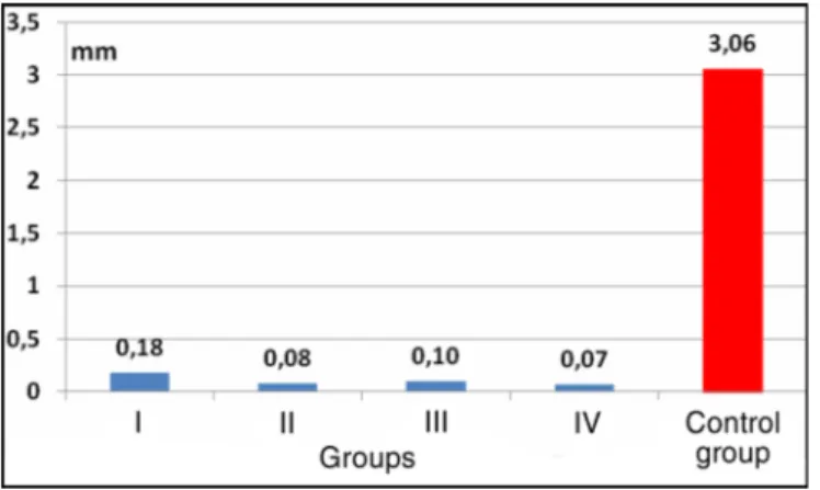
The average micro leakage after apical resection in the control group was greater (3.06 mm) than that of all other groups constituted according to the way of the processing and filling. The differences have significant character (table 2. and fig. 7.)
Table 2. Comparative analysis of microleakage after apical resection by two different apical preparations of two ma-terials (MTA and Biodentine) and control group
Control group Groups
n X SD n X SD p
I 10 0,18 0,16 8 3,06 0,61 <0,001
II 10 0,08 0,09 8 3,06 0,61 <0,001
III 10 0,10 0,13 8 3,06 0,61 <0,001
IV 10 0,07 0,11 8 3,06 0,61 <0,001
Fig. 7. Comparative analysis of microleakage after apical resection of two different apical preparations of two materials (ProRoot MTA and Biodentine) and control group
DISCUSSION:
Hermetic sealing of the apical part of the root canal and the minimum micro leakage of tissue fluids in this area are of great importance for the healing process after the en-dodontic surgery [17, 18, 19, 20].
The quality of apical seal obtained by root end filling materials has been assessed by the degree of penetration. There are several methods – chemical compounds (silver ni-trate), bacterial penetration, fluid filtration techniques and radioisotope penetration. But the most common ones use dyes (methylene blue, fuchsin, rhodamine B, fluorescent dyes). Using dyes is a simple and safe method for studying micro leakage, which is why we use it in current in-vitro study. This method has also been used by many researchers in their stud-ies [2, 3, 11, 19, 20, 21, 22].
measurement of microleakage in four different materials for retrograde filling - gray MTA, Adhesor, Astralloy, Adseal at an angle of 45 degrees and 3 mm apical resection using a traditional technique. They established that the lowest microleakage was observed for MTA Angelus - from 0.34 to 0.67 mm, while in amalgam the microleakage was most ex-pressed - from 2.8 to 0.44 mm [17].
In current study we have established that microleakage for MTA is 0.18 mm after apical preparation from 0 to 10 degrees by using stainless steel straight fissure bur and 0.08 mm for concave shape of apical cavity preparation. This con-firms the relation between apical preparation and the mate-rials used for retrograde filling as key to success.
Shashi S. et al. have received similar results. They compared the microleakage of methylene blue in MTA-white and gray and Portland cement – white and gray. They estab-lished minimal penetration in white MTA - from 0.18 mm to ±0.31 mm, which isn’t statistically important.
1. Radeva E, Usunov Ts. Endodon-tic surgical treatment-traditional and modern technique. Dental medicine. 2013; 95(2):186-192 (in Bulgarian).
2. Erkut S, Tanyel RC, Keklikoglu N, Yildirim S, Katiboglu AB. A com-parative study of retrograde filling ma-terials. Turk J Med Sci. 2006; 36(2): 113-120.
3. Harican J, Kavitha D, Narayanan L. SEM evaluation of two different root-end preparations and a compara-tive microleakage evaluation of three different retrofilling materials using two different root-end preparations by dye penetration method- an in vitro study. J Ind Aca Dent Spec. 2010 Jul-Sep;1(3):1-6.
4. Kokate SR, Pawar AM. An in vitro comparative stereomicroscopic evaluation of marginal seal between MTA, glass ionomer cement& biodentine as root end filling materials using 1% methylene blue as tracer. Endod. 2012 Dec;24(2):36-42.
5. Tsurumachi T. Current strategy for successful periradicular surgery. J
surgery with root-end filling: A meta-analysis. J Endod. 2010 Jun;36(6):957-973. [PubMed] [CrossRef]
7. Kim S, Kratchman S. Modern endodontic surgery concepts and prac-tice: a reviåw. J Endod. 2006 Jul; 32(7):601-623. [PubMed] [CrossRef]
8. Pop I. Oral surgery: part 2. En-dodontic surgery. Br Dent J. 2013 Sep; 215(6):279-286. [PubMed] [CrossRef] 9. Dammaschke T. Root-end filling with a new bioactive cement. Inside Dentistry. 2012 Mar;8(3).
10. McDonald NJ, Dumsha TC. Evaluation of the retrograde apical seal using dentine bonding materials. Int Endod J. 1990 May;23(3):156-162. [PubMed]
11. Shahi S, Yavari HR, Rahimi S, Eskandarinezhad M, Shakouei S, Unchi M. Comparison of the sealing ability of mineral trioxide aggregate and Portland cement used as root-end filling materials. J Oral Sci. 2011 Dec; 53(4):517-522. [PubMed] [CrossRef] 12. Xavier CB, Weismann R, de Oliveira MG, Demarco FF, Pozza DH. REFERENCES:
13. Caron G, Azerad J, Faure MO, Machtou P, Boucher Y. Use a new ret-rograde filling material (Biodentine) for endodontic surgery: two case re-ports. Int J Oral Sci. 2014 May 9. [PubMed] [CrossRef]
14. Soundappan S, Sundaramurthy JL, Natanasabapathy V. Biodentine ver-sus Mineral trioxide aggregate verver-sus Intermediate restorative material for retrograde root end filling: an in vitro study. J Dent (Tehran). 2014 Mar; 11(2):143-149. [PubMed]
15. Madfa AA, Sanabani FA, Al-Qudami Al-Kudami NH. Endodontic repair filling materials: A review arti-cle. Br J Med Med Res. 2014; 4(16): 3059-3079. [CrossRef]
16. Pawar AM, Kokate SR, Shah RA. Management of a large periapical lesion using Biodentine(™) as retro-grade restoration with eighteen months evident follow-up. J Conserv Dent. 2013 Nov;16(6):573-575. [PubMed] [CrossRef]
17. Kuzmanova Y, Nikiforova H. Microleakage of four root-end filling During the study, it has been established from the X-ray after retrograde filling that radio opacity for Biodentine is lower in comparison with MTA. The content of the radio pacifier - Zirconium oxide in Biodentine differs from the Bis-muth oxide as a radio pacifier in MTA [8, 9].
In current study MTA sealing abilities have been con-firmed and those of Biodentine have been proved in differ-ent cavity preparations after apical resection.
CONCLUSION:
Different apical cavity preparations in both types of material have led to the microleakage dye, but to varying de-grees. According to our results Biodentine can be more ef-fective material for retrograde filling comparing to MTA. Within the limit of this study we can conclude that the api-cal preparation with concave shape and cavity along the root canal in depth of 3 mm after apical resection is important for apical microleakage reduction.
Acknowledgements
The study was carried out at the laboratory for experimental investigations of dental materials of the Faculty of Den-tal Medicine at the Medical University, Sofia
Address for correspondence: Dr Elka Radeva, PhD
Department of Conservative dentistryFaculty of Dental Medicine, Medical University - Sofia;
1, St. Georgi Sofiyski blvd., 1431 Sofia, Bulgaria; Mobile phone: +359 888 319 813
around dental restorations. Dental medicine. 2012; 94(1):39-47 (in Bul-garian).
19. Saini D, Nadig G, Saini R. A comparative analysis of microleakage of three root end filling materials-an in vitro study. Arch Orofac Sci. 2008, 3(2):43-47.
20. Sharifian MR, Motahhari P,
Shahsia S. The effect of bevel angle on apical microleakage following the use of amalgam and MTA. Journal of Den-tistry. 2006; 19(2):43-48.
21. Post LK, Lima FG, Xavier CB, Demarco FF, Gerhardt-Oliveira M. Sealing ability of MTA and amalgam in different root-end preparations and
resection bevel angles: an in vitro evaluation using marginal dye leakage. Braz Dent J. 2010; 21(5):416-419. [PubMed] [CrossRef]
22. Fogel HM, Peikoff MD. Microleakage of root-end filling mate-rials. J Endod. 2001 Jul;27(7):456-459. [PubMed] [CrossRef]
Please cite this article as: Radeva E, Uzunov T, Kosturkov D. Microleakage associated with retrograde filling after root end resection (in vitro study). J of IMAB. 2014 Jul-Sep;20(3):578-583.
DOI: http://dx.doi.org/10.5272/jimab.2014203.578