Action Pathways and Survival in locoregionally Advanced
Nasopharyngeal Carcinoma Treated with
Chemoradiotherapy
Huai Liu
1,2☯, Bin Qi
3☯, Xiang Guo
1,2, Lin-Quan Tang
1,2, Qiu-Yan Chen
1,2, Lu Zhang
1,2, Ling Guo
1,2, Dong-Hua
Luo
1,2, Pei-Yu Huang
1,2, Hao-Yuan Mo
1,2, Yan-Qun Xiang
1,2, Fang Qiu
1,2, Rui Sun
1,2, Ying Zhang
1,4,
Ming-Yuan Chen
1,2, Yi-Jun Hua
1,2, Xing Lv
1,2, Lin Wang
1,2, Chong Zhao
1,2, Ka-Jia Cao
1,2, Chao-Nan Qian
1,2,
Ming-Huang Hong
1,5, Hai-Qiang Mai
1,2*1 State Key Laboratory of Oncology in South China, Guangzhou, P. R. China, 2 Department of Nasopharyngeal Carcinoma, Sun Yat-sen University Cancer Center, Guangzhou, P. R. China, 3 Department of Radiotherapy, Affilated Tumor Hospital of Guangzhou Medical College, Guangzhou, P. R. China, 4 Tumor Resources Bank, Sun Yat-sen University Cancer Center, Guangzhou, P. R. China, 5 Department of Epidemiology, Clinical Trial Study Center, Sun Yat-sen University Cancer Center, Guangzhou, P. R. China
Abstract
Background and Purpose: Treatment outcomes vary greatly in patients with nasopharyngeal carcinoma (NPC). The purpose of this study is to evaluate the influence of radiation and chemotherapy drug action pathway gene polymorphisms on the survival of patients with locoregionally advanced NPC treated with cisplatin- and fluorouracil-based chemoradiotherapy.
Material and Methods: Four hundred twenty-one consecutive patients with locoregionally advanced NPC were prospectively recruited. We utilized a pathway approach and examined 18 polymorphisms in 13 major genes. Polymorphisms were detected using the LDR-PCR technique. Multifactor dimensionality reduction (MDR) analysis was performed to detect potential gene-gene interaction.
Results: After adjustment for clinicopathological characteristics, overall survival was significantly decreased in patients with the MPO rs2243828 CT/CC genotype (HR=2.453, 95% CI, 1.687-3.566, P<0.001). The ERCC1
rs3212986 CC (HR=1.711, 95% CI, 1.135-2.579, P=0.010), MDM2 rs2279744 GT/GG (HR=1.743, 95% CI, 1.086-2.798, P=0.021), MPO rs2243828 CT/CC (HR=3.184, 95% CI, 2.261-4.483, P<0.001) and ABCB1 rs2032582 AT/AA (HR=1.997, 95% CI, 1.086-3.670, P=0.026) genotypes were associated with poor progression-free survival. Prognostic score models based on independent prognostic factors successfully classified patients into low-, intermediate-, and high-risk groups. Furthermore, MDR analysis showed no significant interaction between polymorphisms.
Conclusions: Four single nucleotide polymorphisms were associated with survival in patients with locoregionally advanced NPC treated with cisplatin- and fluorouracil-based chemoradiotherapy. Combining clinical prognostic factors with genetic information was valuable in identifying patients with different risk.
Citation: Liu H, Qi B, Guo X, Tang L-Q, Chen Q-Y, et al. (2013) Genetic Variations in Radiation and Chemotherapy Drug Action Pathways and Survival in locoregionally Advanced Nasopharyngeal Carcinoma Treated with Chemoradiotherapy. PLoS ONE 8(12): e82750. doi:10.1371/journal.pone.0082750
Editor: Xiaoping Miao, MOE Key Laboratory of Environment and Health, School of Public Health, Tongji Medical College, Huazhong University of Science and Technology, China
Received August 20, 2013; Accepted October 27, 2013; Published December 10, 2013
Copyright: © 2013 Liu et al. This is an open-access article distributed under the terms of the Creative Commons Attribution License, which permits unrestricted use, distribution, and reproduction in any medium, provided the original author and source are credited.
Funding: This research was supported by grants from the National Natural Science Foundation of China (No: 81072226, http://www.nsfc.gov.cn), the 863 Project (No: 2012AA02A501, http://www.863.gov.cn/), the National Key Basic Research Program of China (No: 2013CB910304, http://www.973.gov.cn), the Sci-Tech Project Foundation of Guangdong Province (No: 2011B080701034, http://gdsf.gdstc.gov.cn), the Sci-Tech Project Foundation of Guangzhou City (No: 2011J4300100, http://www.gzsi.gov.cn) and the Fundamental Research Funds for the central universities (http://www.sysu.edu.cn). The funders had no role in study design, data collection and analysis, decision to publish, or preparation of the manuscript.
Competing interests: The authors have declared that no competing interests exist. * E-mail: maihq@mail.sysu.edu.cn
Introduction
Prevalent in the south of China, nasopharyngeal carcinoma (NPC) has an ethnic and geographic distribution pattern that is distinctive from other head and neck cancers [1,2]. NPC is highly responsive to radiotherapy and systemic chemotherapy. Based on the results from several large prospective clinical trials using cisplatin- and fluorouracil (5-FU)-based chemoradiotherapy (CRT) [3-5], the combined treatment has become the standard regimen for locoregionally advanced NPC. However, even with this multimodal approach, approximately 30% of patients experience treatment failure within 5 years [4], and treatment outcomes vary widely, even among patients of the same clinical stage. Therefore, prospectively identifying patients who will have better or worse outcomes after CRT would aid in designing appropriate treatment strategies, highlighting the need for improved predictive markers.
The expression and activity levels of the critical enzymes related to radiation and chemotherapy drug response could affect treatment outcomes. Nucleotide excision repair (NER) and base excision repair (BER) are two important mechanisms involved in the repair of non-specific DNA damage induced by radiation and chemotherapy. The major enzymes involved are ERCC1, ERCC2 (NER), and XRCC1, hOGG1, APEX1, ADPRT (BER). Common genetic variations of above genes have been widely studied in the prognosis and susceptibility of various cancers [6-8]. For example, polymorphisms of the XRCC1
gene, such as rs25487 (Arg399Gln), are capable of altering the phenotype of the XRCC1 protein, thus causing a deficiency in DNA repair [9] that is significantly related to patient survival [6].
Drug metabolism and transportation are important in the response of chemotherapy. MTHFR is a key enzyme in the folate metabolism pathway that regulates the intracellular folate pool for the synthesis and methylation of DNA [10]. High level of pre-treatment MTHFR expression was correlated with favorable response to fluorouracil-based chemotherapy [11]. GSTP1 is a primary enzyme responsible for the detoxification of platinum agents. It has been demonstrated to be a predictive marker of overall survival (OS) in cancer patients treated with cisplatin-based chemotherapy [12]. The ABCB1 gene which was also known as MDR1 gene encodes the multi-drug efflux pump P-glycoprotein (P-gp) which is involved in the transport of a wide range of anti-cancer drugs including cisplatin and fluorouracil [13]. The increase expression of ABCB1 was related with multidrug resistance and a poor response to chemotherapy for increased drugs efflux pathways [14]. Polymorphisms of these genes such as MTHFR rs1801131,
GSTP1 rs1138272 and ABCB1 rs1045642 were associated with treatment outcomes in patients receiving chemotherapy using 5-Fu and cisplatin [6].
VEGF, as a critical angiogenic factor, plays an important role in cell growth and survival of endothelial cells and tumor cells. Moreover, VEGF production is a potential predictive marker for chemotherapy. It was significantly higher in cisplatin-resistant cancer cells than that in cisplatin-sensitive parental cells [15]. Genetic variation of VEGF was reported to be related with clinical outcome of patients treated with platinum-based
chemotherapy [16]. The FGF-FGFR family also plays a critical role in cancer development because of its action in angiogenesis [17]. It has been demonstrated that FGFR4
polymorphism is related with resistance to adjuvant therapy in primary breast cancer [18].
P53 plays an important role in cellular processes including cell-cycle arrest, DNA repair, and apoptotic cell death in response to cellular stress including chemotherapy [19]. MDM2 is a major negative regulator of p53. It directly binds to and inhibits p53 by regulating its location, stability, and activity as a transcriptional activator [20]. Previous study showed that
MDM2 polymorphism (rs2279744) located in the first intron of the MDM2 promoter was an independent prognostic factor for cancer patients [21].
MPO is released by neutrophils and macrophages. It is a major enzyme involved in generating highly cytotoxic hypochlorous acid and other reactive oxygen species (ROS) [22], which result in oxidative stress-mediated apoptosis [23]. MPO is also extremely important in drug metabolism. With the ability to oxidize a wide variety of compounds and a broad range of functional groups [24], MPO may enhance the effect of chemotherapy. Functional polymorphism which influences the expression of MPO could impact on survival in patients receiving chemotherapy [25].
Because CRTs exert their effects through multistep, multigenic cascades, it is unlikely that any single SNP would have such a dramatic effect that it could serve as a sole predictive marker for response [6]. Therefore, in the current study, we use a pathway approach to investigate the impact of genetic variations in radiation and chemotherapy drug action pathway genes on the survival of patients with locoregionally advanced NPC treated with standard CRT. A comprehensive panel of 18 SNPs in 13 major genes involved in NER, BER, drug metabolism, drug disposition, oxidative stress reaction, the p53 pathway, FGFR4 and VEGF were selected. We analyzed the association of each polymorphism with patient OS and progression-free survival (PFS) individually; then, according to multivariate analysis results, we built prognostic score models (PSMs) with genotypes and clinical characteristics to define different prognostic risk groups. Furthermore, the interaction between polymorphisms was also investigated.
Materials and Methods
Ethic statement
This study was approved by the independent Institute Research Ethics Committee at the Sun Yat-sen University Cancer Center (SYSUCC, Guangzhou, P. R. China), and written consents were obtained from all participants.
Patient cohort
staging system] histological confirmed NPC. Other eligibility criteria included an age of 18 to 65 years, Han Chinese ethnicity, an Eastern Cooperative Oncology Group (ECOG) performance status of 0 or 1, WHO type II or III NPC, and treatment with concurrent CRT using cisplatin with or without sequential chemotherapy (induction or adjuvant). Patients who had other concomitant malignant diseases or who were previously treated with radiotherapy or chemotherapy were excluded.
Treatment and follow-up
All patients were prospectively included in a disease-specific database. The pretreatment evaluation included a complete physical examination, magnetic resonance imaging (MRI) of the nasopharynx and neck region, fiber optic nasopharyngoscopy, chest X-ray, abdominal ultrasound, bone scan by emission computed tomography, complete blood count and liver and renal biochemistries.
All patients received definitive radiotherapy with 6-MV photons. Uniform radiotherapy protocols for conventional two-dimensional radiotherapy (2D-CRT) and intensity-modulated radiotherapy (IMRT) at the SYSUCC were followed as previously described [26,27]. Radiotherapy was administered 5 times per week at 2 Gy/d for 2D-CRT and at 2.27 Gy/d for IMRT. The accumulated radiation doses were 68-72 Gy to the primary tumor, 60-62 Gy to the involved areas of the neck and 50 Gy to the uninvolved areas.
Concurrent chemotherapy was administered to all patients. Altogether, 28.7% and 25.9% of the patients received induction and adjuvant chemotherapy, respectively (Table 1). For induction chemotherapy, 2 cycles of PF chemotherapy were administered [cisplatin 100 mg/m2 i.v. drip on day 1 and 5-FU 1,000 mg/(m2·d) continuous i.v. for 120 hours, repeated every 3 weeks]. For concurrent chemotherapy, cisplatin was administered at 100 mg/m2 on days 1, 22, and 43 during radiotherapy or at 40 mg/m2 weekly during radiotherapy. For adjuvant chemotherapy, concurrent CRT followed by a combination of cisplatin (80 mg/m2) plus 5-FU [1000 mg/(m2·d)] was administered by 96-hour infusion every four weeks for three cycles.
Patients were assessed at the completion of treatment, at least once every three months over the first three years and at least once every six months thereafter. The patient evaluation included a clinical examination, nasopharyngeal endoscopy, MRI of the nasopharynx and neck area, chest X-ray and abdominal ultrasound. Follow-up ended on September 12, 2012, with a median follow-up time of 62.0 (range from 5 to 125) months (Table 1). During the follow-up period, 116 (27.6%) and 136 (32.3%) patients died or experienced disease progression, respectively. Five-year OS and PFS rates for the entire patient cohort were 74.3% and 67.8%, respectively.
DNA extraction and genotyping
Blood for genotyping was prospectively collected at the time of enrollment. All sample collection and storage procedures were standardized. Genomic DNA was extracted from lymphocytes using the QIAamp DNA Blood Midi Kit (Qiagen, Hilden, Germany) according to the manufacturer’s protocol.
The genes involved in drug action pathways were identified using the Pharmacogenomics Knowledge Base (http:// www.pharmgkb.org/). The genes involved in radiation response pathways were identified by a search of the published literature. SNPs with a minor allele frequency of 0.05 or more in Asian populations were included for evaluation. Overall, we selected 18 SNPs from 13 major genes (Table 2). Genotyping
Table 1. Demographic and clinical characteristics of patients.
Patient characteristics N=421 (%)
Age, years
Median 45
Range 18-65
≤45y 215 (51.1)
>45y 206 (48.9)
Gender
Male 322 (76.5)
Female 99 (23.5)
Histology
WHO type II 20 (4.8) WHO type III 401 (95.2) T stage a
T1 8 (1.9)
T2 139 (33.0)
T3 184 (43.7)
T4 90 (21.4)
N stage a
N0 77 (18.3)
N1 156 (37.1)
N2 137 (32.5)
N3 51 (12.1)
Stage a
III 284 (67.5)
IVa-b 137 (32.5)
EBV DNA
<4000 copies/ml 235 (55.8) ≥4000 copies/ml 186 (44.2) Treatment
CCRT 191 (45.4)
IC+CCRT 121 (28.7)
CCRT+AC 109 (25.9)
RT technique
2D-CRT 323 (76.7)
3D-CRT 14 (3.3)
IMRT 84 (20.0)
Follow-up time (months)
Median 62.0
Range 5-125
a 2002 American Joint Committee on Cancer/International Union Against Cancer
staging system.
Abbreviation: CCRT, Concurrent Chemoradiotherapy; IC, Induction Chemotherapy; AC, Adjuvant Chemotherapy; RT, Radiotherapy; 2D-CRT, Conventional Two-Dimensional Radiotherapy; 3D-CRT, Three-Two-Dimensional Conformal Radiotherapy; IMRT, Intensity-Modulated Radiotherapy.
of the SNPs was performed by the Shanghai BioWing Applied Biotechnology Company (BioWing, Shanghai, China) using the ligase detection reaction- polymerase chain reaction (LDR-PCR) technique [28,29]. To validate the genotyping results, 10% of the samples were randomly selected for genotyping by a second investigator; the agreement rate was 100%.
Statistical analysis
The primary endpoint for this study was OS, defined as the time from the date of enrollment to the date of the last follow-up visit or death from any cause. The second endpoint was PFS, calculated from the date of enrollment to the date of the first failure at any site, death from any cause or last follow-up visit.
Clinicopathological characteristics were dichotomized as follows: age (≤45 y vs. >45 y), gender (male vs. female), histology (WHO type II vs. III), T stage (T1-2 vs. T3-4), N stage (N0-1 vs. N2-3), clinical stage (III vs. IVa-b), and plasma Epstein-Barr virus (EBV) DNA level (<4000 copies/ml vs. ≥4000 copies/ml). Associations of the genotypes with clinicopathological characteristics were evaluated by χ2 or Fisher’s exact tests. The Hardy-Weinberg equilibrium was determined for each SNP using a goodness-of-fit χ2 test. The impact of the polymorphisms on OS and PFS were examined using the Cox proportional hazard model with the calculation of hazard ratios (HRs) and 95% confidence intervals (CIs). To account for multiple comparisons in the SNP-based analysis, Q
values set at 0.20 were computed using model-free test (two degrees of freedom) P values to quantify the probability that a P value may be a false positive [30], accepting a false discovery rate (FDR) of 20%. The survival end points were analyzed and estimated using the Kaplan-Meier method. The significance of the differences among survival curves was compared using the log-rank test. Multivariate analyses using the Cox proportional hazard model were used to detect independent prognostic factors, including genotypes and clinicopathological characteristics. The regression coefficient (“n” in the Cox regression equation HR=en) of each independent prognostic factor was then transformed into an integral number to build a PSM [31]. We evaluated the predictive value of the PSMs and clinical stage by receiver operating characteristic (ROC) curve analysis [32]. The areas under curves (AUC) were compared between PSMs and overall stage.
To detect potential interactions among polymorphisms, the multifactor dimensionality reduction (MDR) analysis was performed (MDR software v3.0.2; available on http:// sourceforge.net/projects/mdr/). The non-parametric MDR method is described in detail elsewhere [33,34]. Patients with missing data for polymorphisms were excluded from the analysis. We assumed that patients with beneficial or unfavorable genetic profiles have a survival much longer or shorter than the median. Therefore, we chose patients in the top and bottom quartiles of survival (OS and PFS, respectively) in the gene-gene interaction analysis to increase discriminating power [35,36]. In the interaction analysis, the ratio between patients in the top and bottom survival quartile for each genotype combination was evaluated. Combinations with more patients in top quartile than in the bottom quartile were
considered high chance of favorable survival and vice versa. This procedure was carried out across 10-fold cross-validation samples to avoid over-fitting and was repeated for all possible combinations of two to four polymorphisms. The best combination was considered, if it had minimal prediction error and maximal cross-validation consistency (CVC). Statistical significance was further evaluated by a 1000-time permutation test to compare observed testing accuracies with those expected under the null hypothesis of no association (MDR permutation test module v1.0 beta 2; available on http:// sourceforge.net/projects/mdr/files/mdrpt/).
All statistical tests were two-sided, and a P value of less than 0.05 was required for statistical significance. All the statistical analyses except those specifically mentioned were performed using SPSS 16.0 software (SPSS Inc., Chicago, IL).
Results
Patient characteristics and distribution of genotypes
Patient characteristics are presented in Table 1. For each SNP, two to seven samples could not be genotyped (98.1-99.5% call rate). Because only the C allele was detected for rs1138272 in our patient cohort, it was excluded from the analysis. Except for rs3212986 (P<0.001), rs1799793 (P=0.009), rs2032582 (P=0.020), rs2279744 (P=0.021) and rs833061 (P=0.021), all SNPs were tested at the Hardy-Weinberg equilibrium (P>0.05).
Polymorphisms and clinicopathological characteristics
Table S1 displays the associations between genotypes and clinicopathological characteristics. The distributions of XRCC1
rs25478 and MTHFR rs1801133 were not in equilibration at overall stage (P =0.018) and EBV DNA level (P =0.043), respectively. For rs25478, genotype GG was significantly more frequently observed in patients with stage III (59.2%) vs. stage IV disease (47.7%). The rs1801133 genotype CC was more frequently observed in patients with high level of plasma EBV DNA than in patients with low EBV DNA (51.9% vs. 39.5%, respectively).
Polymorphisms and survival
Table 2 illustrates the associations between polymorphisms and survival in locoregionally advanced NPC patients.
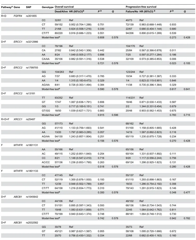
MPO rs2243828 and MDM2 rs2279744 SNPs were significantly associated with patient OS (model-free test,
P<0.001, Figure 1A and P=0.036, Figure 1B, respectively, Table 2). Patients with a variant C allele (CT/CC) at rs2243828 and a variant G allele at rs2279744 (GT/GG) had significantly increased death risks relative to those patients with the wild-type genowild-type (rs2243828: HR=2.561, 95% CI, 1.776-3.713,
P<0.001, Figure 1C; rs2279744: HR=1.778, 95% CI, 1.074-2.943, P=0.025, Figure 1D).
We found that the ERCC1 rs3212986, MDM2 rs2279744,
MPO rs2243828 and ABCB1 rs2032582 SNPs were significantly associated with patient PFS (model-free test,
Table 2. Radiaton and chemotherapy pathway gene polymorphisms and survival in patients with NPC treated with cisplatin/5-FU based CRT.
PathwayaGene SNP Genotype Overall survival Progression-free survival
Dead/Alive HR (95%CI)b P b Q Failure/No HR (95%CI) b P b Q
R+D FGFR4 rs351855
CC 32/81 Ref 40/73 Ref
CT 59/152 0.952 (0.704-1.288) 0.751 72/139 0.983 (0.668-1.448) 0.933 TT 21/68 0.824 (0.558-1.216) 0.330 22/67 0.680 (0.404-1.144) 0.680 CT/TT 80/220 0.916 (0.686-1.223) 0.551 94/206 0.890 (0.615-1.289) 0.539
Model-free testc 0.608 0.578 0.273 0.428
D+F ERCC1 rs3212986
CC 78/196 Ref 104/170 Ref
CA 27/82 0.842 (0.542-1.306) 0.442 25/84 0.567 (0.366-0.878) 0.011 AA 8/24 1.049 (0.505-2.177) 0.898 7/25 0.597 (0.277-1.284) 0.186 CA/AA 35/106 0.882 (0.591-1.316) 0.538 32/109 0.573 (0.385-0.853) 0.006
Model-free testc 0.721 0.578 0.023 0.105
D+F ERCC2 rs1799793
GG 104/263 Ref 123/244 Ref
AG 7/37 0.683 (0.317-1.470) 0.785 10/34 0.727 (0.381-1.387) 0.333 AA 1/4 1.315 (0.183-9.473) 0.329 1/4 0.823 (0.115-5.901) 0.846 AG/AA 8/41 0.726 (0.353-1.494) 0.384 11/38 0.735 (0.396-1.364) 0.329
Model-free testc 0.590 0.578 0.617 0.641 D+F ERCC2 rs13181
TT 93/252 Ref 114/231 Ref
GT 17/47 1.067 (0.636-1.791) 0.806 18/46 0.871 (0.530-1.433) 0.587 GG 1/3 0.717 (0.100-5.151) 0.741 2/2 1.344 (0.331-5.454) 0.679 GT/GG 18/50 1.039 (0.627-1.721) 0.883 20/48 0.903 (0.562-1.453) 0.675
Model-free testc 0.915 0.596 0.783 0.715 R+D+F XRCC1 rs25487
GG 57/173 Ref 68/162 Ref
AG 41/110 1.133 (0.759-1.693) 0.541 51/100 1.158 (0.805-1.665) 0.428 AA 13/20 1.797 (0.983-3.285) 0.057 14/19 1.587 (0.892-2.823) 0.116 AG/AA 54/130 1.243 (0.857-1.804) 0.251 65/119 1.230 (0.875-1.728) 0.234
Model-free testc 0.156 0.576 0.270 0.428
F MTHFR rs1801131
AA 55/166 Ref 65/156 Ref
AC 49/115 1.252 (0.851-1.840) 0.254 60/104 1.331 (0.937-1.892) 0.111 CC 8/21 1.148 (0.547-2.410) 0.716 9/20 1.117 (0.556-2.244) 0.756 AC/CC 57/136 1.236 (0.853-1.790) 0.263 69/124 1.298 (0.925-1.823) 0.131
Model-free testc 0.518 0.578 0.280 0.428
F MTHFR rs1801133
CC 47/145 Ref 55/137 Ref
CT 52/119 1.300 (0.876-1.930) 0.193 61/110 1.293 (0.898-1.863) 0.167 TT 12/39 0.948 (0.502-1.789) 0.857 18/33 1.286 (0.754-2.192) 0.356 CT/TT 64/158 1.216 (0.834-1.773) 0.310 79/143 1.291 (0.915-1.823) 0.146
Model-free testc 0.350 0.578 0.348 0.477
D+F ABCB1 rs1045642
CC 44/106 Ref 48/102 Ref
CT 51/151 0.895 (0.597-1.343) 0.593 66/136 1.064 (0.734-1.543) 0.744 TT 19/48 1.083 (0.631-1.859) 0.771 22/45 1.064 (0.642-1.762) 0.811 CT/TT 70/199 0.940 (0.643-1.374) 0.748 88/181 1.064 (0.748-1.512) 0.730
Model-free testc 0.742 0.578 0.942 0.782
D+F ABCB1 rs2032582
GG 30/78 Ref 35/73 Ref
Table 2 (continued).
PathwayaGene SNP Genotype Overall survival Progression-free survival
Dead/Alive HR (95%CI)b P b Q Failure/No HR (95%CI) b P b Q
AG 10/17 1.279 (0.623-2.623) 0.502 8/19 0.870 (0.403-1.878) 0.723 AT 7/11 1.608 (0.704-3.673) 0.259 10/8 2.398 (1.185-4.853) 0.015 AA 0/5 2/3 1.150 (0.276-4.785) 0.848 GT/TT/AG/AT/AA 0.953 (0.627-1.449) 0.822 0.996 (0.678-1.464) 0.984 Others vs. AT/AA 1.207 (0.560-2.598) 0.631 2.132 (1.176 -3.863) 0.013
Model-free testc 0.591 0.578 0.046 0.157 D MPO rs2243828
TT 63/245 Ref 71/237 Ref
CT 39/59 2.131 (1.428-3.178) 2.09E-4 53/45 2.779 (1.946-3.970) 1.91E-8 CC 11/1 8.994 (4.720-17.136) 2.42E-11 12/0 12.041 (6.457-22.454) 5.03E-15 CT/CC 50/60 2.561 (1.776-3.713) 7.14E-7 65/45 3.226 (2.302-4.521) 1.01E-11
Model-free testc 1.88E-11 2.08E-10 4.03E-17 5.52E-16 R hOGG1 rs1052133
GG 39/106 Ref 44/101 Ref
CG 57/148 1.045 (0.695-1.572) 0.832 76/129 1.257 (0.886-1.825) 0.228 CC 16/49 0.899 (0.502-1.609) 0.720 15/50 0.725 (0.404-1.304) 0.283 CG/CC 73/197 1.009 (0.683-1.490) 0.964 91/179 1.121 (0.781-1.608) 0.536
Model-free testc 0.867 0.596 0.114 0.312
R APEX1 rs1130409
TT 47/107 Ref 52/102 Ref
GT 51/151 0.824 (0.554-1.225) 0.338 64/138 0.962 (0.667-1.387) 0.835 GG 14/44 0.805 (0.443-1.462) 0.476 19/39 1.011 (0.597-1.709) 0.969 GT/GG 65/195 0.820 (0.563-1.193) 0.278 83/177 0.973 (0.688-1.376) 0.875
Model-free testc 0.582 0.578 0.970 0.782
R ADPRT rs1136410
TT 40/89 Ref 46/83 Ref
CT 51/156 0.796 (0.526-1.204) 0.280 63/144 0.834 (0.570-1.220) 0.349 CC 22/56 0.928 (0.552-1.562) 0.779 27/51 1.023 (0.636-1.646) 0.926 CC/CT 73/212 0.832 (0.565-1.223) 0.349 90/195 0.883 (0.618-1.260) 0.491
Model-free testc 0.544 0.578 0.541 0.618
D MDM2 rs2279744
TT 18/80 Ref 21/77 Ref
GT 49/134 1.586 (0.923-2.722) 0.095 61/122 1.735 (1.056-2.851) 0.030 GG 46/89 2.041 (1.183-3.521) 0.010 54/81 2.127 (1.284-3.524) 0.003 GT/GG 95/223 1.778 (1.074-2.943) 0.025 115/203 1.899 (1.192-3.026) 0.007
Model-free testc 0.036 0.199 0.014 0.096
D VEGF rs2010963
GG 45/113 Ref 48/110 Ref
CG 47/146 0.877 (0.583-1.321) 0.530 61/132 1.069 (0.732-1.561) 0.729 CC 20/45 1.084 (0.640-1.836) 0.764 25/40 1.254 (0.773-2.035) 0.359 CG/CC 67/191 0.930 (0.637-1.358) 0.708 86/172 1.117 (0.784-1.591) 0.539
Model-free testc 0.687 0.578 0.655 0.641
D VEGF rs833061
TT 58/167 Ref 70/155 Ref
CT 49/124 1.144 (0.782-1.673) 0.489 60/113 1.159 (0.821-1.637) 0.401 CC 5/12 1.119 (0.448-2.794) 0.809 4/13 0.721 (0.263-1.975) 0.524 CT/CC 54/136 1.141 (0.788-1.673) 0.485 64/126 1.117 (0.796-1.568) 0.523
Model-free testc 0.783 0.578 0.519 0.618 D VEGF rs3025039
CC 79/212 Ref 100/191 Ref
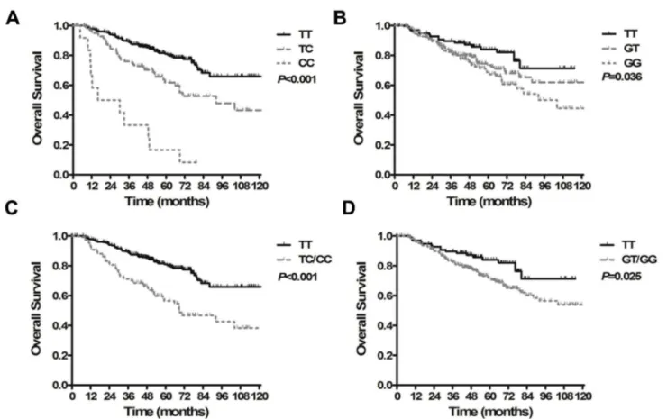
the ERCC1 rs3212986 (C8092A) genotypes CA/AA had significantly improved PFS compared with the wild-type homozygote CC (HR=0.573, 95% CI, 0.385-0.853, P=0.006, Figure 2B). Patients with the MDM2 rs2279744 (SNP209TG) genotype GT/GG had a 1.8-fold greater progression risk than those patients with the TT genotype (HR=1.899, 95% CI, 1.192-3.026, P=0.007, Figure 2D). Compared to carriers of the
MPO rs2243828 (T-764C) TT genotype, the 5-year PFS rate was significantly lower for CT/CC carriers (HR=3.226, 95% CI, 2.302-4.521, P<0.001, Figure 2F). Patients with the ABCB1
rs2032582 AT/AA genotype had significantly increased
progression risk relative to patients with other genotypes (GG/GT/TT/AG vs. AT/AA, HR=2.132, 95% CI, 1.176-3.863,
P=0.013, Figure 2H).
Multivariate analysis
Multivariate analysis identified that age>45 y (HR=1.898, 95% CI, 1.293-2.785, P=0.001), N2-3 (HR=1.593, 95% CI, 1.068-2.376, P=0.022), EBV DNA≥4000 copies/ml (HR=1.579, 95% CI, 1.075-2.320, P=0.020) and MPO rs2243828 CT/CC genotype (HR=2.453, 95% CI, 1.687-3.566, P<0.001) were
Table 2 (continued).
PathwayaGene SNP Genotype Overall survival Progression-free survival
Dead/Alive HR (95%CI)b P b Q Failure/No HR (95%CI) b P b Q
Model-free testc 0.630 0.578 0.281 0.428
a R, Radiation; D, DDP; F, 5-Fluorouracil
b HR, 95%CI and P values were calculated by using Cox proportional hazard model with no adjustment. c The association analysis was done based on the genetic model-free test (2 degrees of freedom)
doi: 10.1371/journal.pone.0082750.t002
Figure 1. Kaplan-Meier curves for overall survival in patients with locoregionally advanced nasopharyngeal carcinoma treated with cisplatin- and fluorouracil-based chemoradiotherapy. (A) Survival time based on the MPO rs2243828 TT, CT and CC genotypes. (B) Survival time based on the MDM2 rs2279744 TT, GT and GG genotypes. (C) Survival time based on the MPO rs2243828 TT and CT/CC genotypes. (D) Survival time based on the MDM2 rs2279744 TT and GT/GG genotypes.
Figure 2. Kaplan-Meier curves for progression-free survival in patients with locoregionally advanced nasopharyngeal carcinoma treated with cisplatin- and fluorouracil-based chemoradiotherapy. (A) Survival time based on the ERCC1 rs3212986 CC, CA and AA genotypes. (B) Survival time based on the ERCC1 rs3212986 CC and CA/AA genotypes. (C) Survival time based on the MDM2 rs2279744 TT, GT and GG genotypes. (D) Survival time based on the MDM2 rs2279744 TT and GT/GG genotypes. (E) Survival time based on the MPO rs2243828 TT, CT and CC genotypes. (F) Survival time based on the MPO rs2243828 TT and CT/CC genotypes. (G) Survival time based on the ABCB1 rs2032582 GG, GT, TT, AG, AT and AA genotypes. (H) Survival time based on the ABCB1 rs2032582 GG/GT/TT/AG and AT/AA genotypes.
negative independent factors for OS (Table 3). This analysis also indicated that EBV DNA≥4000 copies/ml (HR=1.512, 95% CI, 1.059-2.160, P=0.023), ERCC1 rs3212986 CC genotype (HR=1.711, 95% CI, 1.135-2.579, P=0.010), MDM2 rs2279744 GT/GG genotype (HR=1.743, 95% CI, 1.086-2.798, P=0.021),
MPO rs2243828 CT/CC genotype (HR=3.184, 95% CI, 2.261-4.483, P<0.001) and ABCB1 rs2032582 AT/AA genotype (HR=1.997, 95% CI, 1.086-3.670, P=0.026) were negative independent factors for PFS (Table 3).
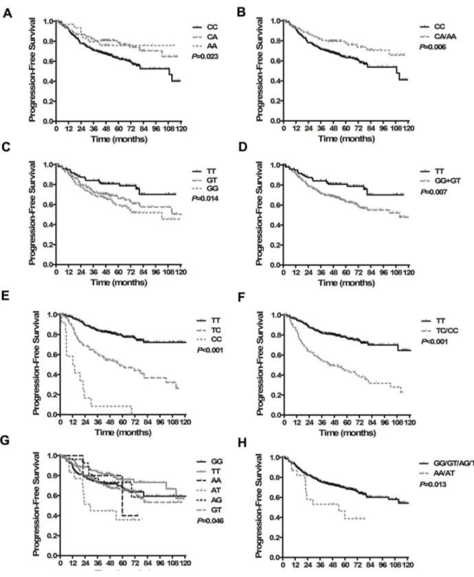
Prognostic score models
To build systemic PSMs for OS and PFS, an integral score was derived from the regression coefficients of each independent prognostic factor. If the factor was absent, a score of zero was recorded. If the factor was present, a score of 1 to 3 was recorded according to the n value (Table 4). The maximum scores for OS and PFS were 5 and 8, respectively. The overall score of the PSM for each patient was calculated as the total of the scores of each independent factor. All patients were then categorized into three groups according to cut-off points at the 25th and 75th percentiles of the score distribution as follows: low-risk group (total score 0-1 for OS, 0-2 for PFS), intermediate-risk group (total score 2-3 for OS, 3-4 for PFS) and high-risk group (total score 4-5 for OS, 5-8 for PFS). For OS, 235, 102 and 84 patients were in the low-, intermediate- and high-risk groups, respectively, with 5-year OS rates of 88.0%, 71.8% and 37.2%, respectively (P<0.001, Figure 3A). Referring to PFS, 176, 193 and 52 patients were in the low-, intermediate- and high-risk groups, respectively, and the 5-year PFS rates were 81.8%, 58.9% and 40.4%, respectively (P<0.001, Figure 3B). Figures 4A and 4B show the
Table 3. Multivariate analysis of treatment outcomes.
Variables HR 95% CI P
Overall survival
Age (≤45 vs. >45y) 1.898 1.293-2.785 0.001 Gender (Male vs. Female) 0.766 0.479-1.224 0.264 Histology (WHO type II vs. WHO type III) 1.441 0.581-3.574 0.431 T stage (T1-2 vs. T3-4) 1.238 0.832-1.840 0.292 N stage (N0-1 vs. N2-3) 1.593 1.068-2.376 0.022 EBV DNA level (<4000 vs. ≥4000 copies/ml) 1.579 1.075-2.320 0.020
MPO rs2243828 (TT vs. CT/CC) 2.453 1.687-3.566 <0.001
MDM2 rs2279744 (TT vs. GT/GG) 1.550 0.930-2.582 0.092
Progression-free survival
Age (≤45 vs. >45y) 1.040 0.738-1.464 0.823 Gender (Male vs. Female) 0.754 0.495-1.149 0.189 Histology (WHO type II vs. WHO type III) 0.944 0.456-1.955 0.878 T stage (T1-2 vs. T3-4) 1.204 0.840-1.725 0.313 N stage (N0-1 vs. N2-3) 1.415 0.980-2.045 0.064 EBV DNA level (<4000 vs. ≥4000 copies/ml) 1.512 1.059-2.160 0.023
MPO rs2243828 (TT vs. CT/CC) 3.184 2.261-4.483 <0.001
ERCC1 rs3212986 (CA/AA vs. CC) 1.711 1.135-2.579 0.010
MDM2 rs2279744 (TT vs. GT/GG) 1.743 1.086-2.798 0.021
ABCB1 rs2032582 (others vs. AT/AA) 1.997 1.086-3.670 0.026 doi: 10.1371/journal.pone.0082750.t003
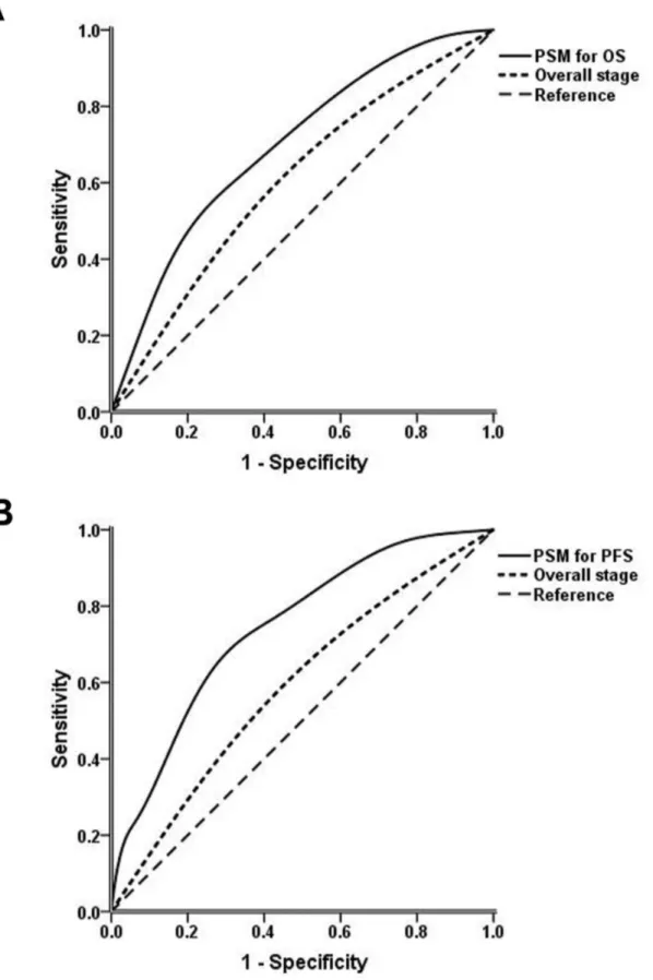
ROC curves for the PSMs and overall stage. The AUCs of the PSMs for OS and PFS were significantly larger than the AUCs for overall stage (OS: 0.702 vs. 0.572, respectively, P=0.002; PFS: 0.745 vs. 0.571, respectively, P<0.001)
Multifactor dimensionality reduction analysis
We further performed the MDR analysis to evaluate potential interactions between polymorphisms. As shown in Table 5, the overall best MDR model was the one factor model using MPO
rs2243828, which had a maximum CVC (10/10 for both OS and PFS) and minimum prediction error (43% for OS and 36% for PFS). Moreover, no potential interactions were found between investigated polymorphisms in this study (permutation test
P>0.05 for the best two to four polymorphisms combinations).
Discussion
In this study, we used a pathway approach to systematically investigate the associations between the genotypes of 18 SNPs in 13 major genes from radiation and chemotherapy response pathways and survival in patients with locoregionally advanced NPC treated with standard CRT. Following adjustment for clinicopathological characteristics, we found that the MPO rs2243828 SNP was independently significant associated with OS and PFS and that the ERCC1 rs3212986,
MDM2 rs2279744, and ABCB1 rs2032582 SNPs were independently significant associated with PFS. Moreover, we performed MDR analysis and found no significant interaction between polymorphisms. We believe our results to be biologically plausible for the following evidences.
As mentioned above, MPO could oxidize a wide variety of compounds and a broad range of functional groups [24], then enhance the effect of chemotherapy. MPO rs2243828 (T-764C) is a functional promoter polymorphism that is linked with rs2333227 (G-463A) [6]. The wild-type alleles (T and G) are associated with higher expression levels of MPO [37,38]. Previous studies have found that low-activity MPO genotypes are associated with poor survival in esophageal and breast cancer patients treated with chemotherapy [6,25]. We also showed that patients with a variant allele had approximately
Table 4. Prognostic score models.
Overall survival Progression-free survival
Factors n (HR=en)ScoreFactors n (HR=en)Score
Age (>45y) 0.64 1 EBV DNA (≥4000
copies/ml) 0.41 1
N stage (N2-3) 0.47 1 MPO rs2243828
(CT/CC) 1.16 3 EBV DNA (≥4000
copies/ml) 0.46 1 ERCC1 rs3212986 (CC) 0.54 1
MPO rs2243828
(CT/CC) 0.90 2
MDM2 rs2279744
(GT/GG) 0.56 1
ABCB1 rs2032582
2.5-fold greater death risks and 3.2-fold greater progression risks than wild-type homozygous patients. To the best of our knowledge, this is the first study focusing on the relationship between the polymorphisms of MPO and NPC prognosis.
However, some studies have found that the variant allele has a protective effect against cancer [39,40]. We hypothesize that this contradictory finding is a result of the variations in ROS activity during the different phases of cancer. In the early Figure 3. Kaplan-Meier survival curves for the three different risk groups classified by prognostic score models. (A) Overall survival. (B) Progression-free survival.
Figure 4. Receiver operating characteristic curves of prognostic score models (PSMs) and overall stage. (A) PSM and overall stage for overall survival. (B) PSM and overall stage for progression-free survival.
phases of carcinogenesis, ROS are important molecules that are involved in damaging normal cells and thus contribute to the induction of carcinogenesis. On the contrary, in the context of cancer treatment, ROS play a different role by contributing to tumor cell death mechanisms.
High expression of MDM2 has been detected in several cancers and is related to decreased response to treatment and poor prognosis [41]. MDM2 rs2279744 (SNP309 T>G) is located in the first intron of the MDM2 promoter and therefore has the potential to influence the expression of MDM2. Previous studies have shown that cells carrying the GG genotype have an increased binding affinity for the transcriptional activator Sp1, which subsequently results in higher expression levels of MDM2 mRNA and protein and elicits a decreased response to DNA-damaging agents [42]. Several studies have also demonstrated that the GG genotype in patients correlates with poor survival [21]. Liu et al found that
MDM2 T309G (rs2279744) genotypes were not related with OS and PFS in patients with advanced non-small cell lung cancer (NSCLC) treated with platinum-based chemotherapy, however, the combined analysis showed that significant shorter survival was in patients with the p53 Pro/Pro and MDM2 GG genotype [43]. With respect to NPC, rs2279744 is associated with the risk of developing the disease [44]; however, no studies have focused on the clinical outcome of NPC related to this SNP. In the present study, we found that patients with the variant allele G had significant lower PFS than those patients with the wild-type homozygote. This association between genowild-types and PFS could be explained by the attenuated activity of p53; this attenuation is related to the poor chemo/radio-sensitivity of the tumor [45,46].
The NER pathway is important in the repair of the DNA adducts which were typically caused by cytotoxic drugs such as cisplatin. ERCC1 is a major component of the NER complex
Table 5. MDR analysis for the survival prediction in patients with locoregionally advanced nasopharyngeal carcinoma.
The best combination TBA CVC P value a
Overall survival
rs2243828 0.57 10/10 0.30 rs2243828, rs2032582 0.56 8/10 0.42 rs2032582, rs2279744, rs1136410 0.44 3/10 NR b rs2032582, rs1136410, rs1801131, rs2010963 0.43 3/10 NR b
Progression-free survival
rs2243828 0.64 10/10 0.06 rs2243828, rs1801131 0.62 6/10 0.11 rs2243828, rs1801131, rs2032582 0.53 6/10 0.67 rs2243828, rs2032582, rs2279744, rs351855 0.45 4/10 NR b The above SNPs were specified as following: MPO rs2243828, ABCB1 rs2032582,
MDM2 rs2279744, ADPRT rs1136410, VEGF rs2010963, MTHFR rs1801131,
FGFR4 rs351855.
P value for 1000-fold permutation test.a b. Negative result, P value nearly equals to 1.00.
Abbreviation: MDR, Multifactor dimensionality reduction; TBA, Testing balanced accuracy; CVC, Cross-validation consistency.
doi: 10.1371/journal.pone.0082750.t005
and is the rate-limiting enzyme in the pathway. Previous studies have found that over expression of ERCC1 is associated with resistance to cisplatin-based chemotherapy in various cancers [47], including NPC [48]. A common polymorphism of the ERCC1 gene, rs3212986 (C8092A) is located in the 3’ untranslated region and therefore may affect mRNA stability and result in a decreased expression level [49]. However, the association of the rs3212986 genotypes and cancer prognosis has been inconsistent. In the present study, we found that patients with the wild-type homozygous allele had significantly lower PFS rates than did those patients with CA/AA genotypes. Our result is consistent with study using cisplatin-based treatment for gastric cancers [50]. However, other studies have reported the opposite results in NSCLC and ovarian cancer patients who were treated with platinum-based chemotherapy [51,52]. These contradictory results may be ascribed to differences in patient populations, tumor characteristics or treatment combinations. Altough ERCC2 is another major component of NER, we did not find any association between ERCC2 genotypes and survival in NPC patients. A previous study also found ERCC2 polymorphism was not related with survival in NSCLC patients receiving platinum-based chemotherapy [53].
The ABCB1 is closely related to clinical multidrug resistance. The mechanism has multiple aspects, including reduced drug accumulation, altered drug metabolism, increased tolerance of cellular damage and diminished apoptotic signaling [54]. The rs2032582 (G>T/A) SNP is located in exon 21 of the ABCB1
gene and generates an amino acid change from Ala to Thr/Ser. This SNP is often at linkage disequilibrium with another SNP at exon 26, rs1045642 (3435 C/T) [55]. The phenotypes of both rs2032582 and rs1045642 are associated with the clinical outcomes of several cancers [56,57]. In the present study, we did not find any correlation between the rs1045642 genotypes and patient survival but did find that patients harboring the rs2032582 AT/AA genotypes had 2.13-fold greater progression risks than other genotypes. Although the exact biological reason is unclear, the change in activity of P-gp may account for the change in chemotherapy sensitivity.
Additionally, we found that the XRCC1 rs25478 and MTHFR
rs1801133 genotypes were correlated with clinical stage and EBV DNA level, respectively. As mentioned above, XRCC1 is a key protein that is directly involved in the repair of DNA base damage, and the Arg399Gln amino acid variant may alter the phenotype of the XRCC1 protein, causing deficient DNA repair [9]. XRCC1 Arg399Gln was reported to be associated with susceptibility and prognosis of various cancers [6,8].The rs1801133 (C667T) variant is a common polymorphism of the
MTHFR gene that leads to an amino acid substitution and decreased enzyme activity [58]. A previous study showed that patients with the variant allele CT/TT have improved survival compared with patients with the CC allele [59]. However, the reasons for the association of these two SNPs with the identified NPC characteristics are still unclear and require intensive investigation.
Liu et al performed a multi-loci analysis in NSCLC patients and they found that interactions among XRCC1 Arg194Trp,
with sensitivity to platinum-based chemotherapy [60]. However, the overall best MDR model was the one factor model using
MPO rs2243828 and permutation test P values for the best two to four polymorphisms combinations were over 0.05 in the present study, revealing no significant interactions among polymorphisms.
PSMs were built for OS and PFS containing the significant SNPs described above. These PSMs categorized patients into three risk groups according to their prognosis and demonstrated certain value in predicting survival.
There are some limitations to this study, however. First, as with any study of modest size, this study may lack a degree of generalizability. Second, 18 SNPs were examined in our study, which could lead to false results due to multiple comparisons; to address this concern, we used a Q value to maintain the FDR under 20%. Finally, our results require validation with a large patient cohort prior to clinical application. Therefore, an additional prospective multicenter study should be conducted to further validate our results within the NPC patient population. In conclusion, this study used a pathway approach to demonstrate that genetic variations within MPO, MDM2,
ERCC1 and ABCB1 were associated with survival in patients with locoregionally advanced NPC treated with cisplatin- and 5-FU-based CRT. Furthermore, our PSMs demonstrated that genetic polymorphisms in combination with clinical prognostic
factors showed certain value in identifying patients from different risk groups. With prospective validation, our results have the potential to provide valuable information for individualized treatment.
Supporting Information
Table S1. Distribution of genotypes in patient clinical characteristics.
(DOC)
Acknowledgements
The authors thank Professor Qing Liu for statistical assistance and the Tumor Resources Bank of Sun Yat-sen University Cancer Center for providing blood samples.
Author Contributions
Conceived and designed the experiments: HQM HL BQ. Performed the experiments: HL BQ LQT QYC LZ DHL PYH. Analyzed the data: XG LG HYM YQX FQ RS. Contributed reagents/materials/analysis tools: YZ MYC YJH XL LW CZ KJC CNQ MHH. Wrote the manuscript: HL BQ HQM.
References
1. Cao SM, Simons MJ, Qian CN (2011) The prevalence and prevention of nasopharyngeal carcinoma in China. Chin J Cancer 30: 114-119. doi:10.5732/cjc.010.10377. PubMed: 21272443.
2. Wee JT, Ha TC, Loong SL, Qian CN (2010) Is nasopharyngeal cancer really a "Cantonese cancer"? Chin J Cancer 29: 517-526. doi:10.5732/ cjc.009.10329. PubMed: 20426903.
3. Al-Sarraf M, LeBlanc M, Giri PG, Fu KK, Cooper J, et al. (1998) Chemoradiotherapy versus radiotherapy in patients with advanced nasopharyngeal cancer: phase III randomized Intergroup study 0099. J Clin Oncol 16: 1310-1317.
4. Lee AW, Tung SY, Chua DT, Ngan RK, Chappell R et al. (2010) Randomized trial of radiotherapy plus concurrent-adjuvant chemotherapy vs radiotherapy alone for regionally advanced nasopharyngeal carcinoma. J Natl Cancer Inst 102: 1188-1198. doi: 10.1093/jnci/djq258. PubMed: 20634482.
5. Wee J, Tan EH, Tai BC, Wong HB, Leong SS et al. (2005) Randomized trial of radiotherapy versus concurrent chemoradiotherapy followed by adjuvant chemotherapy in patients with American Joint Committee on Cancer/International Union against cancer stage III and IV nasopharyngeal cancer of the endemic variety. J Clin Oncol 23: 6730-6738. doi:10.1200/JCO.2005.16.790. PubMed: 16170180. 6. Wu X, Gu J, Wu TT, Swisher SG, Liao Z et al. (2006) Genetic
variations in radiation and chemotherapy drug action pathways predict clinical outcomes in esophageal cancer. J Clin Oncol 24: 3789-3798. doi:10.1200/JCO.2005.03.6640. PubMed: 16785472.
7. Shiraishi K, Kohno T, Tanai C, Goto Y, Kuchiba A et al. (2010) Association of DNA repair gene polymorphisms with response to platinum-based doublet chemotherapy in patients with non-small-cell lung cancer. J Clin Oncol 28: 4945-4952. doi:10.1200/JCO. 2010.30.5334. PubMed: 20940192.
8. Miao X, Zhang X, Zhang L, Guo Y, Hao B et al. (2006) Adenosine diphosphate ribosyl transferase and x-ray repair cross-complementing 1 polymorphisms in gastric cardia cancer. Gastroenterology 131: 420-427. doi:10.1053/j.gastro.2006.05.050. PubMed: 16890595. 9. Lunn RM, Langlois RG, Hsieh LL, Thompson CL, Bell DA (1999)
XRCC1 polymorphisms: effects on aflatoxin B1-DNA adducts and glycophorin A variant frequency. Cancer Res 59: 2557-2561. PubMed: 10363972.
10. Das PM, Singal R (2004) DNA methylation and cancer. J Clin Oncol 22: 4632-4642. doi:10.1200/JCO.2004.07.151. PubMed: 15542813.
11. Langer R, Specht K, Becker K, Ewald P, Bekesch M et al. (2005) Association of pretherapeutic expression of chemotherapy-related genes with response to neoadjuvant chemotherapy in Barrett carcinoma. Clin Cancer Res 11: 7462-7469. doi: 10.1158/1078-0432.CCR-05-0042. PubMed: 16243820.
12. Shiga H, Heath EI, Rasmussen AA, Trock B, Johnston PG et al. (1999) Prognostic value of p53, glutathione S-transferase pi, and thymidylate synthase for neoadjuvant cisplatin-based chemotherapy in head and neck cancer. Clin Cancer Res 5: 4097-4104. PubMed: 10632346. 13. Orina JN, Calcagno AM, Wu CP, Varma S, Shih J et al. (2009)
Evaluation of current methods used to analyze the expression profiles of ATP-binding cassette transporters yields an improved drug-discovery database. Mol Cancer Ther 8: 2057-2066. doi: 10.1158/1535-7163.MCT-09-0256. PubMed: 19584229.
14. Fung KL, Gottesman MM (2009) A synonymous polymorphism in a common MDR1 (ABCB1) haplotype shapes protein function. Biochim Biophys Acta 1794: 860-871. doi:10.1016/j.bbapap.2009.02.014. PubMed: 19285158.
15. Pyaskovskaya ON, Dasyukevich OI, Kolesnik DL, Garmanchouk LV, Todor IN et al. (2007) Changes in VEGF level and tumor growth characteristics during lewis lung carcinoma progression towards cis-DDP resistance. Exp Oncol 29: 197-202. PubMed: 18004244. 16. Oh SY, Kwon HC, Kim SH, Lee S, Lee JH et al. (2013) The relationship
of vascular endothelial growth factor gene polymorphisms and clinical outcome in advanced gastric cancer patients treated with FOLFOX: VEGF polymorphism in gastric cancer. BMC Cancer 13: 43. doi: 10.1186/1471-2407-13-43. PubMed: 23374220.
17. Basilico C, Moscatelli D (1992) The FGF family of growth factors and oncogenes. Adv Cancer Res 59: 115-165. PubMed: 1381547. 18. Thussbas C, Nahrig J, Streit S, Bange J, Kriner M et al. (2006) FGFR4
Arg388 allele is associated with resistance to adjuvant therapy in primary breast cancer. J Clin Oncol 24: 3747-3755. doi:10.1200/JCO. 2005.04.8587. PubMed: 16822847.
19. Hrstka R, Coates PJ, Vojtesek B (2009) Polymorphisms in p53 and the p53 pathway: roles in cancer susceptibility and response to treatment. J Cell Mol Med 13: 440-453. doi:10.1111/j.1582-4934.2008.00634.x. PubMed: 19379143.
21. Gryshchenko I, Hofbauer S, Stoecher M, Daniel PT, Steurer M et al. (2008) MDM2 SNP309 is associated with poor outcome in B-cell chronic lymphocytic leukemia. J Clin Oncol 26: 2252-2257. doi: 10.1200/JCO.2007.11.5212. PubMed: 18467716.
22. Klebanoff SJ (2005) Myeloperoxidase: friend and foe. J Leukoc Biol 77: 598-625. doi:10.1189/jlb.1204697. PubMed: 15689384.
23. Nakazato T, Sagawa M, Yamato KJ, Xian MJ, Yamamoto T et al. (2007) Myeloperoxidase is a key regulator of oxidative stress-mediated apoptosis in myeloid leukemic cells. Clinical Cancer Research 13: 5436-5445. doi:10.1158/1078-0432.CCR-07-0481. PubMed: 17875773. 24. Hofstra AH, Uetrecht JP (1993) Myeloperoxidase-mediated activation of xenobiotics by human leukocytes. Toxicology 82: 221-242. doi: 10.1016/0300-483X(93)90066-2. PubMed: 8236277.
25. Ambrosone CB, Barlow WE, Reynolds W, Livingston RB, Yeh IT et al. (2009) Myeloperoxidase genotypes and enhanced efficacy of chemotherapy for early-stage breast cancer in SWOG-8897. J Clin Oncol 27: 4973-4979. doi:10.1200/JCO.2009.21.8669. PubMed: 19752340.
26. Xiao WW, Huang SM, Han F, Wu SX, Lu LX et al. (2011) Local control, survival, and late toxicities of locally advanced nasopharyngeal carcinoma treated by simultaneous modulated accelerated radiotherapy combined with cisplatin concurrent chemotherapy: long-term results of a phase 2 study. Cancer 117: 1874-1883. doi:10.1002/ cncr.25754. PubMed: 21509764.
27. Ma J, Mai HQ, Hong MH, Cui NJ, Lu TX et al. (2001) Is the 1997 AJCC staging system for nasopharyngeal carcinoma prognostically useful for Chinese patient populations? Int J Radiat Oncol Biol Phys 50: 1181-1189. doi:10.1016/S0360-3016(01)01537-1. PubMed: 11483327. 28. Khanna M, Cao W, Zirvi M, Paty P, Barany F (1999) Ligase detection
reaction for identification of low abundance mutations. Clin Biochem 32: 287-290. doi:10.1016/S0009-9120(99)00020-X. PubMed: 10463822. 29. Wen YF, Qi B, Liu H, Mo HY, Chen QY et al. (2011) Polymorphisms in
the endothelin-1 and endothelin a receptor genes and survival in patients with locoregionally advanced nasopharyngeal carcinoma. Clin Cancer Res 17: 2451-2458. doi:10.1158/1078-0432.CCR-10-2264. PubMed: 21487064.
30. Storey JD, Tibshirani R (2003) Statistical significance for genomewide studies. Proc Natl Acad Sci U S A 100: 9440-9445. doi:10.1073/pnas. 1530509100. PubMed: 12883005.
31. Jin Y, Cai XY, Cai YC, Cao Y, Xia Q et al. (2012) To build a prognostic score model containing indispensible tumour markers for metastatic nasopharyngeal carcinoma in an epidemic area. Eur J Cancer 48: 882-888. doi:10.1016/j.ejca.2011.09.004. PubMed: 22030451. 32. Zweig MH, Campbell G (1993) Receiver-operating characteristic (ROC)
plots: a fundamental evaluation tool in clinical medicine. Clin Chem 39: 561-577. PubMed: 8472349.
33. Hahn LW, Ritchie MD, Moore JH (2003) Multifactor dimensionality reduction software for detecting gene-gene and gene-environment interactions. Bioinformatics 19: 376-382. doi:10.1093/bioinformatics/ btf869. PubMed: 12584123.
34. Moore JH, Gilbert JC, Tsai CT, Chiang FT, Holden T et al. (2006) A flexible computational framework for detecting, characterizing, and interpreting statistical patterns of epistasis in genetic studies of human disease susceptibility. J Theor Biol 241: 252-261. doi:10.1016/j.jtbi. 2005.11.036. PubMed: 16457852.
35. Pander J, Wessels JA, Gelderblom H, van der Straaten T, Punt CJ et al. (2011) Pharmacogenetic interaction analysis for the efficacy of systemic treatment in metastatic colorectal cancer. Ann Oncol 22: 1147-1153. doi:10.1093/annonc/mdq572. PubMed: 21048041. 36. Sladek R, Rocheleau G, Rung J, Dina C, Shen L et al. (2007) A
genome-wide association study identifies novel risk loci for type 2 diabetes. Nature 445: 881-885. doi:10.1038/nature05616. PubMed: 17293876.
37. Kumar AP, Piedrafita FJ, Reynolds WF (2004) Peroxisome proliferator-activated receptor gamma ligands regulate myeloperoxidase expression in macrophages by an estrogen-dependent mechanism involving the -463GA promoter polymorphism. J Biol Chem 279: 8300-8315. PubMed: 14668325.
38. Winterbourn CC, Vissers MC, Kettle AJ (2000) Myeloperoxidase. Curr Opin Hematol 7: 53-58. doi:10.1097/00062752-200001000-00010. PubMed: 10608505.
39. Ahn J, Gammon MD, Santella RM, Gaudet MM, Britton JA et al. (2004) Myeloperoxidase genotype, fruit and vegetable consumption, and breast cancer risk. Cancer Res 64: 7634-7639. doi: 10.1158/0008-5472.CAN-04-1843. PubMed: 15492293.
40. Lu W, Xing D, Qi J, Tan W, Miao X et al. (2002) Genetic polymorphism in myeloperoxidase but not GSTM1 is associated with risk of lung squamous cell carcinoma in a Chinese population. Int J Cancer 102: 275-279. doi:10.1002/ijc.10712. PubMed: 12397651.
41. Rayburn E, Zhang R, He J, Wang H (2005) MDM2 and human malignancies: expression, clinical pathology, prognostic markers, and implications for chemotherapy. Curr Cancer Drug Targets 5: 27-41. doi: 10.2174/1568009053332636. PubMed: 15720187.
42. Luo JL, Xiao JY, Tian YQ (2000) MDM2 gene amplification and overexpression in nasopharyngeal carcinoma. Hunan Yi Ke da Xue Xue Bao 25: 18-20. PubMed: 12212235.
43. Liu L, Wu C, Wang Y, Zhong R, Duan S et al. (2011) Combined effect of genetic polymorphisms in P53, P73, and MDM2 on non-small cell lung cancer survival. J Thorac Oncol 6: 1793-1800. doi:10.1097/JTO. 0b013e3182272273. PubMed: 21841506.
44. Zhou G, Zhai Y, Cui Y, Zhang X, Dong X et al. (2007) MDM2 promoter SNP309 is associated with risk of occurrence and advanced lymph node metastasis of nasopharyngeal carcinoma in Chinese population. Clin Cancer Res 13: 2627-2633. doi:10.1158/1078-0432.CCR-06-2281. PubMed: 17473193.
45. Reles A, Wen WH, Schmider A, Gee C, Runnebaum IB et al. (2001) Correlation of p53 mutations with resistance to platinum-based chemotherapy and shortened survival in ovarian cancer. Clin Cancer Res 7: 2984-2997. PubMed: 11595686.
46. Gudkov AV, Komarova EA (2003) The role of p53 in determining sensitivity to radiotherapy. Nat Rev Cancer 3: 117-129. doi:10.1038/ nrc992. PubMed: 12563311.
47. Olaussen KA, Dunant A, Fouret P, Brambilla E, André F et al. (2006) DNA repair by ERCC1 in non-small-cell lung cancer and cisplatin-based adjuvant chemotherapy. N Engl J Med 355: 983-991. doi: 10.1056/NEJMoa060570. PubMed: 16957145.
48. Sun JM, Ahn MJ, Park MJ, Lee HY, Ahn JS et al. (2011) Expression of excision repair cross-complementation group 1 as predictive marker for nasopharyngeal cancer treated with concurrent chemoradiotherapy. Int J Radiat Oncol Biol Phys 80: 655-660. doi:10.1016/j.ijrobp. 2010.02.061. PubMed: 21621119.
49. Chen P, Wiencke J, Aldape K, Kesler-Diaz A, Miike R et al. (2000) Association of an ERCC1 polymorphism with adult-onset glioma. Cancer Epidemiol Biomarkers Prev 9: 843-847. PubMed: 10952103. 50. Park SR, Kong SY, Nam BH, Choi IJ, Kim CG et al. (2011) CYP2A6
and ERCC1 polymorphisms correlate with efficacy of S-1 plus cisplatin in metastatic gastric cancer patients. Br J Cancer 104: 1126-1134. doi: 10.1038/bjc.2011.24. PubMed: 21364592.
51. Zhou W, Gurubhagavatula S, Liu G, Park S, Neuberg DS et al. (2004) Excision repair cross-complementation group 1 polymorphism predicts overall survival in advanced non-small cell lung cancer patients treated with platinum-based chemotherapy. Clin Cancer Res 10: 4939-4943. doi:10.1158/1078-0432.CCR-04-0247. PubMed: 15297394.
52. Krivak TC, Darcy KM, Tian C, Armstrong D, Baysal BE et al. (2008) Relationship between ERCC1 polymorphisms, disease progression, and survival in the Gynecologic Oncology Group Phase III Trial of intraperitoneal versus intravenous cisplatin and paclitaxel for stage III epithelial ovarian cancer. J Clin Oncol 26: 3598-3606. doi:10.1200/ JCO.2008.16.1323. PubMed: 18640939.
53. Liu L, Yuan P, Wu C, Zhang X, Wang F et al. (2011) Assessment of XPD Lys751Gln and XRCC1 T-77C polymorphisms in advanced non-small-cell lung cancer patients treated with platinum-based chemotherapy. Lung Cancer 73: 110-115. doi:10.1016/j.lungcan. 2010.11.004. PubMed: 21129812.
54. Gottesman MM (2002) Mechanisms of cancer drug resistance. Annu Rev Med 53: 615-627. doi:10.1146/annurev.med.53.082901.103929. PubMed: 11818492.
55. Hoffmeyer S, Burk O, von Richter O, Arnold HP, Brockmöller J et al. (2000) Functional polymorphisms of the human multidrug-resistance gene: multiple sequence variations and correlation of one allele with P-glycoprotein expression and activity in vivo. Proc Natl Acad Sci U S A 97: 3473-3478. doi:10.1073/pnas.97.7.3473. PubMed: 10716719. 56. Gréen H, Söderkvist P, Rosenberg P, Horvath G, Peterson C (2006)
mdr-1 single nucleotide polymorphisms in ovarian cancer tissue: G2677T/A correlates with response to paclitaxel chemotherapy. Clin Cancer Res 12: 854-859. doi:10.1158/1078-0432.CCR-05-0950. PubMed: 16467099.
57. Wu H, Kang H, Liu Y, Tong W, Liu D et al. (2012) Roles of ABCB1 gene polymorphisms and haplotype in susceptibility to breast carcinoma risk and clinical outcomes. J Cancer Res Clin Oncol 138: 1449-1462. doi:10.1007/s00432-012-1209-z. PubMed: 22526155. 58. Weisberg I, Tran P, Christensen B, Sibani S, Rozen R (1998) A second
genetic polymorphism in methylenetetrahydrofolate reductase (MTHFR) associated with decreased enzyme activity. Mol Genet Metab 64: 169-172. doi:10.1006/mgme.1998.2714. PubMed: 9719624. 59. Martin DN, Boersma BJ, Howe TM, Goodman JE, Mechanic LE et al.
survival. BMC Cancer 6: 257. doi:10.1186/1471-2407-6-257. PubMed: 17069650.
60. Liu L, Wu J, Zhong R, Wu C, Zou L et al. (2013) Multi-loci analysis reveals the importance of genetic variations in sensitivity of