Abstract
Cherubism is a rare hereditary non-neoplastic bone disease characterized by clinically evident bilateral, pain-less enlargements of the jaws, said to give the patient a cherubic appearance. Here we report the case of cherubism in a 5 year-old girl who complained of painless bilateral swelling, noticed by her parents three months back. Pano-ramic radiography and CT images exhibited bilateral mul-tilocular radiolucent areas with cortical expansion. Histopathologic examination revealed proliferating fibrous connective tissue containing numerous multinucleated giant cells.
Keywords:Cherubism, autosomally dominant, familial.
INTRODUCTION
Cherubism, or familial intraosseous fibrous expansion of the mandible, is a genetically-medi-ated disorder first reported in 1933. The word ‘Cherubism’ refers to the spherical facial appea-rance of angels painted in the Renaissance era.
[1,2] It is characterised by the presence of giant
cells and fibrous tissue proliferation. Cherubism is a non-neoplastic hereditary bone lesion cha-racterized by a spherical and symmetrical chubby facial appearance, bilateral painless swelling of the maxilla and mandible, resulting in a fullness of the cheeks and retraction of the lower eyelids, giving an upward turned appearance of the eyes – comparable to a cherub angel. It is an autoso-mal dominant inheritance in 80% of familial pat-terns. Typically, the mandible is primarily affected and, in 60% of cases, maxilla is also involved. Children are normal at birth. At the age of 14 months to 5 years, a symmetric enlar-gement of the jaws begins, progressing until
puberty. [3-5]
Here we report a case of 5year-old girl with a history of bilateral swelling of the mandible.
CASE REPORT
The parents of a 5year-old child came to the department of Oral Medicine and Radiology of Oxford Dental College, Bangalore, with com-plain of bilateral swelling in the cheek region since 3 months. As the baby was chubby since birth, they could not make out the difference in the present swelling and there was no history of trauma. Three months back, parents and friends started noticing differences in the face of the baby. There was no increase in the size of swelling, but they felt hardness in swelling. Parents visited a local physician who advised radiological investigations and biopsy for swelling. They were apprehensive about teeth eruption and came for a second opinion to our college. Family history reveals that girl’s father had similar painless bilateral mandibular swelling during his childhood, and later on, at the age of 13, surgical intervention was carried out for aesthetic purposes.
The general physical examination detected no abnormality in any system. On extra-oral exami-nation, bilateral swelling was noticed in the lower 3rd of the face, causing fullness of the
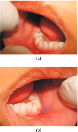
cheeks. Skin over the swelling appeared normal (Fig. 1). On palpation, it was bony-hard in con-sistency and non-tender. Bilateral submandibu-lar lympnodes were ensubmandibu-larged and non-tender. Intra-orally, bilateral obliteration of the buccal vestibule was noticed, extending from the 1st
molar to the retro molar region, due to buccal
CLINICO-RADIOLOGIC FEATURES OF BILATERAL MANDIBULAR
SWELLING – A CASE REPORT OF CHERUBISM AND
LITERATURE REVIEW
Dysanoor Sujatha1, Naik Shobha2
1. Prof. PhD, The Oxford Dental College and Hospital, Hosur Road, Bommanahalli Bangalore
2. P.G. student
A CASE REPORT OF CHERUBISM AND LITERATURE REVIEW
cortical plate expansion (Fig. 2). Based on the family history and on classical findings, provisi-onal diagnosis of Familial Cherubism of mandi-ble was arrived at.
Fig. 1. Extra-oral photograph showing bilateral facial swelling
(a)
(b)
Fig. 2. Intra-oral photograph revealing buccal cortical plate expansion
OPG and PA mandible revealed bilateral well-defined multilocular radiolucency, extending from the distal aspect of the 1st molar till the
sub-condylar region, with buccal cortical expansion.
Tooth buds of permanent 1st molars were
embed-ded within the radiolucency, giving a floating tooth appearance (Fig. 3). CT also revealed the same features in the mandible without involving maxilla (Fig. 4). According to the radiographic grading given by Ramon and Engelberg [11],
dia-gnosis of grade I Cherubism was given. Serolo-gical investigations revealed increase in alkaline phosphatase level (165 IU/ml) and normal serum calcium levels. Incisional biopsy confirmed the clinical diagnosis of Cherubism.
(a)
(b)
Fig. 3. (a) Panoramic radiography, (b) PA mandible
showing multilocular radiolucency with floating tooth appearance extending upto the condyles
After a vivid clinical review, the parents of the child were informed about the benign nature of the swelling, about its familial pattern, possible regression and delayed eruption, crowding and missing of permanent teeth, and advised yearly
(a)
(b)
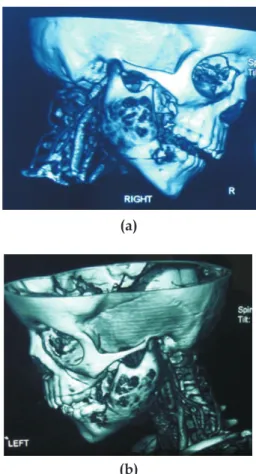
Fig. 4. 3D Computer tomography – right side (a) and left side (b) view of a 3D image showing
the deformity in the body and ramus region
DISCUSSION
The first description of Cherubism was made by Jones, who analyzedthe familial occurrence of painless enlargement of the jaws in three siblings. [1] According to the World Health
Organization, Cherubism, belonging to a group of non-neoplastic bony lesions affecting only the jaws, is an autosomal dominant inheritance, with100% occurrence in males and 50 to 70% – in females. [2,3] The molecular gene pathogenesis
proposed demonstrated SH3BP2 gene mutati-ons, mapped to locus 4p16.3, and causing dis-function of the Msx-1 gene, involved in the regulating mesenchymal interaction in cranio-facial morphogenesis.[2] The SH3BP2 mutation
is thought to lead to parathyroid hormone receptor (PTHr) signaling and Msx-1 activation. No spatial partitioning takes placein the cap stage of the second and third molars, which is necessary for normal dental development, lea-ding to disfunction of mesenchymal bone
formation, development of multinucleated giant cells and abundant deposition of fibrohistiocytic tissues. [6,7] Disfunction of Msx1 stops in the end
of molar development, leading to remineraliza-tion of lesions. [2] Perivascular fibrosis leading
to mesenchymal disorders and reduced oxyge-nation is the most widely accepted theory evi-dencing the pathogenesis of cherubism. [8]
CLINICAL CHARACTERISTICS
It is a benign, self-limiting fibro-osseous
disor-der characterized by bilateral expansion of the mandible, maxilla, or of both. The lesions are usually symmetrical and painless. Frequently, Cherubism is accompanied by dental arch and dental eruption abnormalities. The rounded facies and occasional upward cast of the eyes, with exposure of the sclera below the pupil, is due to enlargement of orbital floor and fragile support of the lower lids, with a superior globe displacement and sclera exposition, giving the “eyes-to-heaven appearance”. This classical cli-nical characteristic is not observed in every pati-ent. Our case, for example, had only mandible involvement. [3,5] Affected children appear
phy-sically and mentally normal at birth, with no cli-nically or radiographically evident disease until 14 months to 3 years of age, when bilateral jaw expansion begins. [9] Typically, the earlier the
lesion appears, the more rapidly it advances, becoming progressively larger until puberty. Lesion regression is expected to occur spontane-ously by the end of puberty, being solved by middle age. [2]
Dental abnormalities
Early exfoliation of deciduous teeth, impac-tion or displacement of teeth, ectopic tooth erup-tion, agenesis of the permanent teeth, mainly (second and third molars) due to involution of their germs, delayed eruption of the perma-nent teeth and, in severe cases, root resorption of teeth occurs. [7,9,11] All these features results
in malocclusion, as well as in problems of pho-nation and swallowing, the latter being exacer-bated by flattening or inversion of the palatal
A CASE REPORT OF CHERUBISM AND LITERATURE REVIEW
Biochemical parameters
The haematological parameters, such as serum calcium and phosphorus concentrations, are usually within normal limits, only the alkaline phosphatase levels may be elevated. [1,7,9,10,16]
Our case revealed a high level of alkaline pho-sphatase and normal serum calcium levels.
Radiographic features
Radiographically, lesions appear as cystic multilocular radiolucencies clearly bounded by cortical bone and distributed bilaterally in the posterior quadrants of the mandibleand/orma-xilla, often beginning near the angle of the man-dible and spreading to the mandibular ramus and body. Maxillary lesions may concomitantly occur, usually in the maxillary tuberosity region.
[1,7,11] Our case demonstrated lesions confined
to mandible body and ramus. Frequently, teeth appear as displaced and impacted, and root resorption is observed; frequently, the mandibu-lar canal is also displaced. [7]
Radiographic grading of Cherubism
Several grading systems have been proposed to describe the severity of cherubism. Arnott suggested radiographic staging for the lesions of
Cherubism according to their location and degree of expansion.[12] Ramon and Engelberg
modi-fied grading based on the area of involvement, as follows: [8]
• Grade I: involvement of both mandibular
ascending rami.
• Grade II: involvement of both maxillary
tuberosities, as well as of the mandibular ascending rami.
• Grade III: massive involvement of the whole
maxilla and mandible, except the coronoid process and the condyles, resulting in
consi-derable facial deformity.
• Grade IV: grade 3 plus the involvement of
the floor of the orbits, causing orbital com-pression.
The grade may change, dependingon the results of follow-up examination.
Differential diagnosis of Cherubism
[8,10,16]
Cytogenetic and molecular studies, such as fluorescence, in situ hybridization and quantita-tive analysis of Msx-1 expression in different tissues, are used in cherubism diagnosis. [13]
TREATMENT
Cherubism is generally a self-limiting lesion, which spontaneously regresses with age. Jaw remodelling continues through the third decade of life, at the end of which the clinical abnorma-lity may be subtle. The frequency of remodelling is unknown, since most of the recorded cases have been surgically treated before reaching puberty. Conventional treatment of cherubism includes jaw contouring, curettage of the lesions, and management of dental disharmony. Curet-tage alone or in combination with surgical con-touring has been considered the treatment of choice, and some authors have reported a mas-sive growth of the lesion after surgery, especially when performed during the active growth phase. Liposuction has been used to change the contour of the jaws in patients with cherubism. [3-5]
Some authors point medical therapy in the form of calcitonin, as a possibility to curtail the disease and obviate the need for surgery. Calcitionin has been shown to cause inhibition of bone resor-ption by multinucleate cells in cherubic tissue in vitro. [14] The daily use of 200 IU salmon
calci-tonin via nasal spraying for cherubism has been
recently reported. [13] Based on the genetic
mutations related to the disease, gene therapy is expected to play a role in future treatments. [2]
CONCLUSIONS
Cherubism is a rare, giant-cell-containing lesion of the jaw bones. In cases of suspicion of cherubism, radiographic examination is essen-tial, since the clinical presentation and the loca-tion and distribuloca-tion of lesions may define the diagnosis. Histopathological examination is complementary. Nowadays, genetic tests should be used for final diagnosis of cherubism. Knowledge of the clinical and radiographic
alterations observed in patients with cherubism is important, since the dentist might be the first professional representative to diagnose this disease.
References
1. P. Prabal, S. Singh, J. Singh. Cherubism: a case report and review of literature, Int. J. Dent. Case Reports 2011; 1(2):61-72.
2. C.S. Edgard, C.S. Guilberme, C.V. Tainab. Cheru-bism: Clinicoradiographic Features, Treatment, and Long-Term Follow-Up of 8 Cases, J. Maxillofacial Sur-gery, 2007; 65:517-522.
3. T. Victor Perez, D. Rogerio A., G. Andre, B. Alexan-der Augusto, C. Miranda Franca. Cherubism: case report and literature Review, Rev. de Clín. Pesq. Odon-tol., 2004; 1(1).
4. Kozakiewicz M., Perczynska-Partyka W., Kobos J. Cherubism – clinical picture and treatment, Oral Dis., 2001; 7:123-30.
5. Pulse C.L., Moses M.S., Greenman D., Rosenberg
S.N., Zegarelli D.J. Cherubism: case reports and litera-ture review, Dent Today, 2001; 20:100-103.
6. Eduardo C., Raposo A., Marcelo de Campos Guidi et al. Two-Stage Surgical Treatment of Severe Cheru-bism, Annals of Plastic Surgery, June 2007; 58(6).
7. Morais Gouvêa G. Lima, Janete Dias Almeida et al. Cherubism: Clinicoradiographic Features and Treatment, J. Oral Maxillofac. Res., 2010; vol. 1(2).
8. M.H. Atalar, E. Albayrak, P. Erdinc, S. Bulut. Cheru-bism as a Rare Cause of Bilateral Expansion of the Man-dible: Radiological Manifestations, J. Hong Kong Coll. Radiol., 2008; 11:76-80.
9. R. Ongole, R. Pillai, K.M. Pai. Cherubism in Siblings: A Case Report, J. Can. Dent. Assoc., 2003; 69(3):150-4.
10. Greenberg M.S., Glick M., Ship J.A. (2008), Burket’s Oral medicine, Eleventh edition, BC Decker, p. 146. 11. Stuart C. White, M. Pharoah. Oral Radiology –
prin-ciples & interpretation, Elsevier 6th edition, p. 446. 12. P. Miguel, B. Jaime, M.M. Juan, B. Jose Vicente,
V. Francisco. Cherubism: A Clinical, Radiographic, and Histopathologic Comparison of 7 Cases, J. Oral Maxillo-fac. Surg., 2006; 64:924-930.
13. A. Osman Etoz, Dogan D., Gunhan O. Treatment of Cherubism with Salmon Calcitonin: A Case Report, Eur. J. Dent., 2011; 5:486-491.
14. Meng X.-M. Clinicopathologic study of 24 cases of che-rubism, Int. J. Oral Maxillofac. Surg., 2005; 34:350-356. 15. Freny R. Karjodkar. Text book of dental & Maxillofacial
radiology, 2nd edition, Jaypee, pp. 644-645.