Vol. 23, No.1, Winter-2011
Hypoglossal Schowannoma:
A Case Report and Review of Literature
*Masoud Naghibzadeh1, Mohammad Faraji2, Shima Kamandi3
Abstract Introduction:
Schawannomas (neuromas, neurilemmomas) are benign tumors originating from showann cells or nerve fiber sheet cells. They are solitary, encapsulated tumors usually attached to, or surrounded by a nerve.
Case Report:
We present a case of left hypoglossal nerve schwannoma in a 19 year old man who was admitted with progressive left tongue atrophy.
Conclusion:
Schwannoma of the hypoglossal nerve usually develops in the intracranial and extracranial portion or both in the intracranial and extracranial components forming a dumbbell shape tumor .The peripheral hypoglossal schwannomas are extremely rare.
Keywords:
Hypogloss, Schawannomas, Tumur
Received date: 8 May 2010 Accepted date: 26 Aug 2010
1
Ear, Nose, Throat, Head and Neck surgery Research Center,Mashhad University of Medical Sciences, Mashhad, Iran
2
Department of neurosurgery, Mashhad University of Medical Sciences, Mashhad, Iran
3
Department of otorhinolaryngology, Mashhad University of Medical Sciences, Mashhad, Iran *Corresponding author:
Ghaem Hospital, Mashhad University of Medical Sciences, Mashhad, Iran Email: naghibzadehm@mums.ac.ir Tel: +985118413492
Introduction
Schawannomas (neuromas, neurilemmomas) are benign tumors originating from showann cells or nerve fiber sheet cells. They represent about 8% of all intracranial tumors. They are solitary, encapsulated tumors usually attached to, or surrounded by a nerve, and can be associated with von Recklinghausens disease.
Schowannomas located at the
craniocervical region are rare.
The clinical presentation is variable, depending on location and can be associated with neurofibromatosis (1). We would like to report a case with a schwannoma presented with hypoglossal nerve palsy and treated surgically with excellent results.
Case Report
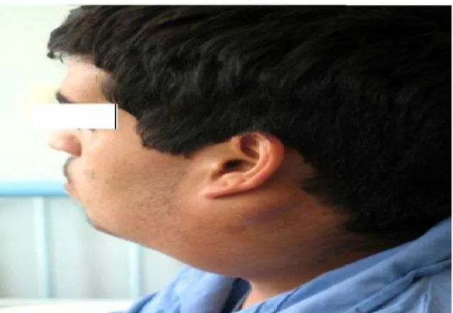
A 19 year- old right- handed male patient complaining of cervical mass since 13 years ago was admitted to the hospital (Fig 1). He had a six – month history of dysphonia and hot potato voice.
The preoperative examination revealed left hypoglossal nerve palsy, unilateral tongue atrophy and fasciculation, bulging of lateral wall of the oropharynx on the left side and shifting of uvula to the right side, asymmetry and atrophy in the left shoulder and partial paralysis of the accessory nerve on the left side, and left cervical mass 10 by 13cm in level 2,3 nontender, soft ,immobile and without inflammation and erythema (Fig 2).
Radiological Findings
Magnetic resonance imaging (MRI) revealed huge, heterogeneous and abnormal signal intensities of the mass effect with some border irregularities probably with left CP angleorigin after intravenous Gadolinium injection demonstrated mild tumural enhancement. Axial CT scan revealed a left paraspinal and retropharyngeal heterogeneous mass that extended from nasopharynx to
submandibular space, putting pressure on pharynx and larynx (Fig 3,4).
In left carotid and vertebral angiography, the internal and external carotid arteries were deviated and stretched by the cervical mass lesion. The artries were not encased. The mass lesion was moderately hypervascular with arterial supply from the external carotid artery.
Fig1: Photograph of the patient with cervical mass
Fig 2: Hypoglossal palsy is shown
Fig 4: Gadolinium-enhanced magnetic resonance images, axial views, showing the
intracranial portion of tumor.
Surgical Findings
The tumor was totally removed via two approaches (Fig 5).
First incision made from mastoid tip and was extend in the neck then the subplatismal flap was elevated to reveal and then cut SCM muscle from mastoid tip .The carotid sheet was exposed. The internal jugular vein and carotid artery on the mass were separated from the mass gently. The mass was encapsulated and extended to the skull base. The extracranial part was cut and removed and then the intracranial part of the mass was removed from the retro sigmoid approach.
Fig 5:Resected mass
Histopatological Findings
Histopathological examination showed typical features of a schwannoma: hyper cellular and encapsulated tumor with cellular fascicles and bandles in different directions. Tumoral cells showed spindle, wavy and hyperchromic nuclei and acidophile cytoplasm with mild pleomorphism, occasionally with palisade and verocay bodies with atopy and contained vessels with hyalinized walls. Postoperatively, the patient experienced aspiration and mild paresis of seventh cranial nerve that resolved in the postoperative follow up period.
Discussion
Neuromas are benign, slow- growing tumors of the myelin-producing Schwann’s cells in the peripheral sensory- motor nervous system. Over 90% of cases are presented as acoustic neuromas. Neuromas stem very rarely from primarily motor nerves. The purely motor hypoglossal nerve emerges from the medulla oblongata between the pyramid and the olive, passes extracranially through the hypoglossal canal, and describes a wide ventral curve between the internal jugular vein and the internal carotid artery to the floor of the mouth.
It provides the motor supply to the internal musculature as well as the converging muscles of the ipsilateral side of the tongue. Therefore, lesions of the nerve characteristically lead to ipsilateral atrophy of the tongue and deviation of the tongue to the side of the lesion (2).The most common initial symptom is a headache, often suboccipital, exacerbated with movements of the head and neck. Other symptoms and signs are related to increased intracranial pressure, such as vomiting and papilledema (3).
myxoid consistency. In addition, hemorrhage from adjacent tissue, necrosis, hyalinization and cystic degeneration may also occur (2).
On MRI these appear as clearly circumscribed, partly cystic, space-occupying lesions that produce an inhomogeneous signal. After the administration of contrast agent they show irregular contrast enhancement, often around the margins of the lesion. Dumbbell- shaped neuromas grow along the nerves extracranially from their intracranial origin, extending through and destroying the hypoglossal canal (4). The treatment of choice is enucleation of the tumor while preserving the nerve. In the majority of the case reports a conventional midline or modified suboccipital craniotomy was performed. Smith PG et al. recommend transcranial excising of hypoglossal schwannomas through a transcondylar approach as this improves the visualization of the hypoglossal canal (5). In our patient the extracranial tumor mass was so large that two surgical procedures were taken into our consideration.
Postoperatively, a considerable number of patients suffered from respiratory complications and inappropriate gag reflex; therefore, it was suggested that a prophylactic tracheostomy should be performed at the end of surgery in order to prevent such complications (6).
The first report of neurofibroma of the hypoglossal nerve was released in 1935 with Friedman and Eisenberg. A female, aged thirty with a slowly growing, painless swelling on the right side of the neck, at the angle of the jaw. Conscious of some extent of dysphagia with no interference in mastication;no difference in the thickness or size of the right side of the tongue, no motor disturbance of tongue, or any deviation of tongue when protruded (7). In 1999 Giuseppe Mariniello and his colleagues reported a rare case of cellular
schwannoma of the hypoglossal nerve, with intraspinal extension, presenting with no recognizable hypoglossal nerve dysfunction (8).
In 2002 Johanna Rachinger and his colleagues reported a dumbbell-shaped neuromas of the 12th cranial nerve extending intra- and extracranially. A 32-year-old patient who presented with hypoglossal nerve palsy and a two-year history of headache. MRI showed
inhomogeneous contrast agent
enhancement in a tumor that was partly cystic, partly solid, in the cerebellopontine cistern. The tumor, with its main lesion mass located in the parapharyngeal space, had extended along the canal of the hypoglossal nerve. The tumor was excised by two-stage suboccipitalosteoclastic craniotomy and later through a cervical approach. Pathohistology showed a grade I of schwannoma and the patient was discharged symptom- free without any further therapy (9).
In 2005 Kimitoshi Sato and his colleagues reported a 54-year-old woman presented with gradually worsening left hypoglossal nerve palsy. There was a tumor lying in the left hypoglossal canal and paraspinal region and the findings were consistent with hypoglossal schwannoma. Subtotal intracapsular removal of the tumor was performed via transcervical approach. The symptoms improved, and no additional symptoms were noted (10).
C1-2 laminectomy, including opening of the dura mater and gross-total removal of the lesion. The patient had hypoglossal nerve palsy and mild hemiparesis on the left side which had regressed almost totally at the 3-month follow-up. The far-lateral approach with the patient in the sitting position is very important and facilitates the total removal of the schwannoma. Simple suboccipitalcraniectomy provided enough exposure for total removal in this case (11).
Conclusion
References:
1. Leal Filho MB, Borges G, Ferreira A, Franca D, Mello P. Schwannoma of the craniocervical junction: Surgical approach of two cases. Arq Neuropsiquiatr2003; 61: 639-41.
2. Batsakis JG. Tumors of the head and neck.Clinical and pathological considerations. 2nd ed. Baltimore: Williams and Wilkins; 1979: 313-33.
3. Odake G. Intracranial hypoglossal neurinoma with extracranial extension: Review and case report. Neurosurgery 1989; 24: 583-7.
4. Sato M, Kanai N, Fukushima Y. Hypoglossal neurinoma extendingintra-and extracranially: Case report. Surg Neurol 1996; 45: 172-5.
5. Smith PG, Backer RJ, Kletzker GR, Mishler ET, Loosmore JL, Leonetti JP, et al. Surgical management of transcranial hypoglossal schwannomas. Am J Otol 1995; 16(4): 451-6.
6. White W, Shiu MH, Rosenblum MK. Cellular schwannoma: A clinical study of 57 patients and 58 tumors. Cancer1990; 66: 1266-75.
7. Friedman L, Eisenberg AA. Neurofibromaof the hypoglossal nerve. Ann Surg 1935; 101(3): 834-8.
8. Giuseppe M, Horvat A, Popovic M, Vinko V. Cellular dumbbell schwannoma of the hypoglossal nerve presenting without hypoglossal nerve palsy. Clin Neuro Neurosurg 2000; 102: 40-3.
9. Rachinger J, Fellner FA, Trenkler J. Dumbbell- shaped hypoglossal schwannoma: A case report. Magnet Reson Imag 2003; 21: 155-8.
10. Sato K, Shimizu S, Oka H, Nakahara K, Utsuki S, Fujii K. Usefulness of transcervical approach for surgical treatment of hypoglossal
schwannoma with paraspinal extension: case report. Surgical Neurology 2006; 65: 397- 401.
11. Kabatas S, Cansever T, Yilmaz C, Demiralay E, Celebi S, Caner H. Giant