Acute Kidney Injury in patients infected by H1N1 - clinical
histological correlation in a series of cases
Authors
Gabriela Sevignani1 Maria Fernanda Soares1 Gustavo Lenci Marques1 Ana Karyn Ehrenfried de Freitas1
Arthur Gentili1
Domingos Candiota Chula1 Marcelo Mazza do Nascimento1
1 Federal University of Paraná (UFPR).
Submitted on: 10/31/2012. Approved on: 05/14/2013.
Correspondence to:
Gabriela Sevignani. University Hospital - Federal University of Paraná (UFPR). Rua General Carneiro, nº 411, Alto da Glória, Curitiba - PR, Brazil. CEP: 80060-150
E-mail: [email protected]
Introduction: Influenza A (H1N1) virus was first reported on April 2009 and, since then, several studies have reported the characteristics concerning the clinical presentation and pulmonary involvement. However, accurate information about the acute kidney injury (AKI) and kidney histopathological findings in these patients remain scarce. Objective: To describe the kidney histopathological findings of 6 patients with H1N1 who developed AKI and underwent kidney biopsy, correlating them with clinical features. Methods: We studied six patients admitted to Hospital de Clínicas UFPR with a PCR-confirmed diagnosis of H1N1who developed ARF and underwent kidney biopsy. We reviewed their medi-cal file and the microscopy findings of the biopsy. Results: Clinical and/or laboratory evidence of AKI was present in all cases, and only one did not present oliguria. Kidney tissues revealed glomerular lesions in two patients: one patient, with systemic lupus erythematosus, showed changes consistent with lupus nephritis class III A-C according to the ISN/RPS 2003 and focal thrombotic microangiopathy; the other one had inter-capillary nodular glomerulosclerosis, but without clinical or laboratory evidence of diabetes. Vacuolar degenerative tubular changes were present in all cases, with focus of oxalosis in two cases. Mild to moderate atherosclerosis was found in two patients. Conclusion: In this study, varying degrees of vacuolar degenerative tubular changes were present in all patients, but there were no signs of acute tubular necrosis. It seems that in the present study a prerenal cause of acute renal failure was the main involved mechanim to explain the cause of renal failure in these patients.
A
BSTRACTKeywords: acute kidney injury; biopsy; influenza A virus, H1N1 subtype.
I
NTRODUCTIONThe influenza A (H1N1) was first identified in early April of 2009, quickly reaching global proportions and causing a pandemic in June 2009.1,2 It is estimated that the mortality related to this new infec-tion is similar to that found in seaso-nal influenza, differing from it by the fact that severe forms of the disease are not restricted to patients with co-morbidities and age extremes.2
Acute renal failure (ARF) has been reported as a frequent occur-rence in critically ill patients with H1N1, with an incidence of up to 66% in studies conducted in Canada, Argentina and Brazil.3-5 In addition, ARF and the need for hemodialysis have adverse developments in these patients, being related to a prolonged hospital stay and greater mortality.3-7 Several studies have reported the re-levant characteristics to the clinical presentation and the pulmonary in-volvement of the disease. However, accurate information regarding ARF and kidney histopathological chan-ges in these patients is scarce.
The lack of information regarding the pathophysiology of ARF in these patients imposes limitations to the development of therapeutic strategies to allow for better management of these cases. The aim of this study was to describe the clinical and labora-tory findings, and the pathology of
patients who had proven infection by H1N1 and who developed ARF during the viral out-break which occurred in the city of Curitiba in 2009.
M
ETHODSWe retrospectively evaluated six patients who were admitted to the University Hospital of the Federal University of Paraná (HC-UFPR) in 2009 with a diagnosis of influenza A (H1N1) and who were submitted to kidney biopsy. The study was approved by the Ethics Committee on Human Research of the University Hospital of the UFPR.
The H1N1 infection was confirmed by the polymerase chain reaction (PCR) in combined viral samples from nasal and oropharyngeal secretions obtained by "Rayon swab" from each site. The samples were then submitted to reverse transcriptase reaction followed by polymerase chain reaction (PCR) in real time, being processed in the 7500 Real Time PCR Systems (Applied Biosystems, Foster, CA, USA) equipment, avai-lable in the Molecular Biology Section of the Central Laboratory of Paraná (PR-LACEN). Biochemical tests (serum creatinine, creatine phosphokinase (CPK) and urea) were performed by interfaced spectrophotometry (Abbott, IL, USA). ARF was defined by the abrupt increase (within 48 hours) in serum creatinine ≥ 50% or ≥ 0.3 mg/dL from the baseline value, accor-ding to the AKIN criteria.8 Regarding urinary output, we assessed only the presence or absence of oliguria, defined as the reduction in urinary volume to less than 400 mL.9,10
Histopathological analysis of kidney tissue was obtained by ultrasound-guided needle biopsy in five patients, three biopsies post-mortem. A sixth patient underwent a full autopsy. All specimens were subjected to his-tological processing, following the steps of dehydration and diaphanization in a rotary auto-technical embedment in paraffin and 2 mm thick microtome. The sections were fixed on slides and stained by hematoxylin-eosin, PAS, Masson's trichrome and PAMS. In both antemortem biopsies, we also obtained material
to study the immunoglobulins (IgA, IgG, and IgM) and C3 fraction of the complement system by immunofluorescence.
Clinical history was based on the analysis of records pertaining to clinical and laboratory data and the biopsy slides.
R
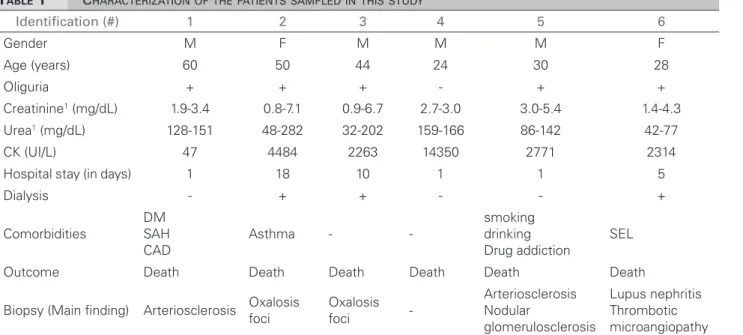
ESULTSWe investigated six patients with confirmed H1N1 infection. The clinical, laboratory and histopathological findings are summarized on Table 1.
All cases investigated depicted clinical and laboratory data of ARF and only one did not have oliguria. Five patients had elevated CPK. All pa-tients presented with acute respiratory failure re-quiring mechanical ventilation and vasoactive dru-gs. Three patients underwent hemodialysis, and the other three patients died quickly before dialysis could be started. Hypervolemia-related oliguria was the main dialysis indication in these patients. All patients received oseltamivir, and died.
Figure 1. Kidney sample from case #6, HE with 400x: glomerulus with endocapillary glomerulonephritis, leucocyte infiltration and microthrombi.
Figure 2. Kidney sample from case #5, PAS with dig 100x: glomerulus with intercapillary nodular glomerulosclerosis.
Figure 3. Kidney sample from case #5, PAS with dig 400x: proximal tubules with vacuolar degenerative alterations.
Figure 4. Lung sample from case #6, HE 100x: lung tissue with septal lymphomononuclear inflammation, hemorrhage and hyaline membrane.
TABLE 1 CHARACTERIZATIONOFTHEPATIENTSSAMPLEDINTHISSTUDY
1 Value upon admission and maximum value reached.
Identification (#) 1 2 3 4 5 6
Gender M F M M M F
Age (years) 60 50 44 24 30 28
Oliguria + + + - + +
Creatinine1 (mg/dL) 1.9-3.4 0.8-7.1 0.9-6.7 2.7-3.0 3.0-5.4 1.4-4.3
Urea1 (mg/dL) 128-151 48-282 32-202 159-166 86-142 42-77
CK (UI/L) 47 4484 2263 14350 2771 2314
Hospital stay (in days) 1 18 10 1 1 5
Dialysis - + + - - +
Comorbidities
DM SAH CAD
Asthma -
-smoking drinking Drug addiction
SEL
Outcome Death Death Death Death Death Death
Biopsy (Main finding) Arteriosclerosis Oxalosis
foci
Oxalosis
foci
-Arteriosclerosis Nodular
glomerulosclerosis
D
ISCUSSIONAcute lung injury has been described as the H1N1 infection hallmark. However, the systemic dysfunction of multiple organs may happen and cause the patient to die.6 Several studies show the involvement of multiple organs and systems: skeletal muscles, the brain, blood and lung.1,2,11,12 Although poorly understood, kidney involvement is often described in the literature, reaching an incidence of up to 66% in H1N1 critically ill patients.3-5 In addition, high mortality rates have been described in the literature, with a significantly higher rate among H1N1 patients with ARF.3-7 The high incidence and mortality rates associated with ARF justifies the importan-ce of assessing the clinical characteristics of these ARF cases.
Abdulkader e colaboradores5 reported a 53% ARF incidence, and a significant association between the need for mechanical ventilation and ARF development (76% of the cases required mechanical ventilation). Other factors such as arterial oxygen concentration/ fraction of inspired oxygen < 200 mmHg, vasopressors and high levels of lactate dehydrogenase also had an impact on morta-lity in this population. The same study found that death was reported only in patients who developed ARF.5 In the present study, all patients required mechanical ventilation and vasoactive drugs, they developed ARF and died. These findings taken together, demons-trate the severity of the clinical context in which ARF is presented, stressing the need for understanding the pathophysiology involved in the development of ARF in patients with H1N1 infection.
Recent studies have reported the virus in kidney cells6 and in urine samples from patients infected with H1N1.5,11 However, these results do not indicate that a virus has caused kidney injury directly. Our findings of acute tubular necrosis (ATN), thrombotic microangiopathy and myoglobin pigments have been reported in the literature.1,6,11 However, the ARF pathophysiology in these
patients is still poorly understood, and some authors suggest a multifactorial mecha-nism: a combination of hypoperfusion, renal vasoconstriction and rhabdomyolysis in the context of a severe systemic inflammatory response syndrome (SIRS).3,6,13
Our results suggest that the major mechanism involved in the development of acute kidney failure in these patients was pre-renal in origin, and may be associated with SIRS developed in most cases. However, there are a number of li-mitations, such as small sample size and the lack of laboratory tests that reinforce this hypothesis, especially urinary sodium and the sodium excretion fraction (SEF), which substantially limit the differential diagnosis between pre-renal ARF and intrinsic renal. Recent markers, such as serum and urinary neutrophil gelatinase asso-ciated lipocaine (NGAL) and the kidney injury molecule 1 (KIM1) could be useful in these pa-tients, because they proved to be more accurate in diagnosing ischemic renal injury in shocked patients - which is the hypothesis considered.14,15 However, these markers were still not available in our hospital at the time the patients had been hospitalized.
A likely explanation for a possible pre-renal etiology in this population of patients with H1N1 infection in intensive care is the possibility of an incorrect estimate of intravas-cular volume adequacy, mainly as a result of pulmonary edema - characteristic of acute lung injury caused by the H1N1 virus. This aspect may have hindered interpretation of the need for adequate fluid replacement and correction of intravascular space depletion. Another possibility to explain this etiology is that a mechanism similar to that of sepsis, wherein the cytokine released by the inflammatory process would cause vasodilation, resulting in a redistribution of blood flow and consequent low kidney input.
nons-hypoxia (hypoxic-ischemic damage) arising from the unfavorable hemodynamic and infectious alterations to which these patients were submitted. Similarly, pulmonary histopa-thological findings are nonspecific, common to multiple organ failure conditions, and also point to a lung tissue response to SIRS. Furthermore, the absence of immune depo-sits upon immunofluorescence signals to the absence of mechanisms mediated by antibo-dies or complement, implicated in the genesis of the kidney injuries found. Therefore, all the findings of our study suggest that the pre-renal mechanism is probably the major determinant of ARF found in H1N1 patients, associated with a severe inflammatory response expected by the severity of the reported cases.
In addition, the association between rhabdomyolysis and ARF development has been described in some studies.16-18 The disproportionate increase in creatinine levels in some patients could be related to muscle lysis. Still, little is known about the mechanism of rhabdomyolysis development in these patients; however, the available data in the literature suggests that it is one of the complications of the viral infection, which may be listed as one of the factors involved in ARF development or worsening.18 In our series of six patients, five had elevated CPK levels, indicating rhab-domyolysis and its clinical relevance in H1N1 infected patients. Even when there were no histopathological indicators of rhabdomyolysis, such as pigmented cylinders in the renal tissue, this intense muscle injury could be a reason for the outcome of patients who had a rapid creati-nine increase in 24 hours and died, as patient # 5, who had 2.4 mg/dL increase in creatinine and 2771 IU/L in CPK within 24h.
C
ONCLUSIONIn our study, all patients had some degree of renal histopathological changes. However, these changes are nonspecific and common in critically ill patients and that could result from the presence of other comorbidities. Thus, these findings suggest that ARF in this population
would likely be secondary to the combination of several factors, predominantly a pre-renal me-chanism. The set of our observations reinforce the need and importance of new studies that seek to clarify and characterize the pathophy-siological events that lead to ARF in patients diagnosed with H1N1.
R
EFERENCES1. Mauad T, Hajjar LA, Callegari GD, da Silva LF, Schout D, Galas FR, et al. Lung pathology in fatal novel human influenza A (H1N1) infection. Am J Respir Crit Care Med 2010;181:72-9. PMID: 19875682
2. Harms PW, Schmidt LA, Smith LB, Newton DW, Pletneva MA, Walters LL, et al. Autopsy findings in eight patients with fatal H1N1 influenza. Am J Clin Pathol 2010;134:27-35. PMID: 20551263
3. Sood MM, Rigatto C, Zarychanski R, Komenda P, Sood AR, Bueti J, et al. Acute kidney injury in critically ill patients infected with 2009 pandemic influenza A(H1N1): report from a Canadian Province. Am J Kidney Dis 2010;55:848-55. PMID: 20303633
4. Trimarchi H, Greloni G, Campolo-Girard V, Giannasi S, Pomeranz V, San-Roman E, et al. H1N1 infection and the kidney in critically ill patients. J Nephrol 2010;23:725-31. 5. Abdulkader RC, Ho YL, de Sousa Santos S, Caires R,
Aran-tes MF, Andrade L. Characteristics of acute kidney injury in patients infected with the 2009 influenza A (H1N1) virus. Clin J Am Soc Nephrol 2010;5:1916-21.
6. Carmona F, Carlotti AP, Ramalho LN, Costa RS, Ra-malho FS. Evidence of Renal Infection in Fatal Cases of 2009 Pandemic Influenza A (H1N1). Am J Clin Pathol 2011;136:416-23.
7. Martin-Loeches I, Papiol E, Rodríguez A, Diaz E, Zaragoza R, Granada RM, et al. Acute kidney injury in critical ill pa-tients affected by influenza A (H1N1) virus infection. Crit Care 2011;15:R66.
8. Mehta RL, Kellum JA, Shah SV, Molitoris BA, Ronco C, Warnock DG, et al. Acute Kidney Injury Network: report of an initiative to improve outcomes in acute kidney injury. Crit Care 2007;11:R31.
9. Uchino S, Kellum JA, Bellomo R, Doig GS, Morimatsu H, Morgera S, et al.; Beginning and Ending Supportive Thera-py for the Kidney (BEST Kidney) Investigators. Acute renal failure in critically ill patients: a multinational, multicenter study. JAMA 2005;294:813-8. PMID: 16106006
10. Klahr S, Miller SB: Acute oliguria. N Engl J Med 1998;338:671-5. PMID: 9486997
11. Gill JR, Sheng ZM, Ely SF, Guinee DG, Beasley MB, Suh J, et al. Pulmonary pathologic findings of fatal 2009 pan-demic influenza A/H1N1 viral infections. Arch Pathol Lab Med 2010;134:235-43. PMID: 20121613
12. He YX, Gao ZF, Lu M, Sui GJ, Ran GW, Cao B, et al. A histopathological study on Influenza A H1N1 infection in humans. Beijing Da Xue Xue Bao 2010;42:137-9. PMID: 20396350
14. Mishra J, Mori K, Ma Q, Kelly C, Yang J, Mitsnefes M, et al. Amelioration of ischemic acute renal injury by neu-trophil gelatinase-associated lipocalin. J Am Soc Nephrol 2004;15:3073-82.
15. Han WK, Bailly V, Abichandani R, Thadhani R, Bonven-tre JV. Kidney Injury Molecule-1 (KIM-1): a novel bioma-rker for human renal proximal tubule injury. Kidney Int 2002;62:237-44. PMID: 12081583
16. Carrillo-Esper R, Ornelas-Arroyo S, Pérez-Bustos E, Sán-chez-Zúñiga J, Uribe-Esquivel M. Rhabdomyolysis and acute renal failure in human influenza A H1N1 media-ted infection. Gac Med Mex 2009;145:519-21. PMID: 20077871
17. Lai CC, Wang CY, Lin HI. Rhabdomyolysis and acute kidney injury associated with 2009 pandemic influenza A(H1N1). Am J Kidney Dis 2010;55:615. PMID: 20189052 18. Parikh M, Dolson G, Ramanathan V, Sangsiraprapha W.