Polymorphisms of Folate Pathway Enzymes
(Methylenetetrahydrofolate Reductase and Thymidylate
Synthase) and Their Relationship with Thymidylate
Synthase Expression in Human Astrocytic Tumors
De´bora Menezes da Costa, Germano Paulo Venceslau de Lima, Ma´rio Henrique Gira˜o Faria, and Silvia Helena Barem Rabenhorst
Two important polymorphisms of folate cycle enzymes, methylenetetrahydrofolate reductase (MTHFR) C677T and thymidylate synthase (TS) enhancer region (TSER) 28-bp tandem repeat, are related to risk of various types of cancer, including brain tumors, although there are few studies on this subject. A case–control study of these two polymorphisms in astrocytomas of different grades was carried out using polymerase chain reaction– restriction fragment length polymorphism, also determining the immunohistochemical expression of TS. The MTHFR677 TT genotype was less associated with astrocytic tumors (odds ratio [OR]=0.00;p=0.0238), but the TSER polymorphism did not show any significant association. Combined genotype TT-double repeats/triple repeats (2R/3R) had a protective effect against astrocytomas (OR=0.00;p=0.0388). Expression of TS protein was observed in the majority of cases, with grade IV tumors being the exception. Moreover, the medianH-score for the pilocytic astrocytomas was significantly higher when compared with that for diffuse tumors. There was an inverse correlation between the 2R/2R genotype and the highest TS-expressing tumors, and 3R/3R was rela-tively more frequent among the tumors grouped in the third and fourth quartiles. Our results provide support for the role of MTHFR and TS polymorphism in gliomagenesis, possibly because of the alteration of DNA methylation and repair status. Moreover, high levels of TS expression were detected in these tumors.
Introduction
T
he carcinogenic process in manytypes of cancer is associated with the interaction of endogenous and ex-ogenous agents, resulting in an abnormal response to DNA damage. In the case of human brain tumors, the data on nonhereditary factors from epidemiological studies include chemical and infectious exposures, but only radiation is currently determined as a risk factor (Wrenschet al., 1993). Genetic factors that contribute to cancer susceptibility in-clude rare, highly penetrant, dominant mutations or more common genetic polymorphisms. Polymorphisms in cancer-related genes have been suggested to be the genetic basis of the individual’s susceptibility to cancer. Although they may confer a small absolute cancer risk, they could be, individ-ually or in combination, responsible for the imbalance of crucial metabolism involved in cancer predisposition (Liuet al., 2010).
Folate is an essential nutrient required for DNA synthe-sis, repair, and methylation. A folate-derived methyl group
is transferred to a series of compounds, and this biological network is known as one-carbon metabolism (Kim et al., 1999). In particular, two enzymes, methylenetetrahy-drofolate reductase (MTHFR) and thymidylate synthase (TS), have an interrelated role (Wajedet al., 2001). MTHFR is a key enzyme in folate metabolism, regulating the flow of folate groups. Its substrate, 5,10-methylenetetrahydofolate, is an intracellular form of folate required for de novo syn-thesis of thymidylate. The product of MTHFR activity, 5-methyltetrahydrofolate, the most important circulating form of folate, is the carbon donor forde novomethionine synthesis and, consequently, DNA methylation. On the other hand, TS competes with MTHFR for 5,10-methyle-netetrahydofolate as a substrate for intracellular conversion of dUMP to dTMP, a rate-limiting step in DNA synthesis and repair (Blountet al., 1997). Therefore, individual genetic variation in these enzymes could influence the general balance between DNA synthesis, repair, and methylation. All these processes seem to be involved in gliomagenesis because of the genetic instability and the presence of
Molecular Genetics Laboratory, Department of Pathology and Forensic Medicine, School of Medicine, Federal University of Ceara´, Fortaleza, Ceara, Brazil.
ªMary Ann Liebert, Inc. Pp. 57–66
DOI: 10.1089/dna.2011.1273
methylated genes described by some studies (Gonzalez-Gomezet al., 2003).
A common polymorphism, MTHFR C677T, substituting alanine for valine at codon 222, in the N-terminal catalytic domain, results in an allozyme with decreased activity. The
MTHFR 677 TT genotype is associated with lower levels of circulating folate (5-methyl-THF), accumulation of 5,10-methylene-THF, and substantially reduced levels of global DNA methylation in peripheral blood leukocytes (Kimet al., 1999; Chung et al., 2010). Also, a tandem 28-bp repeat se-quence in the TS promoter enhancer region (ER), mostly double repeats (2R) or triple repeats (3R), has been identified (Horieet al., 1995). Studiesin vitroandin vivodemonstrated that TS expression was genotype dependent, wherein the 3R allele was associated with higher TS expression (Horieet al., 1995; Kristensenet al., 2010).
Although there are published studies suggesting that the polymorphism of folate cycle enzymes may affect many human cancers, so far few papers relate this to brain tumor risk (Kafadaret al., 2006; Semmleret al., 2006; Bethkeet al., 2008; Sirachainanet al., 2008). All these publications have the
MTHFR C677T polymorphism in common, but only one paper addresses this subject with regard to TSER, without any reference to TS expression. Moreover, the number of astrocytomas was very limited, and no association between the two polymorphisms was shown. Additionally, these studies were from different populations: two from northern European countries, one from Thailand, and the other from Turkey, with one of them only involving glioblastomas. Thus, in the present study, we performed an analysis of
MTHFRC677T and TSER individually and in combination in a case–control study with a series of astrocytic tumors from the state of Ceara´, Brazil. We also investigated the relation-ship between TS genotype and gene expression.
Materials and Methods
The present study was approved by the Ethics Committee of the Hospital Complex of the Federal University of Ceara´ under the protocols no. 32/04 and no. 121/04, according to Resolution 196/96 of the National Council of Health, Ministry of Health, Brazil. We investigated 93 cases of astro-cytic tumors of different grades (World Health Organization [WHO]) (17 grade I, 19 grade II, 14 grade III, and 43 grade IV) from BIOPSE(Biome´dica, Pesquisas e Servic¸os Ltda). All the samples were sectioned at 5mm and processed for
histopath-ological evaluation (hematoxylin–eosin staining) and im-munostaining for TS. Control subjects were cancer-free individuals born and living in the same region as the cases. Among the 492 (general population) controls, 93 were mat-ched by sex and age (–2 years) (matched population).
DNA extraction from paraffin-embedded specimens and blood
All specimens were taken from paraffin-embedded tis-sues. Ten sections from each sample were obtained from the blocks with adequate precaution to prevent contamination between cases, including replacement of blades between each block. Microtome holders were cleaned using xylene between cases to prevent contamination of tissue from one block to the next. Deparaffinization was done through xylol baths at 65C and rehydrated with solutions of decreasing
concentrations of ethanol and deionized water. DNA was extracted using a solution containing 20% Chelex and 10 mg/mL proteinase K, followed by incubation at 55C
overnight. The supernatant containing DNA was collected after centrifugation in a new sterile tube.
Blood samples were obtained from the control population individuals by standard venipuncture in test tubes contain-ing ethylenediaminetetraacetic acid (EDTA) and then geno-mic DNA was extracted using a salting-out method. Agarose gel electrophoresis with ethidium bromide staining was performed to ensure DNA quality. Information on the de-mographic characteristics and familial history of cancer was collected using a questionnaire by a trained interviewer.
Genotype analysis
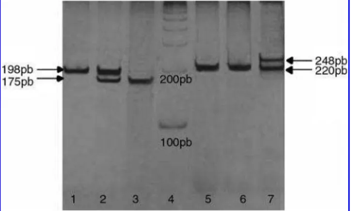
MTHFRC677T: The fragment of 198 bp from exon 4 of the gene was amplified using the primer sequence and condi-tions described by Frosstet al. (1995). The restriction endo-nucleaseHinfI was used to digest the 10-mL polymerase chain
reaction (PCR) product to determine the genotype. The C to T transition at nucleotide 677 (valine variant) creates a new
HinfI site, which generates fragments of 175 and 23 bp. PCR products were verified in a 1% agarose gel stained with ethidium bromide and the restriction digest fragments were visualized in a 7% polyacrylamide gel with silver staining (Fig. 1).
TSER: The presence of a 28-bp repeat polymorphism was determined using the primer sequences and PCR conditions described by Etienneet al.(2002, 2004) except for the addition of 10% of dimethyl sulfoxide. The expected fragment sizes were 220 bp (2R) and 248 bp (3R). PCR products were visu-alized in 7% polyacrylamide gels with silver staining (Fig. 1). Quality control samples were included in all laboratory analyses. Random samples (10% of case and control samples)
were reanalyzed for control of laboratory procedures with the identities unknown to the laboratory staff. Concordance in the analysis was 99.5% for both polymorphisms. For the discordant samples, the genotype assays were repeated by two independent researchers to achieve 100% concordance.
Immunohistochemistry and immunostaining analysis
Immunostaining was performed according to the protocol in an earlier report (Grundaet al., 2006) and is briefly de-scribed here. Antigen retrieval was performed by pretreating deparaffinized sections with 10 mM EDTA (pH 8.0) in a pressure cooker for 5 min. After cooling, the sections were immersed in phosphate-buffered saline (PBS) containing 3% hydrogen peroxide for 10 min to block endogenous peroxi-dase activity. Sections were then incubated in a humidified chamber overnight at 4C with TS primary antibody (clone
TS106; dilution 1:80; Neomarkers). After rinsing with PBS, the slides were incubated with secondary antibody followed by streptavidin–biotin–peroxidase complex (LSAB+ system;
DakoCytomation), both for 30 min at room temperature with a PBS wash between steps. The reaction was revealed with diaminobenzidine–H2O2 (DAB+ system;
DakoCyto-mation), counterstained with Harry’s hematoxylin, and mounted. A confirmed case of TS-positive human breast carcinoma was used as a positive control. Controls for pri-mary antibody specificity included omission of pripri-mary an-tiserum or its substitution by normal bovine serum.
The slides were independently evaluated by three expe-rienced technicians. Immunostaining analysis was carried out using direct light microscopy in 5–10 different fields at 400·magnification. Positivity index (PI) represents the
per-centage of tumors that express TS protein in each group (histological grade); the labeling index (LI) expresses the percentage of positive cells in each tumor sample; the
H-score takes into account the intensity of the TS stain ex-pressed in values ranging from 0 to 3 (0=no stain; 1=weak;
2=moderate; and 3=strong), following the methods
de-scribed by McCartyet al.(1986). For both the LI andH-score, we examined at least 1000 astrocytic cells counted in several fields of the same sample with high-power magnification.
Statistical analyses
Statistical analysis was performed with the use of SPSS version 14.0. Descriptive data were expressed as frequency distributions and medians. Comparison between quantita-tive variables was performed by nonparametric approaches (Mann–Whitney U-test and Spearman’s rank correlation). Hardy–Weinberg equilibrium for case and control popula-tions was assessed using the chi-square test. The odds ratio (OR) with 95% confidence interval (CI), adjusted for age and sex, was calculated to estimate the relative risk of develop-ment of astrocytic tumors for theMTHFRC677T and TSER genotypes. Statistical significance was set atp<0.05.
Results
The frequency of the polymorphic genotypes in the gen-eral population was 7.7% forMTHFR677 TT and 28.9% for TSER 3R/3R. The genetic polymorphisms in this population were in Hardy–Weinberg equilibrium. Comparison between general and matched populations showed that the two
populations did not differ (Supplementary Tables 1–4; Sup-plementary Data are available online at www.liebertonline .com/dna).
Differences in genotype distribution for MTHFR C677T and TSER polymorphisms, as well as the combined genotype of these polymorphisms, were tested between the total cases and control population (n=93). These data are presented in
Tables 1 and 2, respectively. Although the genotypeMTHFR
677 TT was not present in the patients, the heterozygotes were slightly more frequent, which can explain the similar allele frequencies between the two groups. A polyacrylamide gel with representative results from both polymorphisms studied is shown in Figure 1.
TheMTHFR677 TT genotype was absent in the astrocytic tumors. Considering theMTHFR677 CC genotype as refer-ence, despite that the OR cannot be considered because of a lack of TT genotype in the cases generating a zero risk, the significance found by the chi-square test (p=0.0238) is a
strong indication of the association of this genotype with protective effect against the development of astrocytoma.
On the other hand, the TSER polymorphism did not show any significant associations despite a decrease in the inci-dence of the 3R/3R genotype observed in the patients com-pared with the controls (Table 1). However, when the
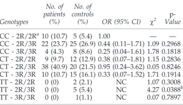
MTHFR C677T and TSER genotypes were combined, a potential protective effect was found with the TT-2R/3R ge-notype in relation to astrocytic tumors (p=0.0388) (Table 2).
Table 3 shows the distribution forMTHFR and TSER ge-notypes according to the histological subtypes of astrocy-toma. No statistically significant differences were found between these polymorphisms with regard to histological classification.
Examples of immunostaining for TS in astrocytic tumors are illustrated in Figure 2. Expression of TS protein was observed in the majority of cases (PI>90%), but grade IV
Table1. Genotype Frequencies
of Methylenetetrahydrofolate ReductaseC677T
and Thymidylate Synthase Enhancer Region Polymorphisms in Astrocytic Tumors
and Matched Controls
Allele
No. of patients
(%)
No. of controls
(%) OR (95% CI) w2
p -Value
MTHFRC677T polymorphism
CCa 36 (38.7) 38 (40.8) 1.00 — — CT 57 (61.3) 47 (50.6) 1.28 (0.67–2.43) 0.43 0.5101 TT 0 (0.00) 8 (8.6) NC 5.10 0.0238b Ca 129 (69.4) 123 (66.1) 1.00 — — T 57 (30.6) 63 (33.9) 0.86 (0.55–1.36) 0.31 0.5791 TSER polymorphism
2R/2Ra 19 (20.4) 19 (20.4) 1.00 — — 2R/3R 60 (64.5) 50 (53.8) 1.20 (0.54–2.68) 0.09 0.7675 3R/3R 14 (15.1) 24 (25.8) 0.58 (0.21–1.61) 0.86 0.3545 2Ra 98 (52.7) 88 (47.3) 1.00 — — 3R 88 (47.3) 98 (52.7) 0.81 (0.53–1.24) 0.87 0.3506
aReference allele. b
p<0.05: statistically significant.
tumors showed a slightly lower PI (*80%). Moreover, the
medianH-scores for the pilocytic tumors (WHO grade I) was significantly higher when compared with the diffuse tumors (Fig. 3). Among the diffuse tumors, no difference was found between grades II and III, but grade IV tumors had signifi-cantly lower medianH-scores.
When these results were correlated to the TS genotype, a negative correlation was found between genotype 3R/3R and TS expression (R= -0.282 andp=0.002). In view of this
contradictory finding, TS expression was categorized into quartiles, based on the LI scores (Fig. 4). This made it possible to demonstrate an inverse correlation between the wild-type homozygous genotype (2R/2R) and the highest TS-expressing tumors as well as a greater presence of the heterozygous genotype (2R/3R) among cases of median TS expression (LI=25%–49%). The distribution of the
polymor-phic homozygote (3R/3R) did not display a clear pattern, although this genotype was relatively more frequent among the tumors grouped in the third (LI=50%–74%) and fourth
(LI=75%–100%) quartiles. Interestingly, the 3R/3R was the
predominant genotype in cases negative for TS expression (LI<5%), which were represented by glioblastomas (grade
IV), except for a single case of grade III tumor.
Discussion
The present study analyzed the risk of the development of astrocytic tumors according to the presence of polymor-phisms of two important enzymes of the folate cycle, MTHFR C677T and TSER, crossing data from DNA somatic tissue (controls) with the archived tumor tissue. This com-parison between different sources of sample was in conso-nance with a previous analysis (Blomekeet al., 1997; Marsh
et al., 2005; Weiss et al., 2007). Moreover, we assessed TS immunoexpression and performed a correlation with its polymorphic status.
In basic cancer research, there is an increasing need to establish factors that predispose to the development of can-cer. In recent years, genetic factors have been increasingly recognized as major contributors to cancer risk (Weber and Nathanson, 2000; Ponder, 2001; Dumitrescu and Cotarla, 2005). Several genetic polymorphisms, including the poly-morphisms of folate pathway enzymes, are reported to have an important role in the modification of cancer susceptibility. Although there are some conflicting results, it seems to de-pend on the cancer type. In relation to brain tumor risk, there are still few studies in the literature.
There are many variations in the genotype frequency of the polymorphismsMTHFRC677T and TSER among world populations. The Hardy–Weinberg equilibrium analyses in the present study showed that the control population was in equilibrium, but not the brain tumor samples, demonstrating an imbalance in these specific patient populations. Analyses ofMTHFRC677T showed that the frequency of genotype 677 TT was similar to that for American Caucasian (Yanget al., 2008), Portuguese (Castro et al., 2003), German (Semmler
et al., 2006), and Finnish (Bethkeet al., 2008) populations, but showing a lower frequency compared with Chinese (Shrub-soleet al., 2004) and higher than in South African (Pegoraro
Table2. Frequencies of Methylenetetrahydrofolate ReductaseC677Tand Thymidylate Synthase
Enhancer Region Genotypes Combination in Astrocytic Tumors and Matched Controls
Genotypes
No. of patients
(%)
No. of controls
(%) OR (95% CI) w2
p -Value
CC - 2R/2Ra10 (10.7) 5 (5.4) 1.00 — — CC - 2R/3R 22 (23.7) 25 (26.9) 0.44 (0.11–1.71) 1.09 0.2968 CC - 3R/3R 4 (4.3) 8 (8.6) 0.25 (0.04–1.61) 1.78 0.1818 CT - 2R/2R 9 (9.7) 12 (12.9) 0.38 (0.07–1.81) 1.15 0.2836 CT - 2R/3R 38 (40.9) 20 (21.5) 0.95 (0.24–3.62) 0.05 0.8246 CT - 3R/3R 10 (10.7) 15 (16.1) 0.33 (0.07–1.52) 1.71 0.1914 TT - 2R/2R 0 (0) 2 (2.1) NC 1.07 0.3008 TT - 2R/3R 0 (0) 5 (5.4) NC 4.27 0.0388b
TT - 3R/3R 0 (0) 1(1.1) NC 0.07 0.7897
a
Reference allele.
bp<0.05: statistically significant.
Table3. Genotype Distribution of Methylenetetrahydrofolate ReductaseC677Tand Thymidylate Synthase Enhancer Region Polymorphisms According to Astrocytic Tumors Histological Classification
(World Health Organization)
Grade OR (95% CI) p OR (95% CI) p OR (95% CI) p
TSER 2R/2R 2R/3R 3R/3R
I–IV (all) 0.680 (0.176–2.626) 0.419 1.389 (0.592–3.259) 0.294 2.542 (0.780–8.287) 0.108 I 1.273 (0.968–1.673) 0.535 12.000 (0.983–153.885) 0.060 4.000 (0.190–84.199) 0.426 II 1.031 (0.601–1.769) 0.664 1.867 (0.283–12.310) 0.430 4.000 (0.388–41.288) 0.272 III — — 0.538 (0.326–0.891) 0.571 1.625 (1.057–2.497) 0.643 IV 1.178 (0.850–1.633) 0.390 0.667 (0.187–2.377) 0.381 3.107 (0.562–17.169) 0.192 II-IV (diffuse) 0.834 (0.206–3.375) 0.552 0.942 (0.370–2.397) 0.554 2.333 (0.645–8.435) 0.168
MTHFRC677T CC CT TT
et al., 2004) and Thai (Sirachainanet al., 2008) populations. The frequencies of TSER polymorphism appear to be sim-ilar to that of a US population (Ulrichet al., 2005), but dif-fering from that of a Chinese population (Zhang et al., 2005), which had higher frequency of the 3R/3R genotype (Table 4).
The present study that with MTHFR C677T, although the OR could not be considered, the significant absence of this genotype in the cases indicated that the genotype 677 TT may confer a potential protective effect, thereby de-creasing the risk of this cancer. The reduction of risk related
to this genotype has been described for other cancers, such as hepatocellular carcinoma (Yuanet al., 2007), colorectal can-cer (Cui et al., 2010), non-Hodgkin lymphoma (Kim 2007), and prostate cancer (Safarinejadet al., 2010).
On the other hand, these findings are contradictory to those of some studies that assessed brain tumors. Sir-achainanet al.(2008) studied 31 glial tumors and did not find any association with MTHFR C677T polymorphism in Thai children; however, only seven of them were astrocytic tumors, and this population had a genotypic frequency distribution that differed from that in the present study.
FIG. 2. Immunohisto-chemistry for thymidylate synthase protein in formalin-fixed, paraffin-embedded astrocytic tumors (·400).(A)
Pilocytic astrocytoma (WHO grade I): diffuse staining (H-score=42, LI=24);(B)
gemistocytic astrocytoma (WHO grade II): moderate staining (H-score=111,
LI=65);(C)anaplastic
astro-cytoma (WHO grade III): slight staining (H-score=25,
LI=20);(D)glioblastoma multiforme (WHO grade IV): diffusely intense staining (H-score=160, LI=88). LI,
labeling index; WHO, World Health Organization.
FIG. 3. MedianH-score at-tributed to thymidylate syn-thase immunostaining according to histopathological classification of the cases studied (n=93). *p<0.05
Kafadar et al. (2006) also did not find any association, but they found a higher frequency of the TT genotype, possibly because these authors analyzed high-grade gliomas without distinction.
A major study on the polymorphism of folate enzymes was conducted by Benthkeet al.(2008) in a northeast Euro-pean population, which provided support for the role of folate metabolism in the development of primary brain tumors. In this study, genotypes associated with increased 5,10-MTHF levels were correlated with elevated risk of de-veloping brain tumors. Although no statistically significant association was found with the folate metabolism polymor-phisms studied, their data indicated a decrease in risk related to the MTHFR 677 TT genotype in the astrocytoma group. Also, theMTHFR677 CC genotype was associated with risk of glioblastoma when in combination with MTHFR 1298 AC/CC polymorphism. It was a well-designed study, matching the case with control from the same origin; the frequency of the MTHFR 677 TT genotype shows large variation among the different studied populations, which can explain the differences relative to our data.
One possible explanation for the protective genotype against astrocytic tumors could be the decrease in methyla-tion of tumor suppressor genes due to the low activity of the polymorphic enzyme MTHFR, compromising the supply of methyl groups. However, this hypothesis is contradictory to the study by Cadieuxet al. (2006), which indicated that in-adequate methyl production promotes genomic hypo-methylation and instability, explaining the increased proliferative activity in these tumors. Another plausible ex-planation for this 677 TTMTHFRprotective effect is the in-creased conversion rate of dUMP to dTMP that prevents dUMP misincorporation into DNA and promotes a more efficient repair and higher genomic stability (Duthie, 2010). The removal of dUMP leads to a temporary strand break, and when it happens repeatedly, repair becomes inefficient, which may cause DNA strand breaks, chromosomal dam-age, and malignant cell transformation (Reidy, 1987; Blount and Ames, 1994).
With regard to the TSER polymorphism, we did not find any correlation with the risk of developing astrocytic tumors. The same was found in the study of Semmleret al.(2006). Studies that analyzed this polymorphism regarding cancer susceptibility showed many variations, according to the localization of the tumor, indicating that there is no tendency toward establishing a consensual role about this polymor-phism in human cancers (Table 5).
We did not observe significant tendencies in the risk an-alyses related to the distribution of the two genotypes in different histological grades. None of the previous studies reported its results considering each astrocytoma WHO grade and few reports performed a combined genotype analysis. Semmler et al. (2006) studied the association of glioblastoma development withMTHFRC677T, methionine synthase (MTR) A2756G, and transcobalamin 2 C776G polymorphisms; these authors found that only grade IV as-trocytomas showed an increased risk related to theMTRG allele. When we performed a genotype association, we found that the TT-2R/3R genotype was significantly less associated with tumor frequency. It is important to observe that the
FIG. 4. TSER genotype distribution according to quartiles of expression, based on labeling index scores of the cases studied (n=93).
Table4. Genotypic Frequencies of Methylenetetrahydrofolate ReductaseC677Tand Thymidylate Synthase Enhancer Region5¢UTRPolymorphisms in Different Populations
Populations Genotypic distribution Reference
C677TMTHFR CC (%) CT (%) TT (%) —
Brazil (CE) 40.8 50.6 8.6 This study USA (Caucasian) 45.8 42.6 11.7 Yanget al., 2008 Portugal 43.6 46.2 10.3 Castroet al., 2003 Germany 46 46 9 Semmleret al., 2006 Finland 61 32.4 6.5 Bethkeet al., 2008 Brazil (RS) 45 41 14 Brandalizeet al., 2007 China 33.4 49.7 16.9 Shrubsoleet al., 2004 South Africa 88 11 1 Pegoraroet al., 2004 Thailand 72.7 25.9 1.5 Sirachainanet al., 2008
TSER 2R/2R (%) 2R/3R (%) 3R/3R (%) —
Brazil (CE) 20.4 53.8 25.8 This study
USA 22 50 28 Ulrichet al., 2005
China 3.7 33.3 63 Zhanget al., 2005
combinations ofMTHFR677 TT and TSER 3R/3R genotypes were in low frequency among the tumor cases studied, demonstrating the complexity of genotypic combinations owing to genetic interactions and nutritional habits. The potential protective effect related to this genotype association can be due to a decreased activity of MTHFR in association with an increased activity of TS. This association would favor DNA synthesis and repair processes in detriment to meth-ylation, promoting a more efficient repair and a higher genomic stability (Duthie, 2010).
The high TS immunohistochemical levels in the majority of tumor cases indicate the importance of this enzyme in the maintenance of genomic stability due to DNA synthesis, al-though some differences were observed when the number
(LI) or intensity (H-score) of stained cells were assessed. The significant expression in pilocytic astrocytomas (grade I), differing from the other grades (II, III, and IV), could be explained by the common benign behavior of these cases associated with the well-differentiated status. We hypothe-size that other genetic factors associated with malignancy, absent in grade I tumors, could be activated in diffuse tumors (especially in grade IV), perhaps inhibiting, at least partially, TS expression (Cadieuxet al., 2006).
On the other hand, some studies that analyzed thein vitro
correlation between genotype and TS expression demon-strated a higher enzyme expression associated with the ge-notype 3R/3R (Horie et al., 1995; Yu et al., 2008). In the present study, when immunohistochemical data were cros-sed with genotypes, a negative correlation was found bet-ween 3R/3R and TS expression. However, when the genotype was distributed according to expression score quartiles, we found that the 2R/2R genotype showed an inverse correlation with TS expression. Conversely, despite a variation in TS expression associated with the 3R3R geno-type, its frequency was high in the higher quartiles. This lack of a clear pattern associated with the TS expression may be due to the presence of a single-nucleotide polymorphism (C-G) in the second repeat of 3R alleles, wherein the presence of 3G sequence has three to four times greater efficiency than the other sequences (2C, 2G, 3C) (Kawakami and Watanabe, 2003; Mandolaet al., 2003).
Curiously, the few cases with negative TS expression were observed only among astrocytic tumors with 2R/3R and, notably, 3R/3R genotypes. Moreover, all these cases were high-grade tumors (predominantly grade IV), which are ex-pected to show more genetic alterations. Thus, we suggest that the accumulation of molecular alterations could, at least partially, block TS expression, resulting in low DNA repair and predispose to more genetic instability. The slight de-crease in TS expression observed in glioblastomas compared with other grades, demonstrated in the present study, also supports this hypothesis. These findings are in accordance with the extensive knowledge about glioblastomas, which show lower repair rates and marked genetic instability due to the sum of multiple genetic alterations that characterize their molecular and clinical malignancy (Collins, 2004).
The high levels of TS expression also raise speculation about the use of this enzyme as a target in chemotherapy for astrocytoma. Some studies have shown success in treating brain tumors with fluoropyrimidines, especially for radio-sensitization of glioblastomas (5-fluorouracil and capecita-bine) (Meneiet al., 2005; Grundaet al., 2010). Recently, novel folate-based TS inhibitors derived from quinazolines (ralti-trexed, peme(ralti-trexed, and nolatrexed) demonstrated a great potential in the treatment of gliomas (Van Triestet al., 2000; Clarkeet al., 2010). A better understanding of the molecular pathway of gliomagenesis and translational studies will en-hance our knowledge about these tumors and will certainly lead to further rapid improvements in clinical outcome.
In conclusion, our results provide support for the role of polymorphisms of the folate pathway enzymes MTHFR and TS in astrocytic tumorigenesis, possibly because of an alter-ation in methylalter-ation and repair status. Although the sample had a reasonable size, the lack of MTHFR677 TT indicates the need for further analysis, with a higher number of cases to confirm these findings. Moreover, the high level of TS
Table5. Studies That Analyzed the Relationship Between Thymidylate Synthase Enhancer Region Polymorphism and Risk to Cancer Development
Cancer type Results Reference
Acute lymphoblastic leukemia
No association was found between TS 5¢UTR polymorphism
and the ALL risk in children.
Lauten
et al., 2003
Colorectal adenoma
The 3R/3R genotype was associated with an increased risk when the alcohol consumption was elevated.
Chenet al., 2003
Gastric cancer The 3R allele was associated with increase risk to GC.
Graziano
et al., 2004 Gastroesophagic
cancer and esophagic carcinoma
The TS lower expression increased the risk of cancer.
Tanet al., 2005
Gastric cancer TS 5¢UTR 2R and
TS 3¢-UTR 6 bp in
association can act in GC etiology (Chinese population).
Zhanget al., 2005
Head and neck carcinoma
The 2R/3R genotype was associated with the increased risk of cancer, but no association was found with 2R/2R genotype.
Zhanget al., 2004
Breast cancer No association was found.
Stevens
et al., 2007 T-cell lymphoma The 2R/2R genotype
was associated with the increased risk to this lymphoma.
Kimet al., 2008
Non-Hodgkin lymphoma
The 2R/3R genotype was associated with the increased risk to NHL.
Lightfoot
et al., 2005
Pancreatic cancer No association was found.
Wanget al., 2005
expression detected in these tumors, despite not being di-rectly related to TSER, shows a potential targeted therapy, notably in high-grade astrocytomas. Unfortunately, we did not measure the plasma levels or dietary intake of folate and the homocysteine in our population as well as the MTHFR and TS enzyme activity in brain tumor tissues. However, it invites new speculation that would certainly warrant ex-ploration in future studies.
Acknowledgment
This study was financially supported by CNPq.
Disclosure Statment
No competing financial interests exist.
References
Bethke, L., Webb, E., Murray, A., Schoemaker, M., Feychting, M., Lo¨nn, S., Ahlbom, A., Malmer, B., Henriksson, R., Auvi-nen, A., Kiuru, A., SalmiAuvi-nen, T., Johansen, C., Christensen, H.C., Muir, K., McKinney, P., Hepworth, S., Dimitropoulou, P., Lophatananon, A., Swerdlow, A., and Houlston, R. (2008). Functional polymorphisms in folate metabolism genes influ-ence the risk of meningioma and glioma. Cancer Epidemiol Biomarkers Prev17,1195–1202.
Blomeke, B., Bennett, W.P., Harris, C.C., and Shields, P.G. (1997). Serum, plasma and paraffinembedded tissues as sources of DNA for studying cancer susceptibility genes. Carcinogenesis
18,1271–1275.
Blount, B.C., and Ames, B.N. (1994). Analysis of uracil in DNA by gas chromatography-mass spectrometry. Anal Biochem
219,195–200.
Blount, B.C., Mack, M.M., Wehr, C.M., Mac Gregor, J.T., Hiatt, R.A., Wang, G., Wickramasinghe, S.N., Everson, R.B., and Ames, B.N. (1997). Folate deficiency causes uracil mis-incorporation into human DNA and chromosome breakage: Implications for cancer and neuronal damage. Proc Natl Acad Sci USA94,3290–3295.
Brandalize, A.P.C., Bandinelli, E., Borba, J.B., Fe´lix, T.M., Roisenberg, I., and Schu¨ler-Faccini, L. (2007). Polymorphisms in genes MTHFR, MTR and MTRR are not risk factors for cleft lip/palate in South Brazil. Braz J Med Biol Res40,
787–791.
Cadieux, B., Ching, T., Vandenberg, S.R., and Costello, J.F. (2006). Genome-wide hypomethylation in human glioblasto-mas associated with specific copy number alteration, methy-lenetetrahydrofolate reductase allele status, and increased proliferation. Cancer Res1,8469–8473.
Castro, R., Rivera, I., Ravasco, P., Jakobs, C., Blom, H.J.,
Camilo, M.E., and de Almeida, I.T. (2003).
5,10-Methylenetetrahydrofolate reductase 677CT and 1298AC mutations are genetic determinants of elevated homo-cysteine. Q J Med 96, 297–303.
Chen, J., Hunter, D.J., Stampfer, M.J., Kyte, C., Chan, W., Wet-mur, J.G., Mosig, R., Selhub, J., and Ma, J. (2003). Poly-morphism in the thymidylate synthase promoter enhancer region modifies the risk and survival of colorectal cancer. Cancer Epidemiol Biomarkers Prev12,958–962.
Chung, C.J., Pu, Y.S., Su, C.T., Chen, H.W., Huang, Y.K., Shiue, H.S., and Hsueh, Y.M. (2010). Polymorphisms in one-carbon metabolism pathway genes, urinary arsenic profile, and ur-othelial carcinoma. Cancer Causes Control21,1605–1613.
Clarke, J., Butowski, N., and Chang, S. (2010). Recent advances in therapy for glioblastoma. Arch Neurol67,279–283. Collins, V.P. (2004). Brain tumours: classification and genes. J
Neurol Neurosurg Psychiatry75,2–11.
Cui, L., Shin, M.H., Kweon, S.S., Kim, H.N., Song, H.R., Piao, J.M., Choi, J.S., Shim, H.J., Hwang, J.E., Kim, H.R., Park, Y.K., and Kim, S.H. (2010). Methylenetetrahydrofolate reductase C677T polymorphism in patients with gastric and colorectal cancer in a Korean population. BMC Cancer10,2362–2368. Dumitrescu, R.G., and Cotarla, I. (2005). Understanding breast
cancer risk—where do we stand in 2005? J Cell Mol Med9,
208–221.
Duthie, S.J. (2010). Folate and cancer: how DNA damage, repair and methylation impact on colon carcinogenesis. J Inherit Metab Dis34,101–109.
Etienne, M-C., Chazal, M., Laurent-Puig, P., Magne´, N., Rosty, C., Formento, J.L., Francoual, M., Formento, P., Rene´e, N., Chamorey, E., Bourgeon, A., Seitz, J.F., Delpero, J.R., Letou-blon, C., Pezet, D., and Milano, G. (2002). Prognostic value of tumoral thymidylate synthase and p53 in metastatic colorectal cancer patients receiving fluorouracil-based chemotherapy: phenotypic and genotypic analyses. J Clin Oncol 20,2832– 2843.
Etienne, M-C., Ilc, K., Formento, J.L., Laurent-Puig, P., For-mento, P., Cheradame, S., Fischel, J.L., and Milano, G. (2004). Thymidylate synthase and methylenetetrahydrofolate reduc-tase gene polymorphisms: relationships with 5-fluorouracil sensitivity. Br J Cancer90,526–534.
Frosst, P., Blom, H.J., Milos, R., Goyette, P., Sheppard, C.A., Matthews, R.G., Boers, G.J.H., den Heijer, M., Kluijtmans, L.A.J., van den Heuve, L.P., and Rozen, R. (1995). A candidate genetic risk factor for vascular disease: a common mutation in methylenetetrahydrofolate reductase. Nat Genet10,111–113. Gonzalez-Gomez, P., Bello, J., Arjona, D., Lomas, J., Alonso,
M.E., De Campos, J.M., Vaquero, J., Isla, A., Gutierrez, M., and Rey, J.A. (2003). Promoter hypermethylation of multiple genes in astrocytic gliomas. Int J Oncol22,601–608.
Graziano, F., Kawakami, K., Watanabe, G., Ruzzo, A., Humar, B., Santini, D., Catalano, V., Ficarelli, R., Merriman, T., Panunzi, S., Testa, E., Cascinu, S., Bearzi, I., Tonini, G., and Magnani, M. (2004). Association of thymidylate synthase polymorphisms with gastric cancer susceptibility. Int J Cancer
112,1010–1014.
Grunda, J.M., Fiveash, J., Palmer, C.A., Cantor, A., Fathallah-Shaykh, H.M., Nabors, L.B., and Johnson, M.R. (2010). Ra-tionally designed pharmacogenomic treatment using concur-rent capecitabine and radiotherapy for glioblastoma; gene expression profiles associated with outcome. Clin Cancer Res
16,2890–2898.
Grunda, J.M., Nabors, L.B., Palmer, C.A., Chhieng, D.C., Steg, A., Mikkelsen, T., Diasio, R.B., Zhang, K., Allison, D., Grizzle, W.E., Wang, W., Gillespie, G.Y., and Johnson, M.R. (2006). Increased expression of thymidylate synthetase (TS), ubiquitin specific protease 10 (USP10) and survivin is associated with poor survival in glioblastoma multiforme (GBM). J Neu-rooncol80,261–274.
Horie, N., Aiba, H., Oguro, K., Hojo, H., and Takeishi, K. (1995). Functional analysis and DNA polymorphism of the tandemly repeated sequences in the 5¢-terminal regulatory region of the human gene for thymidylate synthase. Cell Struct Funct20,
191–197.
(MTHFR) in meningiomas and high-grade gliomas. Antic-ancer Res26,2445–2449.
Kawakami, K., and Watanabe, G. (2003). Identification and functional analysis of single nucleotide polymorphism in the tandem repeat sequence of thymidylate synthase gene. Cancer Res.63,6004–6007.
Kim, H.N., Lee, I.K., Kim, Y.K., Tran, H.T., Yang, D.H., Lee, J.J., Shin, M.H., Park, K.S., Shin, M.G., Choi, J.S., and Kim, H.J. (2008). Association between folate-metabolizing pathway polymorphism and non-Hodgkin lymphoma. Br J Haematol.
140,287–294.
Kim, D. (2007). The interactive effect of methyl-group diet and polymorphism of methylenetetrahydrofolate reductase on the risk of colorectal cancer. Mutat Res622,14–18.
Kim, Y.I. (1999). Folate and carcinogenesis: evidence, mecha-nisms, and implications. J Nutr Biochem10,66–88.
Kristensen, M.H., Weidinger, M., Bzorek, M., Pedersen, P.L., and Mejer, J. (2010). Correlation between thymidylate synthase gene variants, RNA and protein levels in primary colorectal adenocarcinomas. J Int Med Res38,484–497.
Lauten, M., Asgedon, G., Welte, K., Schrappe, M., and Stanulla, M. (2003). Thymidylate synthase gene polymorphism and its association with relapse in childhood B-cell precursor acute lymphoblastic leukemia. J Hematology88,353–354.
Lightfoot, T.J., Skibola, C.F., Willett, E.V., Skibola, D.R., Allan, J.M., Coppede, F., Adamson, P.J., Morgan, G.J., Roman, E., and Smith, M.T. (2005). Risk of non–hodgkin lymphoma as-sociated with polymorphisms in folate-metabolizing genes. Cancer Epidemiol Biomarkers Prev14,2999–3003.
Liu, Y., Shete, S., Hosking, F.J., Robertson, L.B., Bondy, M.L., and Houlston, R.S. (2010). New insights into susceptibility to glioma. Arch Neurol67,275–278.
Marsh, S., Mallon, M.A., Goodfellow, P., and McLeod, H.L. (2005). Concordance of pharmacogenetic markers in germline and colorectal tumor DNA. Pharmacogenomics6,873–877. Mandola, M.V., Stoehlmacher, J., Muller-Weeks, S., Cesarone,
G., Yu, M.C., Lenz, H.J., and Ladner, R.D. (2003). A novel single nucleotide polymorphism within the 5¢tandem repeat polymorphism of the thymidylate synthase gene abolishes USF-1 binding and alters transcriptional activity. Cancer Res.
63,2898–2904.
McCarty, K.S., Jr., Szabo, E., Flowers, J.L., Cox, E.B., Leight, G.S., Miller, L., Konrath, J., Soper, J.T., Budwit, D.A., Creasman, W.T., Seigier, H.F., and McCarty, K.S., Sr. (1986). Use of a monoclonal anti-estrogen receptor antibody in the immuno-histochemical evaluation of human tumors. Cancer Res46,
4244–4248.
Menei, P., Capelle, L., Guyotat, J., Fuentes, S., Assaker, R., Ba-taille, B., Franc¸ois, P., Dorwling-Carter, D., Paquis, P., Bauchet, L., Parker, F., Sabatier, J., Faisant, N., and Benoit, J.P. (2005). Local and sustained delivery of 5-fluorouracil from biode-gradable microspheres for the radiosensitization of malignant glioma: a randomized phase II trial. Neurosurgery56, 242– 248.
Pegoraro, R.J., Chikosi, A., Rom, L., Roberts, C., and Moodley, J. (2004). Methylenetetrahydrofolate reductase gene polymor-phisms in black South Africans and the association with pre-eclampsia. Acta Obstet Gynecol Scand83,449–454.
Ponder, B.A.J. (2001). Cancer genetics. Nature411,336–341. Reidy, J.A. (1987). Folate- and deoxyuridine-sensitive chromatid
breakage may result from DNA repair during G2. Mutat Res
192,217–219.
Safarinejad, M.R., Shafiei, N., and Safarinejad, S. (2010). Re-lationship between three polymorphisms of
methylenete-trahydrofolate reductase (MTHFRC677T, A1298C, and G1793A) gene and risk of prostate cancer: a case-control study. Prostate
70,1645–1657.
Semmler, A., Simon, M., Moskau, S., and Linnebank, M. (2006). The methionine synthase polymorphism c.2756A > G alters susceptibility to glioblastoma multiforme. Cancer Epidemiol Biomarkers Prev15,2314–2316.
Shrubsole, M.J., Gao, Y.T., Cai, Q., Shu, X.O., Dai, Q., He´bert, J.R., Jin, F., and Zheng, W. (2004). MTHFR polymorphisms, dietary folate intake, and breast cancer risk: results from the shanghai breast cancer study. Cancer Epidemiol Biomarkers Prev13,190–196.
Sirachainan, N., Wongruangsri, S., Kajanachumpol, S., Pakaka-sama, S., Visudtibhan, A., Nuchprayoon, I., Lusawat, A., Phudhicharoenrat, S., Shuangshoti, S., and Hongeng, S. (2008). Folate pathway genetic polymorphisms and susceptibility of central nervous system tumors in Thai children. Cancer Detect Prev32,72–78.
Stevens, V.L., McCullough, M.L., Pavluck, A.L., Talbot, J.T., Feigelson, H.S., Thun, M.J., and Calle, E.E. (2007). Association of polymorphisms in one-carbon metabolism genes and postmenopausal breast cancer incidence. Cancer Epidemiol Biomarkers Prev16,1140–1147.
Tan, W., Miao, X., Wang, L., Yu, C., Xiong, P., Liang, G., Sun, T., Zhou, Y., Zhang, X., Li, H., and Lin, D. (2005). Significant increase in risk of gastroesophageal cancer is associated with interaction between promoter polymorphisms in thymidylate synthase and serum folate status. Carcinogenesis 26, 1430– 1435.
Ulrich, C.M., Curtin, K., Potter, J.D., Bigler, J., Caan, B., and Slattery, M.L. (2005). Polymorphisms in the reduced folate carrier, thymidylate synthase, or methionine synthase and risk of colon cancer. Cancer Epidemiol Biomarkers Prev14,2509– 2516.
Van Triest, B., Pinedo, H.M., Giaccone, G., and Peters, G.J. (2000). Downstream molecular determinants of response to 5-fluorouracil and antifolate thymidylate synthase inhibitors. Ann Oncol11,385–391.
Wajed, A.S., Laird, P.W., and DeMeester, T.R. (2001). DNA methylation: an alternative pathway to cancer. Ann Surg234,
10–20.
Wang, L., Miao, X., Tan, W., Lu, X., Zhao, P., Zhao, X., Shan, Y., Li, H., and Lin, D. (2005). Genetic polymorphisms in methy-lenetetrahydrofolate reductase and thymidylate synthase and risk of pancreatic cancer. Clin Gastroenterol Hepatol3,743– 751.
Weber, B.L., and Nathanson, K.L. (2000). Low penetrance genes associated with increased risk for breast cancer. Eur J Cancer
36,1193–1199.
Weiss, J.R., Baer, M.R., Ambrosone, C.B., Blanco, J.G., Hutson, A., Ford, L.A., and Moysich, K.B. (2007). Concordance of pharmacogenetic polymorphisms in tumor and germ line DNA in adult patients with acute myeloid leukemia. Cancer Epidemiol Biomarkers Prev16,1038–1041.
Wrensch, M.R., Bondy, M.L., Wiencke, J., and Yost, M. (1993). Environmental risk factors for primary malignant brain tu-mors: a review. J Neuro-oncol17,47–64.
Yu, K.H., Wang, W.X., Ding, Y.M., Li, H., and Wang, Z.S. (2008). Polymorphism of thymidylate synthase gene associated with its protein expression in human colon cancer. World J Gas-troenterol14,617–621.
Yuan, J.M., Lu, S.C., Van Den Berg, D., Govindarajan, S., Zhang, Z.Q., Mato, J.M., and Yu, M.C. (2007). Genetic polymorphisms in the methylenetetrahydrofolate reductase and thymidylate synthase genes and risk of hepatocellular carcinoma. Hepa-tology46,749–758.
Zhang, Z., Shi, Q., Sturgis, E.M., Spitz, M.R., Hong, W.K., and Wei, Q. (2004). Thymidylate synthase 5¢- and 3¢-untranslated region polymorphisms associated with risk and progression of squamous cell carcinoma of the head and neck. Clin Cancer Res10,7903–7910.
Zhang, Z., Xu, Y., Zhou, J., Wang, X., Wang, L., Hu, X., Guo, J., Wei, Q., and Shen, H. (2005). Polymorphisms of thymidylate synthase in the 5¢- and 3¢-untranslated regions associated with
risk of gastric cancer in South China: a case–control analysis. Carcinogenesis26,1764–1769.
Address correspondence to: