Saudi J Kidney Dis Transpl 2017;28(3):645-647
© 2017 Saudi Center for Organ Transplantation
Case Report
Oliguric Acute Kidney Injury as Initial Presentation of
Renal Non-Hodgkin
’s
Lymphoma Infiltration
Tacyano T. Leite1, Alexandre B. Libório1,2, Geraldo B. Silva Junior2,3, Elizabeth De Francesco Daher1,2
1
Division of Nephrology, Hospital Geral de Fortaleza,2Department of Internal Medicine, Post-Graduation Program in Medical Sciences, Federal University of Ceará,3School of Medicine,
Master in Collective Health, University of Fortaleza, Fortaleza, Ceará, Brazil
ABSTRACT.We report a case of a 20-year-old man presented to the emergency department with oliguria and renal failure requiring urgent dialysis. An ultrasound revealed enlarged kidneys, and a renal biopsy showed non-Hodgkin’s lymphoma, subtype diffuse large B-cell.
Introduction
Secondary involvement of the kidney by lymphomatous disease is well known, but it is not documented due to the low rate of kidney biopsies performed in such patients.1 Renal manifestations of kidney lymphomas are rare, occurring in only 10% of non-Hodgkin’s
lymphoma and <1% in primary lymphoma of the kidney. Oliguric acute kidney injury as the first manifestation of renal evolvement is even rarer.2 In this paper, we report a case of small bowel non-Hodgkin’s lymphoma with secon-dary kidney involvement presenting as oliguric and severe acute kidney injury requiring emergency dialysis.
Correspondence to:
Prof. Elizabeth De Francesco Daher, Division of Nephrology,
Hospital Geral de Fortaleza, Fortaleza, Ceará, Brazil. E-mail: ef.daher@uol.com.br
Case Report
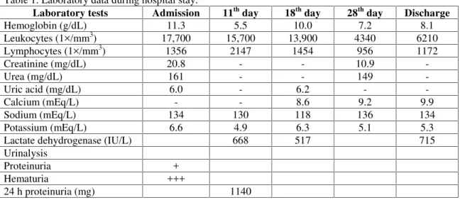
A 20-year-old man presented to the emer-gency department with fever, dry cough, sub-costal pain, nausea and vomiting, oliguria, and dark-colored urine. One day after admission, he had a generalized tonic–clonic convulsion. Physical examination was unremarkable, ex-cept for lower and upper limbs edema and hypertension (180/120 mm Hg). Cranial com-puted tomography (CT) was normal. Labora-tory tests showed a serum creatinine of 20.8 mg/dL and urea of 161 mg/dL. Laboratory data during hospital stay are shown in Table 1. Emergency hemodialysis was started. An ultrasound showed enlarged kidneys (right kidney: 18.1 × 10.3 cm, left kidney: 17.8 × 10.1 cm). These findings were confirmed by abdominal CT (Figure 1a).
He had a prior history of an intestinal tumor mass resection five months ago in another hospital. The histopathological analysis of the intestinal mass was suggestive of
lymphoproli-Saudi Journal
of Kidney Diseases
and Transplantation
ferative lesion.
A renal biopsy revealed large lymphoid cells proliferation, atypical, pleomorphics, with evi-dent nucleus, surrounded by a small amount of lymphocytes, without atypias, arranged as nodular infiltrate in the renal parenchyma (Figure 1b). The immunohistochemical pattern was 70% positive for Ki-67 antibody and also for bcl-6 and CD20. The final diagnosis was non-Hodgkin’s lymphoma, subtype diffuse large B-cell lymphoma (DLBCL).
Chemotherapy was started with rituximab,
cyclophosphamide, doxorubicin, vincristine, and prednisone (R-CHOP schema). The pa-tient became clinical stable and was dis-charged to the hematology outpatients’ clinics, but with no renal function recovery and con-tinuing hemodialytic treatment.
Discussion
Renal involvement has been reported frequent in non-Hodgkin’s lymphoma.3 Flank pain, he-maturia, weight loss, and palpable abdominal
Table 1. Laboratory data during hospital stay.
Laboratory tests Admission 11th day 18th day 28th day Discharge
Hemoglobin (g/dL) 11.3 5.5 10.0 7.2 8.1
Leukocytes (1×/mm3) 17,700 15,700 13,900 4340 6210
Lymphocytes (1×/mm3) 1356 2147 1454 956 1172
Creatinine (mg/dL) 20.8 - - 10.9
-Urea (mg/dL) 161 - - 149
-Uric acid (mg/dL) 6.0 - 6.2 -
-Calcium (mEq/L) - - 8.6 9.2 9.9
Sodium (mEq/L) 134 130 118 136 134
Potassium (mEq/L) 6.6 4.9 6.3 5.1 5.3
Lactate dehydrogenase (IU/L) 668 517 715
Urinalysis
Proteinuria +
Hematuria +++
24 h proteinuria (mg) 1140
(a) (b)
Figure 1. (a) Abdominal computed tomography evidencing large-sized kidneys (right kidney: 18.2 × 8.8cm, left kidney: 18.6 × 9.7cm), with dense images in the periphery; (b) Renal biopsy showing large lymphoid cells proliferation, atypical, pleomorphics, with evident nucleus, surrounded by a small amount of lymphocytes, without atypias, arranged as nodular infiltrate in the renal parenchyma.
646 Leite TT, Libório AB, Silva Junior GB, et al
mass are the most common manifestations. In primary renal lymphoma, most patients present with acute kidney injury.4 Our presented as severe oliguric acute kidney injury requiring dialysis, a rare manifestation of secondary infiltration of the kidney by non-Hodgkin’s lymphoma. In these patients, the renal infiltration is usually diagnosed in post mortem analysis. Tumor lysis syndrome was excluded in this case as his serum uric acid level was normal.
Renal lymphoma in the majority of cases originates from a multisystemic dissemination but can also occur as a primary lymphoma, which is extremely rare.5 Secondary renal involvement is found in 30%–40% of autop-sies from patients with lymphoma5 and pri-mary lymphoma in only 0.7% of extranodal lymphomas.6
Renal lymphomas are indistinguishable from other renal tumors through clinical manifes-tations and radiological findings.7 The lesions can be solitary masses (10%–20%) or multiple (60%). They are generally bilateral and its extension occurs through contiguity (25%–
30%), diffuse infiltration (20%), or perirenal involvement (10%). Radiological findings fre-quently indicate renal involvement with mul-tiple nodules and help elucidate diagnosis. Renal lymphoma is generally represented by a bilateral nodular infiltrate associated with diffuse kidneys enlargement.8 Non-Hodgkin’s
DLBCL is the most common type of lym-phoma in adults, and it is responsible for 30%–
40% of non-Hodgkin’s lymphomas.9
Imaging is very important to diagnose renal lymphomas as the clinical presentation is often unspecific. It is more probable that kidneys are involved in diffuse forms of lymphomas.1 The diagnosis is confirmed with histopathological examination and immunohistochemistry. Renal biopsy is the best method to establish the diagnosis of renal involvement and has a high sensitivity and specificity.6 Treatment is deter-mined according to the classification of lym-phoma subtype.
Conflict of interest:None declared.
References
1. Martina MN, Solé M, Massó E, Pérez N, Campistol JM, Quintana LF. Mixed cryo-globulinaemia not related to hepatitis C virus, mesangiocapillary glomerulonephritis and lymphoplasmocytic lymphoma. Nefrologia 2011;31:743-6.
2. Martín Laborda y Bergasa F, Lozano Lozano D, Gil Fernández JJ, Serrado Pardo R, Fernández Rañada JM. Non-Hodgkin’s lymphoma and urinary tract. About a case reported. Actas Urol Esp 2005;29:427-32. 3. Carvalho JG, Tafarel JR, Carvalho WB,
Azambuja AP, Zenaro ES, Bendlin R. Acute renal failure as first clinical presentation of Burkitt’s renal lymphoma. J Bras Patol Med Lab 2006;42:179-83.
4. Olusanya AA, Huff G, Adeleye O, et al.
Primary renal non-Hodgkins lymphoma
presenting with acute renal failure. J Natl Med Assoc 2003;95:220-4.
5. Jhamb R, Gupta N, Garg S, et al. Diffuse lymphomatous infiltration of kidney presenting as renal tubular acidosis and hypokalemic paralysis: Case report. Croat Med J 2007; 48:860-3.
6. Dash SC, Purohit K, Mohanty SK, Dinda AK. An unusual case of bilateral renal enlargement due to primary renal lymphoma. Indian J Nephrol 2011;21:56-8.
7. Torrecilla García-Ripoll JR, Pascual Samaniego M, Martín Blanco S, Rivera Ferro J, Peral Martínez JI, Fernández del Busto E. Primary renal lymphoma. Actas Urol Esp 2003;27:555-8.
8. Barreto F, Dall'Oglio MF, Srougi M. Renal lymphoma. Atypical presentation of a renal tumor. Int Braz J Urol 2006;32:190-2.
9. Araújo JH, Victorino AP, Melo AC, et al. Linfoma não-Hodgkin de alto grau – Revisão de literatura. Rev Bras Cancerol 2008;54:175-83.
Acute kidney injury due to non-Hodgkin’s lymphoma infiltration 647