University of São Paulo School of Medicine, Department of Gastroenterology, Hepatology Branch.
List of abbreviations used: ADV - Adefovir dipivoxil; ALT - Alanine aminotransferase; HBV - Hepatitis B virus; IFN - Interferon; LAM - Lamivudine; PEG-IFN - Pegylated interferon. Correspondence to: Prof. Luiz Caetano da Silva, IMT2, Av. Dr. Enéas de Carvalho Aguiar 500, 05403-000 São Paulo, SP, Brasil. Tel/fax: 55-11-3064-5132, e-mails:
SIMULTANEOUS QUANTITATION OF SERUM HBV DNA AND HBeAg CAN DISTINGUISH
BETWEEN SLOW AND FAST VIRAL RESPONSES TO ANTIVIRAL
THERAPY IN PATIENTS WITH CHRONIC HEPATITIS B
Luiz Caetano DA SILVA, Maria Luiza da NOVA, Suzane Kioko ONO-NITA, João Renato Rebello PINHO, Roberta SITNIK, Vera Aparecida dos SANTOS & Flair José CARRILHO
SUMMARY
Background: The quantitation of serum HBeAg is not commonly used to monitor viral response to therapy in chronic hepatitis B.
Methods: In this study, 21 patients receiving varying therapies were followed and their viral response monitored by concomitant viral load and HBeAg quantitation in order to study the meaning and the kinetics of both parameters. Results: It was possible to distinguish between three different patterns of viral response. The first was characterized by a simultaneous decrease in serum HBV DNA and HBeAg. The second pattern was characterized by a decrease in serum HBeAg but persistent detection of HBV DNA. The third pattern was characterized by undetectable HBV DNA with persistent HBeAg positivity, which points to a non-response (Pattern III-B) except when HBeAg levels showed a slow but steady drop, characterizing a “slow responder” patient (Pattern III-A). Conclusions: The first pattern is compatible with a viral response. A long-term HBeAg seropositivity with a slow and persistent decrease (Pattern III-A) is also compatible with a viral response and calls for a prolongation of anti-viral treatment.
KEYWORDS: Hepatitis B; Serological markers; Treatment response; Treatment follow-up.
INTRODUCTION
The management of chronic hepatitis B has been the subject of many reviews over the last few years4,11,19,23,24. Updates to the recommendations
for monitoring patients infected with hepatitis B virus (HBV) were recently published20,25, and in them, as well as recent publications on
this subject, serum Hepatitis e antigen (HBeAg) is considered only qualitatively with no evaluation of its expression (quantitation) as a tool for monitoring patients undergoing treatment20,25.
According to KEEFE et al.20, the quantitation of HBeAg has not
been widely used, is expensive, and available data about the predictive value of HBeAg serostatus remains limited. The same authors mention that approximately one third of patients with serum HBV DNA levels lower than 104 copies will seroconvert, suggesting a close relationship
between HBV DNA suppression and the likelihood of seroconversion. HBeAg-positive chronic hepatitis B, considered as the classical form, is characterized by a stable high level viremia (107-1010 HBV copies/mL),
whereas HBeAg-negative hepatitis B is characterized by a less stable and severe viremia, or less than 106 copies/mL30.
Several different types of quantitative assays based on signal or target amplification are available for viral DNA, including high sensitivity
polymerase chain reaction (PCR)-based methods that measure serum
HBV DNA levels with a wide dynamic range1,25. Nowadays, real-time
PCR assays are preferably used25.
The maximum HBV DNA viral load detected in patients with chronic HBV is not well established and may depend on the quantitative technique
used to measure it. Thus, CORDEN et al.5 measured HBV DNA with
the Chiron Amplicor HBV DNA Monitor Assay and found that the viral load was between 4X10² and 1.2X10⁹ copies/mL. In HBeAg-positive
patients, they found HBV DNA levels of up to 109.5 copies/mL. By using
in-house end-point dilution PCR based on Kaneko’s method17,18, our
group found levels of up to 3X10¹² copies/mL6,8. Also, using real-time
PCR, HO et al.14 found median levels of 1.5X10⁷ copies/mL, and JARDI
et al.16 found median a serum HBV DNA viral load of 9.2X10⁸ copies/
mL (range 2.9X10⁵ - 4.8X10¹¹).
For HBeAg-positive chronic hepatitis B patients, the main goals for treatment are HBeAg loss, seroconversion to anti-HBe, normalized alanine aminotransferase (ALT) levels, and the suppression of viremia to undetectable levels25. However, it has been consistently observed that
NEUMANN26. It is worth mentioning that the production of HBeAg is
not dependent on the formation of HBV DNA, but it reflects the level of HBV core/precore gene expression in the liver34.
The role of HBeAg quantitation has already been addressed by some authors13,28. PERILLO et al.28 found that the baseline HBeAg
concentration is the best independent predictor of a patient’s response to interferon-alpha (IFN-α) when compared to baseline HBV DNA levels. Furthermore, it was observed, in this study and another12,13,that a steep
decline in the HBeAg concentration during the first weeks of therapy was predictive of an antiviral response. A long-term study on the serial quantitation of HBeAg demonstrated that a high serum level (> 100 IU/ mL) at week 24 of therapy with peginterferon alpha-2a (Peg-IFN-α2a) had an excellent negative predictive value; less than 5% of patients achieved HBeAg seroconversion at week 7210.
During LAM therapy, different types of changing patterns have been observed in relation to levels of HBeAg pretreatment, and they have been categorized into three groups: “decrescendo” (“falling”), “decrescendo-crescendo” (“falling-rising”), and “no changing” or “fluctuating”27,31. These patterns determined by serial monitoring during
LAM therapy allowed the prediction of the treatment response as well as early recognition of a viral breakthrough. However, a simultaneous determination of HBV DNA was not performed.
It is worth mentioning that, in HBeAg-positive patients, entecavir was found to be more potent than LAM and resulted in undetectable HBV DNA by PCR in 67% of patients at week 48, compared to 36% of patients treated with LAM3. However, the HBeAg seroconversion rate
was similar in the two groups, 21% and 18%. Despite these interesting and valuable observations, serial quantitative determinations of serum HBeAg have not been routinely carried out during the last few years and have not been mentioned in some recent recommendations20,25.
Using serial quantitative PCR for HBV DNA and a simultaneous quantitative HBeAg assay, we previously observed an interesting dissociation between HBV DNA and HBeAg in some patients. Our aim is to show that a simultaneous decrease in HBV DNA and HBeAg levels, in addition to a slow but stepwise decreasing pattern of HBeAg levels in some patients, may predict the negative HBeAg response despite a very prolonged positive response during antiviral therapy. This is particularly important because serum HBV DNA levels may decrease to undetectable levels soon after initiating therapy with LAM or other drugs8,26,30,34 and can not be used further for monitoring the
treatment response.
PATIENTS AND METHODS
Patients: Twenty-one HBeAg-positive patients with chronic hepatitis
B were enrolled in the study (Table 1). Sixteen patients received LAM and five patients received IFN-α plus LAM (Table 2).
In this phase of the study, we detected some patterns of the viral response (described below) in 11 of the 16 patients given LAM and in four of the five patients given IFN-α plus LAM.
Nine patients who did not respond to antiviral therapy, and one who presented with a relapse after LAM withdrawal, were retreated as shown
in Table 3. Three patients (5, 6, and 13) had to receive another series of therapy after the second trial.
Patient ages varied from nine to 65 years (mean 38 yr) and they were studied during a period of 34 to 133 months (Table 1). All patients had Hepatitis B surface antigen (HBsAg) in their serum for at least six months and detectable HBV DNA. Patients were excluded if they were also infected with hepatitis C virus, hepatitis D virus, or human immunodeficiency virus (HIV). Patients who withdrew prematurely from the study or had only minimal lesions (inactive HBsAg carrier state) were also excluded. All patients gave informed consent for the study, which was approved by the Institutional Review Board (Hospital das Clínicas - University of São Paulo School of Medicine).
Treatment schedules and primary endpoints: Sixteen patients
received monotherapy with oral LAM at a dose varying from 150 mg to 300 mg per day for 12 months and 150 mg/day thereafter as previously described8. Three out of four patients with liver cirrhosis received 150
mg/day from the beginning (Table 2). Therapy with IFN-α (3 to 5 MU
three times a week) plus LAM (150 mg/day) was initially given to four patients (Table 2). Another patient was submitted to combined therapy
with Peg-IFN-α2b and LAM because of high serum levels of HBV DNA
and HBeAg and a slight increase in ALT. In our experience, patients with such baseline values are prone to develop resistance to monotherapy with LAM6,8. Such patients were included in the IFN-related group
(Table 2). Overall, 21 series of antiviral therapy were used during this period (Table 2).
Nine LAM-resistant patients with mutations in the YMDD motif of polymerase gene, six with M204V, two with M204I, and one patient with both, were retreated (Table 3). Adefovir (10 mg/day) was given to three patients as a second series and one patient as a third series (Table 3). Pegylated interferon (Peg-IFN) was given to five patients, four received Peg-IFN-α2b (1.5 μg/kg of body weight) and one patient received
Peg-IFN-α2a (180 μg), all in association with LAM. Finally, tenofovir was given at a dose of 300 mg/day to four unresponsive patients as a rescue drug.
As for the two patients who experienced a relapse after withdrawal of LAM, one (patient 2) presented with a spontaneous absence of serum
Table 1
Demographic and baseline data for naïve patients (first phase)
Sex (M/F) 16/5
Age (years) 9 - 65 (± 38)
Caucasians/Asians 14/7
HBV DNA (log) 5.41 - 12.48 (± 9.26)
ALT (xUNL) 0.7 - 8.8 (± 3.24)
Chronic hepatitis/Liver cirrhosis 16/5
First therapy No. of patients
Lamivudine (LAM) 16
IFN + LAM 5
HBeAg but had a persistence of HBV DNA during the non-treatment period and received a second series of LAM with subsequent clearance of the HBV DNA. The other patient (patient 9) was treated with
Peg-IFN-α2b plus LAM and presented with an HBsAg/anti-HBs seroconversion.
Finally, another patient non-responder to LAM (patient 16) received LAM associated to lobucavir as a rescue drug (withdrawn from the market).
As shown in Table 3, more than two therapeutic series had to be used in three patients (patients 5, 6, and 13). Overall, 15 series of antiviral therapy were used in the retreatment, resulting in a total number of 36 therapeutic series for the two phases. The primary endpoints were an inability to detect HBV DNA and the loss of HBeAg with seroconversion to anti-HBe.
Table 2
Demographic, baseline data, and patterns of HBV DNA and HBeAg responses to initial therapy with lamivudine (LAM) in 16 patients or interferon-alpha (IFN-α) plus LAM in five other patients with chronic hepatitis B (CH) or liver cirrhosis (LC)
Patient/
Diagnosis Age/Sex
HBV DNA (log)
ALT* (xUNL)
First therapy
Viral response (VR) / (Mutation)**
Pattern Follow-up
(months) Outcome
1/CH 37/M 10.48 4.5 LAM VR / (NO) I 102 Seroconversion
HBsAg/anti-HBs
2/CH 37/F 6.48 8.8 LAM VR / (NO) I 125 Relapse
Re-treatment
3/CH 45/M 8.63 2.3 LAM VR / (M204V)
(late)
I 125 Breakthrough (late)
Re-treatment
4/LC 65/M 12.48 6.9 LAM VR / (NO) I 109 Seroconversion
HBsAg/anti-HBs
5/CH 48/M 8.48 1.7 LAM BT / (M204V) II 109 Breakthrough
Re-treatment
6/LC 52/M 8.48 1.5 LAM BT / (M204V/I) II 108 Breakthrough
Re-treatment
7/LC 42/M 10.48 3.1 LAM VR / (NO) IIIA 90 Seroconversion
HBeAg/anti-HBe
8/LC 59/M 7.96 8.7 LAM VR / (NO) IIIA 48 Seroconversion
HBeAg/anti-HBe
9/CH 62/M 12.48 1.7 LAM VR / (NO) IIIA 83 VR with relapse
Re-treatment
10/CH 9/F 8.48 3.3 LAM BT / (M204I) IIIB 37 Breakthrough.
Drop out
11/CH 34/M 5.41 2.0 LAM BT / (M204I) IIIB 95 Breakthrough
Re-treatment
12/CH 11/M 10.48 0.7 LAM NR / (M204I) NR 133 Re-treatment
13/CH 30/M 8.48 2.9 LAM NR / (M204V) NR 117 Re-treatment
14/CH 43/M 10.48 1.7 LAM NR / (M204V) NR 86 Re-treatment
15/CH 44/M 10.48 1.2 LAM NR / (M204V) NR 119 Re-treatment
16/CH 49/M 9.48 8.0 LAM NR / (M204V) NR 103 Breakthrough
Re-treatment
17/CH 29/M 12.48 1.5 IFN+LAM VR / (NO) I 85 Seroconversion
HBeAg/anti-HBe
18/CH 21/M 6.48 4.2 IFN+LAM VR / (NO) I 34 Seroconversion
HBeAg/anti-HBe
19/CH 32/F 7.60 2.0 Peg+LAM
***
VR / (NO) IIIA 72 Seroconversion
HBeAg/anti-HBe
20/LC 11/F 6.40 0.7 IFN+LAM VR / (NO) IIIA 62 Seroconversion
HBeAg/anti-HBe
21/CH 12/M 8.70 0.7 IFN+LAM NR / (NO) NR 60 Under treatment.
Steady drop of HBV DNA and HBeAg
Laboratory methods: Serum samples were available for study at the following time points: initial screening (“baseline”), every three to six months during therapy, and after the completion of therapy. The levels of ALT, HBV DNA, and HBeAg were available at each time point. The serum ALT levels were tested using commercially available assays (Abbott Laboratories). Results are expressed as values times the upper normal limit (x UNL). The semi-quantitation of serum HBeAg was performed prospectively and simultaneously with the determination of serum HBV DNA. Because no commercial assay was available for measuring the HBeAg concentration, a micro-particle enzyme immunoassay (MEIA, AxSYM HBe 2.0, Abbott Laboratories, Abbott Park, IL) was used13. The assay is based on two different monoclonal
antibodies, and its reference preparation for quantitation of HBeAg makes use of purified recombinant HBeAg as a control. Results are expressed as an index (sample to cutoff = S/CO) luminescent values ratio27.
In 17 patients, the quantitation of HBV DNA was performed by in-house PCR based on Kaneko’s method17,18 and as previously described6,8.
The sensitivity of the method was reported to be 3x10² copies/mL, and
its dynamic range went up to 3x10¹² copies/mL. Quantitative results were
estimated based on end-point dilutions. In the last four patients (patients 18 to 21), the serum HBV DNA was determined by the Cobas Amplicor HBV Monitor Test (Roche Diagnostics, Branchburg, NJ) with a linear range from 4x10² (lower limit of detection) to 4x10⁷ copies/mL27.We
have recorded the HBV-DNA levels in copies/mL instead of IU/mL as recently reported19.
A virologic response was defined as a decrease in serum HBV
DNA to undetectable levels by PCR and a loss of HBeAg25. Patients
who showed neither of these serological features were considered to be non-responders, and those who presented with only one type of response (HBV DNA or HBeAg) were classified according to the patterns as described below. A sustained response was defined as the persistence of a virologic response for six months after the discontinuation of therapy25. The clearance of HBeAg and HBsAg was defined as the
absence of the particular antigen in two consecutive samples at least one month apart. Viral polymerase, precore/core, and surface genes were sequenced to determine mutations in the YMDD domain as previously described8,32.
Liver biopsies: Liver biopsy was performed with a Tru-cut (Baxter
Health Care, Deerfield, IL, USA) needle in 20 out of 21 patients. The exception was a cirrhotic patient with ascitis and very low platelet counts.
RESULTS
Demographic and baseline characteristics of patients: The
demographics, baseline data, and patterns of initial therapeutic response for the patients studied are shown in Tables 1, 2, and 3. Changes in the HBeAg/anti-HBe system were observed in 15/21 patients, and the behavior of serum HBeAg expression during a long-term follow-up will be further detailed.
As shown in Table 2, a relapse after withdrawal, viral breakthrough,
Table 3
Patterns of HBV DNA and HBeAg responses in chronic hepatitis B patients previously resistant to lamivudine (LAM)* and submitted to a second or a third anti-viral therapy
Patient ** Type of previous resistance
Antiviral therapy (re-treatment)
Pattern of response*** Outcome
2/CH Relapse Lamivudine**** I HBeAg/anti-HBe seroconversion
3/CH M204V Adefovir NR Late HBVDNA negativation and late
HBeAg/anti-HBe seroconversion
5/CH M204V PegIFN-α2b+Lamivudine I Viral response with relapse
Adefovir IIIB HBeAg-non-response
Tenofovir IIIA Profound drop of HBeAg
6/LC M204V/M204I PegIFN-α2b+Lamivudine NR HBVDNA persistently positive
Tenofovir *** Clearance of HBVDNA (See table 2)
9/CH Relapse PegIFN-α2b+Lamivudine I HBsAg/anti-HBs seroconversion
11/CH M204I Adefovir IIIB Breakthrough
12/CH M204I Tenofovir IIIA Under treatment
13/CH M204V PegIFN-α2b+Lamivudine I VR with relapse
Tenofovir I Under treatment
14/CH M204V Adefovir I Under treatment
15/CH M204V PegIFN-α2a+Lamivudine I HBeAg/anti-HBe seroconversion
16/CH M204V Lamivudine+Lobucavir I HBsAg/anti-HBs seroconversion
or non-response was detected in 13/21 (61.9%) patients and in 12/16 (75.0%) patients receiving monotherapy with LAM. Sequencing the YMDD motif in non-responders receiving LAM revealed the M204V mutation in six patients, the M204I mutation in three, and a mixed M204V/I mutation in one patient (Table 2). All but one patient (patient 10) were submitted to re-treatment as stated in Methods (Table 3).
Simultaneous analysis of HBV DNA and HBeAg and characterization of decay patterns: The main goal of our study was to describe the response of HBV to treatment by comparing the patterns of HBV DNA and HBeAg decreases. The results from treated and retreated patients were analyzed together. Overall, 36 series of therapy were obtained.
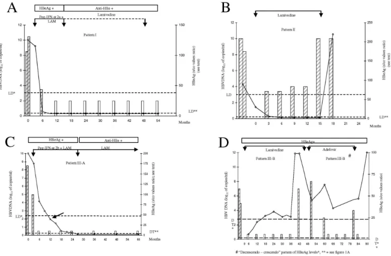
Three different response patterns could be characterized in our patients: Pattern I was characterized by a simultaneous decrease in serum HBV DNA and HBeAg levels until they were absent within a period of six months (Fig. 1A). If the absence of HBV DNA was followed by the
loss of HBeAg in a maximum period of six months, the pattern was still considered as Pattern I.
A concomitant decrease in HBV DNA and HBeAg was observed in 13/36 (36.1%) series as shown in Tables 2 and 3. An example of this pattern is shown in Fig. 1A for patient 15, who had previous resistance to LAM due to the M204V mutation and an excellent response to a combined therapy with Peg-IFN-α2a (180 μg per week) and LAM. Pattern I during LAM or interferon plus LAM was detected in 4/16 patients (15%) and 6/10 patients (60%), respectively (p = 0.10, two- tailed Fisher exact test).
Pattern II was characterized by the persistence of viremia (HBV DNA levels above 1,000 copies/mL) simultaneous with undetectable HBeAg (Fig. 1B) for a period greater than six months.
This pattern was observed in 2/36 (5.6%) series. In one patient (patient5), this pattern was detected during 10 months of therapy with
LAM, but HBeAg became positive thereafter (Fig. 1B). In another patient (patient 6), a loss of HBeAg was observed during therapy with LAM and persisted to be negative throughout despite the high levels of HBV DNA over three years. However, a clearance of serum HBV DNA was detected three months after the administration of tenofovir.
Pattern III was characterized by the persistence of detectable HBeAg despite the absence of HBV DNA for a period greater than six months. In some of these patients, a slow but steady decline of HBeAg was observed and was referred to as Pattern III-A (Fig. 1C).
In other patients, HBeAg decreased followed by an increase in serum levels, or it did not change at all, despite the absence of HBV DNA and was referred to as Pattern III-B (Fig. 1D).
The absence of HBV DNA preceding the loss of HBeAg for more than six months was observed in 11/36 therapeutic series (30.6%).
Pattern III-A was observed in seven patients. Prolonged periods of HBeAg positivity with continuously decreasing levels indicate a good, but slow, viral response (Fig. 1C). One patient (No. 19) with chronic hepatitis B, very high viremia (above 4x10⁷ copies/mL by the Amplicor
Monitor technique), and a high level of HBeAg was submitted to a combined therapy with Peg-IFN-α2b and LAM. As seen in Fig. 1C, HBV DNA became undetectable at the third month. A slow but steady decrease of HBeAg was observed.
Pattern III-B was observed in the other four patients, all of them presenting a viral relapse (Fig. 1D). As seen in Tables 2 and 3, HBeAg/ anti-HBe seroconversion was detected in 5/6 patients who exhibited Pattern III-A, whereas no viral response was seen in the four patients exhibiting Pattern III-B.
In non-responder patients with persistent serum HBV DNA and HBeAg, and in patients presenting with Pattern III-B, the levels of HBeAg varied widely either in a “falling-rising” (“decrescendo-crescendo”) pattern or with fluctuating values27,31.
The small number of patients in each group did not allow a statistical comparison among the different patterns.
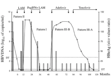
In one patient, all three patterns of viral response were observed, as seen in Fig. 2.
The ALT levels varied widely independently of the described patterns and were not included in this analysis.
DISCUSSION
Based on the simultaneous quantitative determinations of serum HBV DNA and HBeAg, our results suggest that the HBV kinetics can vary widely, depending on the antiviral drugs used for the treatment of HBeAg-positive patients with chronic hepatitis B. The kinetics of HBV have been investigated, but some problems still need clarification22,26.
Our results suggest that patients with positive HBeAg levels are less responsive to LAM or adefovir than combined therapy with interferon and LAM or tenofovir. Thus, Pattern I, which is characterized by the simultaneous decrease of HBV DNA and of HBeAg in a maximum
period of six months, is more frequently found during treatment with interferon plus LAM or during tenofovir treatment. This pattern presents a favorable evolution in most cases, though a relapse or breakthrough is occasionally found. Pattern II was detected in very few cases and must be investigated with a larger number of patients. In our patients a breakthrough was found.
Our patients were followed for periods of time varying from 34 months to 133 months. Such long periods allowed us to observe that different patterns of serum HBeAg changes can be present in the same patient, and it allowed us to note how unpredictable the outcomes of patients on and off antiviral therapy are7. Despite the role of HBV
DNA quantitation, the rapid and dramatic decrease in viremia after the introduction of nucleoside analogues reinforces the necessity to quantify HBeAg levels during the period of HBeAg positivity as an additional marker for monitoring responses. In some patients, a progressive drop in HBeAg levels may be paralleled with an HBV DNA decrease (Pattern I), but in other patients, the decrease of HBeAg is steady and slow, pointing to a late response (Pattern III-A)
A progressive decrease in serum HBeAg has been referred as a “decrescendo” (“falling”) pattern and a decrease in HBeAg followed by a return to high levels as a “decrescendo-crescendo” (“falling-rising”) pattern27,31. The first pattern is predictive of a viral response, whereas most
of the patients who fail to show a continuous decrease in HBeAg levels present with a viral breakthrough or non-response. The characteristics of our study do not allow for a close comparison with the above-mentioned patterns.
Possibly an immune-modulating protein, HBeAg is a nonstructural secreted protein translated from HBV e (precore and core) mRNA. On the other hand, HBcAg, the major protein of the HBV capsid, is
translated from HBV pregenomic mRNA21. Since HBeAg and HBcAg
share a sequence consisting of 149 amino acids, they are collectively called Hepatitis B core-related antigens. The measurement of these antigens would be particularly useful for monitoring the decline of viral
translation activity. It would also allow for a comparison of the efficacies of potent antiviral drugs when a large proportion of patients have HBV DNA below the detection threshold21. ROKUHARA et al.29 reported that
five out of six patients treated with LAM for six to eight months still had detectable levels of core-related antigen. A similar result was also seen in another study34 after 48 weeks of nucleoside analogue therapy; 65%
of the patients who had undetectable serum HBV DNA (< 300 copies/ mL) still had detectable core-related antigen.
Persistent and steady levels of HBeAg for a prolonged period of time, despite the absence of HBV DNA (Pattern III-B) may indicate some degree of resistance and herald a breakthrough, as was observed in some of our patients. This observation led us to switch adefovir for tenofovir in one patient, and a sharp decrease of HBeAg was observed (Fig. 2). On the other hand, a progressive decrease in HBeAg points to a favorable response, even when the persistence of HBeAg is as long as 26 months of therapy. As seen in Fig. 1C, such a long-term persistence might lead to a withdrawal of the combined therapy of Peg-IFN and LAM. As could be predicted by the trend of Pattern III-A in these patients, the loss of HBeAg and seroconversion to anti-HBe was a late event during this therapy.
Previous results6 had also showed that a non-response to LAM
is mainly observed in patients with high viremia, and most of them were positive for HBeAg. This correlation was so significant that a proportion between the probability of non-response and an increase in the logarithm of genome copies per mL could be established. The above-mentioned differences in viral dynamics are not surprising as nucleotide analogues produce a decrease in viral load mainly through a block of viral replication, whereas interferon acts directly through some intracellular antiviral pathways and indirectly through a modulation of the immune system.
Daily LAM doses of 300 mg may lead to an earlier9,15 and more
profound suppression of HBV DNA15. In a previous paper, we showed
a rapid decrease in serum HBV DNA in 27 out of 29 patients (93%) whose quantitation was performed after 12 weeks of LAM treatment6.
This response was demonstrated by three different quantitative methods: endpoint dilution, Amplicor-Monitor (Roche), and branched DNA. The endpoint dilution method using 10-fold serial dilutions was also used by YOTSUYANAGI et al.35 Our data suggests that combination therapy
with PEG-IFN and LAM usually produces a concomitant decrease of HBeAg and HBV DNA, whereas monotherapy with LAM or adefovir may produce a rapid decrease in HBV DNA, but result in a slow, or lack of, decrease of HBeAg in some patients.
Studies in patients with chronic hepatitis B have reported conflicting results regarding the benefits of combining IFN-α or PEG-IFN and LAM, and the role of such combinations in chronic hepatitis B treatment requires further clarification. According to TER BORG et al.33, the HBV DNA
decline after 52 weeks of therapy was significantly higher in patients undergoing combined therapy (PEG-IFN plus LAM) compared to those undergoing monotherapy with PEG-IFN.
The most important limitation of this study relates to the small number of patients and therapeutic series. Despite these limitations, our study provides some evidence that the decrease in serum HBV DNA accompanied by a steady decrease in HBeAg (Pattern I), even when it is
slow (Pattern III-A), points to a favorable outcome. As was also recently proposed for hepatitis C patients2, the use of customized treatment for
each patient may increase the likelihood of a response, and following up on HBV DNA and HBeAg levels helps in choosing the best treatment for each patient as well in predicting treatment response. It must be emphasized that stopping rules based on HBV DNA determination for PEG-IFN treatment could not be established10. Furthermore, our data
suggests that the period of 48 weeks for the discontinuation of PEG-IFN may be too short in some patients.
In conclusion, three different patterns can be observed for HBV DNA and HBeAg levels over time when quantitative PCR and HBeAg (MEIA AxSYM) techniques are used simultaneously. A more detailed study with different therapy schedules and using both methods in a large number of patients with chronic hepatitis B is urgently needed.
RESUMO
A quantificação simultânea do DNA VHB e do AgHBe séricos podem distinguir entre resposta viral lenta e rápida à terapêutica
anti-viral em pacientes com hepatite crônica B
Introdução: A quantificação do AgHBe sérico não é habitualmente utilizada para monitorizar a resposta viral ao tratamento da hepatite crônica B. Métodos: Neste estudo, 21 pacientes sob tratamento com diferentes terapias foram acompanhados e a resposta viral monitorizada pela quantificação concomitante da carga viral e do AgHBe a fim de investigar o significado e a cinética de ambos os parâmetros. Resultados:
Distinguiram-se três diferentes padrões de resposta viral. O primeiro caracterizou-se pela redução simultânea do HBV DNA e AgHBe séricos. O segundo padrão caracterizou-se por uma redução do AgHBe porém com detecção persistente do HBV DNA. O terceiro padrão caracterizou-se por HBV DNA indetectável com positividade persistente do AgHBe, sugerindo ausência de resposta (Padrão III-B), exceto quando os níveis de AgHBe mostraram uma queda lenta porém persistente, caracterizando um “respondedor lento” (Padrão III-A). Conclusões: O primeiro padrão é compatível com resposta viral. Uma seropositividade prolongada do AgHBe porém com uma redução lenta e persistente (Padrão III-A) é também compatível com resposta viral, sugerindo o prolongamento do tratamento anti-viral.
COMPETING INTERESTS
The authors declare that they have no competing interests.
LCDS conceived of the study, participated in its design and coordination, and drafted the manuscript. MLDN participated in its design and drafted the manuscript, SKON participated in its design and drafted the manuscript, JRRP participated in its design and drafted the manuscript, RS participated in drafting the manuscript, VADS carried out the immunoassays, and FJC participated in its design and drafted manuscript. All authors read and approved the final manuscript.
ACKNOWLEDGMENTS
REFERENCES
1. ALBERTI, A. - Can serum HBV-DNA be used as a primary end point to assess efficacy of new treatments for chronic hepatitis B? Hepatology, 38: 18-20, 2003. 2. BUTI, M.; CASADO, M.A. & ESTEBAN, R. - Evaluating the cost of sustained virologic
response in naive chronic hepatitis C patients treated a la carte. Aliment. Pharmacol. Ther., 26: 705-716, 2007.
3. CHANG, T.T.; GISH, R.G.; DE MAN, R. et al. - A comparison of entecavir and lamivudine for HBeAg-positive chronic hepatitis B. New Engl. J. Med., 354: 1001-1010, 2006. 4. CONJEEVARAM, H.S. & LOK, A.S. - Management of chronic hepatitis B. J. Hepat.,
38 (suppl. 1): S90-S103, 2003.
5. CORDEN, S.; BALLARD, A.L.; IJAZ, S. et al. - HBV DNA levels and transmission of hepatitis B by health care workers. J. clin. Virol., 27: 52-58, 2003.
6. DA SILVA, L.C.; DA FONSECA, L.E.; CARRILHO, F.J. et al. - Predictive factors for response to lamivudine in chronic hepatitis B. Rev. Inst. Med. trop. S. Paulo, 42: 189-196, 2000.
7. DA SILVA, L.C.; ONO-NITA, S.K.; PINHO, J.R. & CARRILHO, F.J. - Variable and unpredictable outcome in chronic hepatitis B (CHB) patients during and after therapy with high daily doses of lamivudine (LAM). Hepatology, 36: 638A, 2002. 8. DA SILVA, L.C.; PINHO, J.R.; SITNIK, R.; DA FONSECA, L.E. & CARRILHO, F.J.
- Efficacy and tolerability of long-term therapy using high lamivudine doses for the treatment of chronic hepatitis B. J. Gastroent., 36: 476-485, 2001.
9. DIENSTAG, J.L.; PERRILLO, R.P.; SCHIFF, E.R. et al. - A preliminary trial of lamivudine for chronic hepatitis B infection. New Engl. J. Med., 333: 1657-1661, 1995. 10. FRIED, M.W.; PIRATVISUTH, T.; LAU, G.K. et al. - HBeAg and hepatitis B virus DNA
as outcome predictors during therapy with peginterferon alfa-2a for HBeAg-positive chronic hepatitis B. Hepatology, 47: 428-434, 2008.
11. FUNG, S.K. & LOK, A.S. - Treatment of chronic hepatitis B: who to treat, what to use, and for how long? Clin. Gastroent. Hepat., 2: 839-848, 2004.
12. HEIJTINK, R.A.; JANSSEN, H.L.; HOP, W.C.; OSTERHAUS, A.D. & SCHALM, S.W. - Interferon-alpha therapy in chronic hepatitis B: early monitoring of hepatitis B e antigen may help to decide whether to stop or to prolong therapy. J. viral Hepat., 7: 382-386, 2000.
13. HEIJTINK, R.A.; KRUINING, J.; HONKOOP, P. et al. - Serum HBeAg quantitation during antiviral therapy for chronic hepatitis B. J. med. Virol., 53: 282-287, 1997. 14. HO, S.K.; YAM, W.C.; LEUNG, E.T. et al. - Rapid quantification of hepatitis B virus
DNA by real-time PCR using fluorescent hybridization probes. J. med. Microbiol., 52: 397-402, 2003.
15. HONKOOP, P.; DE MAN, R.A.; NIESTERS, H.G. et al. - Quantitative hepatitis B virus DNA assessment by the limiting-dilution polymerase chain reaction in chronic hepatitis B patients: evidence of continuing viral suppression with longer duration and higher dose of lamivudine therapy. J. viral. Hepat., 5: 307-312, 1998. 16. JARDI, R.; RODRIGUEZ, F.; BUTI, M. et al. - Quantitative detection of hepatitis B virus
DNA in serum by a new rapid real-time fluorescence PCR assay. J. viral Hepat., 8: 465-471, 2001.
17. KANEKO, S.; FEINSTONE, S.M. & MILLER, R.H. - Rapid and sensitive method for the detection of serum hepatitis B virus DNA using the polymerase chain reaction technique. J. clin. Microbiol., 27: 1930-1933, 1989.
18. KANEKO, S, MILLER, R.H.; DI BISCEGLIE, A.M. et al. - Detection of hepatitis B virus DNA in serum by polymerase chain reaction. Application for clinical diagnosis. Gastroenterology, 99: 799-804, 1990.
19. KEEFFE, E.B.; DIETERICH, D.T. HAN, S.H. et al. - A treatment algorithm for the management of chronic hepatitis B virus infection in the United States. Clin. Gastroent. Hepat., 2: 87-106, 2004.
20. KEEFFE, E.B.; ZEUZEM, S.; KOFF, R.S. et al. - Report of an international workshop: roadmap for management of patients receiving oral therapy for chronic hepatitis B. Clin. Gastroent. Hepat., 5: 890-897, 2007.
21. KIMURA, T.; OHNO, N.; TERADA, N. et al. - Hepatitis B virus DNA-negative Dane particles lack core protein but contain a 22-kDa precore protein without C-terminal arginine-rich domain. J. biol. Chem., 280: 21713-21719, 2005.
22. LEWIN, S.R.; RIBEIRO, R.M.; WALTERS, T. et al. - Analysis of hepatitis B viral load decline under potent therapy: complex decay profiles observed. Hepatology, 34: 1012-1020, 2001.
23. LOK, A.S.; HEATHCOTE, E.J. & HOOFNAGLE, J.H. - Management of hepatitis B: 2000--summary of a workshop. Gastroenterology, 120: 1828-1853, 2001. 24. LOK, A.S. & McMAHON, B.J. - Chronic hepatitis B: update of recommendations.
Hepatology, 39: 857-861, 2004.
25. LOK, A.S. & McMAHON, B.J. - Chronic hepatitis B. Hepatology, 45: 507-539, 2007. 26. NEUMANN, A.U. - Hepatitis B viral kinetics: a dynamic puzzle still to be resolved.
Hepatology, 42: 249-254, 2005.
27. PARK, N.H.; SHIN, J.W.; PARK, J.H. et al. - Monitoring of HBeAg levels may help to predict the outcomes of lamivudine therapy for HBeAg positive chronic hepatitis B. J. viral Hepat., 12: 216-221, 2005.
28. PERRILLO, R.; MIMMS, L.; SCHECHTMAN, K.; ROBBINS, D. & CAMPBELL, C. - Monitoring of antiviral therapy with quantitative evaluation of HBeAg: a comparison with HBV DNA testing. Hepatology, 18: 1306-1312, 1993.
29. ROKUHARA, A.; TANAKA, E.; MATSUMOTO, A. et al. - Clinical evaluation of a new enzyme immunoassay for hepatitis B virus core-related antigen; a marker distinct from viral DNA for monitoring lamivudine treatment. J. viral Hepat., 10: 324-330, 2003.
30. SHAW, T.; BOWDEN, S. & LOCARNINI, S. - Chemotherapy for hepatitis B: new treatment options necessitate reappraisal of traditional endpoints. Gastroenterology, 123: 2135-2140, 2002.
31. SHIN, J.W.; PARK N.H.; JUNG, S.W. et al. - Clinical significance of hepatitis B e antigen level measurement during long-term lamivudine therapy in chronic hepatitis B patients with e antigen positive. Wld J. Gastroent., 12: 6693-6698, 2006.
32. SITNIK, R.; PINHO, J.R.; BERTOLINI, D.A. et al. - Hepatitis B virus genotypes and precore and core mutants in Brazilian patients. J. clin. Microbiol., 42: 2455-2460, 2004.
33. TER BORG, M.J.; VAN ZONNEVELD, M.; ZEUZEM, S. et al. - Patterns of viral decline during PEG-interferon alpha-2b therapy in HBeAg-positive chronic hepatitis B: relation to treatment response. Hepatology, 44: 721-727, 2006.
34. WONG, D.K.; TANAKA, Y.; LAI, C.L. et al. - Hepatitis B virus core-related antigens as markers for monitoring chronic hepatitis B infection. J. clin. Microbiol., 45: 3942-3947, 2007.
35. YOTSUYANAGI, H.; YASUDA, K.; IINO, S. et al. - Persistent viremia after recovery from self-limited acute hepatitis B. Hepatology, 27: 1377-1382, 1998.