Activities of Anti-Chagas Disease Lead Compounds Derived from
Tipifarnib
Frederick S. Buckner,aMaria Terezinha Bahia,bPraveen Kumar Suryadevara,cKaren L. White,dDavid M. Shackleford,d Naveen Kumar Chennamaneni,cMatthew A. Hulverson,aJoy U. Laydbak,aEric Chatelain,eIvan Scandale,e
Christophe L. M. J. Verlinde,fSusan A. Charman,dGalina I. Lepesheva,gand Michael H. Gelbc,f
Department of Medicine,a
Department of Chemistry,c
and Department of Biochemistry,f
University of Washington, Seattle, Washington, USA; Department of Biological
Sciences, Federal University of Ouro Preto, Ouro Preto, Minas Gerais, Brazilb
; Center for Drug Candidate Optimisation, Monash Institute of Pharmaceutical Sciences,
Monash University, Parkville, Victoria, Australiad
; Drugs for Neglected Diseases Initiative, Geneva, Switzerlande
; and Department of Biochemistry, School of Medicine,
Vanderbilt University, Nashville, Tennessee, USAg
Chagas disease, caused by the protozoan pathogen
Trypanosoma cruzi, remains a challenging infection due to the unavailability
of safe and efficacious drugs. Inhibitors of the trypanosome sterol 14
␣
-demethylase enzyme (CYP51), including azole antifungal
drugs, are promising candidates for development as anti-Chagas disease drugs. Posaconazole is under clinical investigation for
Chagas disease, although the high cost of this drug may limit its widespread use. We have previously reported that the human
protein farnesyltransferase (PFT) inhibitor tipifarnib has potent anti-T. cruzi
activity by inhibiting the CYP51 enzyme.
Further-more, we have developed analogs that minimize the PFT-inhibitory activity and enhance the CYP51 inhibition. In this paper, we
describe the efficacy of the lead tipifarnib analog compared to that of posaconazole in a murine model of
T. cruzi
infection. The
plasma exposure profiles for each compound following a single oral dose in mice and estimated exposure parameters after
re-peated twice-daily dosing for 20 days are also presented. The lead tipifarnib analog had potent suppressive activity on
para-sitemia in mice but was unsuccessful at curing mice, whereas posaconazole as well as benznidazole cured 3 of 5 and 4 of 6 mice,
respectively. The efficacy results are consistent with posaconazole having substantially higher predicted exposure than that of
the tipifarnib analog after repeat twice-daily administration. Further changes to the tipifarnib analogs to reduce plasma
clear-ance are therefore likely to be important. A crystal structure of a trypanosomal CYP51 bound to a tipifarnib analog is reported
here and provides new insights to guide structure-based drug design for further optimized compounds.
A
lthough Chagas disease is primarily associated with Latin
American countries, the recognition that substantial
num-bers of immigrants to Europe and North America are infected
with
Trypanosoma cruzi
has brought additional attention to this
neglected tropical disease (7,
18). In fact, the first clinical trial in
decades of a new anti-Chagas disease drug candidate,
posacona-zole, is under way in Spain (4). The existing drugs for Chagas
disease, benznidazole and nifurtimox, developed in the 1960s and
1970s, are limited by toxicity and inadequate efficacy (1,
27).
Po-saconazole, developed by Schering-Plough, was approved in 2006
for treating fungal infections. It acts by inhibiting the sterol 14
␣
-demethylase enzyme (CYP51) that is part of the ergosterol
biosyn-thesis pathway. Among the azole antifungal drugs, posaconazole
appears to be the most efficacious in animal models of Chagas
disease, largely due to its potent trypanocidal activity and excellent
pharmacological characteristics (26). If successful in clinical trials
with Chagas disease patients, posaconazole would be a
much-needed addition to the limited armamentarium of anti-Chagas
disease drugs. Unfortunately, the extremely high cost of
po-saconazole may limit its use in resource-limited countries where
the disease is most prevalent (4). If a less-expensive alternative to
posaconazole could be discovered, such a compound could
po-tentially reach larger groups of patients in need of treatment for
Chagas disease.
Previously, we reported that the preclinical anticancer drug
tipifarnib (Fig. 1) has highly potent
in vitro
activity against
T. cruzi
(11–13). Although tipifarnib is an inhibitor of human protein
farnesyltransferase (PFT), we discovered that the target of activity
in
T. cruzi
was CYP51, as demonstrated by inhibition of
endoge-nous sterol biosynthesis (11) and binding to recombinant
T. cruzi
CYP51 (13). Importantly, tipifarnib does not bind the human
CYP51 (13). Since inhibitory activity against human PFT would
lead to undesirable side effects such as bone marrow suppression,
we designed and synthesized analogs of tipifarnib that avoided
PFT-inhibitory activity while retaining CYP51 inhibition. The
best compound, compound 1 (Fig. 1), had subnanomolar
inhib-itory activity on intracellular
T. cruzi
amastigotes and was devoid
of inhibitory activity on human PFT. It was also shown to have
highly potent activity in the murine model of
T. cruzi
infection
(13). However, due to a 2-methyl-phenyl group attached to the
main quinolone ring, compound 1 was compromised by the
exis-tence of stable rotamers (atropisomerism [14]) that we believed
would complicate further drug development. In this study, new
analogs that cannot form atropisomers were designed.
Com-Received27 November 2011Returned for modification3 January 2012
Accepted3 July 2012
Published ahead of print9 July 2012
Address correspondence to Frederick S. Buckner, [email protected].
Supplemental material for this article may be found athttp://aac.asm.org/. Copyright © 2012, American Society for Microbiology. All Rights Reserved.
doi:10.1128/AAC.06244-11
on March 3, 2017 by UNIVERSIDADE FEDERAL DE OURO PRETO
http://aac.asm.org/
pounds 2 and 3 (Fig. 1) were synthesized and shown to be similar
in potency to compound 1 against
T. cruzi
amastigotes (50%
ef-fective concentrations [EC
50] of 0.5 and 0.3 nM, respectively)
(12). Compound 2 proved to have better pharmacokinetic
prop-erties than did compound 3; thus, we performed a murine efficacy
study comparing compound 2 to posaconazole, as described here.
In addition, crystallographic studies were conducted with
tipi-farnib analogs and trypanosomal CYP51, resulting in a
2.0-Å-resolution structure of compound 3 bound in the enzyme active
site. The structure provides insights into the binding mode of the
compound that differ from the previous predictions made by
mo-lecular modeling. As a chemically simple compound, compound 2
appears to have features that are desirable in a new Chagas disease
drug.
MATERIALS AND METHODS
Chiral separation of compound 2.The syntheses of compounds 2 and 3 were previously described (referred to as compounds 12 and 16 in the previous paper) (12). The racemic mixture of compound 2 was separated into its two enantiomers by chiral high-pressure liquid chromatography (HPLC) using a Chiralpak AD-H column (250 by 4.6 mm; Chiral Tech-nologies, Inc., West Chester, PA). The eluent was isopropyl alcohol and hexane (1:20) run at a flow rate of 1 ml/min. The first eluted peak, at a retention time (tR) of 164.3 min, is termed compound 2.1, and a second peak, eluting at atRof 185.4 min, is termed compound 2.2.
Trypanosoma cruzi amastigote growth inhibition assay. Com-pounds were screened against the mammalian stages of the Tulahuen strain ofT. cruzigrown in coculture with 3T3 fibroblast cells using the
-galactosidase reporter system as previously described (2).
PK studies in mice.Pharmacokinetic (PK) studies in mice were per-formed in accordance with the Australian Code of Practice for the Care and Use of Animals for Scientific Purposes, and study protocols were approved by the Monash Institute of Pharmaceutical Sciences Animal Ethics Committee. The experiments were conducted using nonfasted 5-week-old female Swiss outbred mice weighing 20 to 24 g. Mice had access to food and waterad libitumthroughout the pre- and postdose
sampling period. A single dose of compound 2 (40 mg/kg of body weight) or posaconazole (20 mg/kg) was administered orally by gavage in an aque-ous suspension vehicle containing 20% (wt/vol) Trappsol cyclodextrins (CTD, Inc., Alachua, FL) (0.2 ml per mouse). Blood samples were col-lected up to 24 h postdose with up to two samples colcol-lected from each animal at two separate time points, the first via submandibular bleed (approximately 120l; conscious sampling) and the second via terminal cardiac puncture (0.6 ml; under isofluorane anesthesia). Blood was col-lected directly into polypropylene Eppendorf tubes containing heparin, Complete (a protease inhibitor cocktail; Roche), potassium fluoride, and EDTA. Once collected, samples were centrifuged, and plasma was re-moved and stored at⫺20°C until analysis by liquid chromatography-mass spectrometry. The lower limits of quantitation for compound 2 and posaconazole were 0.001M and 0.071M, respectively. A single-com-partment model assuming first-order absorption and first-order elimina-tion was fitted to the measured single-dose data by nonlinear regression (WinNonlin software version 5.2.1; Pharsight Corporation, Mountain View, CA). The broad range of plasma concentrations observed for com-pound 2 necessitated that a weighting scheme of 1/Y2be used, whereas no weighting of data was required to fit the data for posaconazole. To esti-mate the exposure after repeat oral administration in the efficacy model as described below, the fitted single-compartment pharmacokinetic model was used to simulate 20-day repeat-dose (twice-daily [b.i.d.], every-12-h [q12h]) profiles, assuming no changes in the pharmacokinetic properties (i.e., no induction or inhibition of clearance and no change in compound distribution) with repeat dosing. Exposure parameters (maximum con-centration of drug in serum [Cmax] and area under the concentration-time curve from 0 h to infinity [AUC0 –⬁]) and average steady-state
con-centration (Cav,sscalculated as AUC0 –⬁/12 h) for both compounds were
calculated using noncompartmental analysis of the simulated plasma concentration-time profiles after the first administration on day 1 and after the second administration on day 20.
Microsome stability and cytochrome P450 enzyme inhibition as-says.Liver microsome metabolism assays were performed with CD-1 fe-male mouse microsomes or pooled human liver microsomes (BD Biosci-ences, San Jose, CA). The reaction mixtures (400l) contained 0.1 M potassium phosphate buffer (pH 7.4), 3 mM MgCl2, 1 mM EDTA, 1 mM NADP, 5 mM glucose-6-phosphate, 1 U/ml glucose-6-phosphate dehy-drogenase (Sigma), and 0.5 mg/ml liver microsomes. Each reaction mix-ture was incubated at 37°C for 10 min, and then 2l of the test compound (at 300M in dimethyl sulfoxide [DMSO]) was added to give a final concentration of 1.5M. At each time point, samples of the reaction mixtures were stopped with 3⫻the volume of acetonitrile. An extraction standard was added to determine extraction yields. The samples were then centrifuged to remove microsomal solids, and the supernatants were dried under a vacuum. These samples were redissolved in acetonitrile and injected into an Agilent C18column using a standard reverse-phase gra-dient followed by mass spectrometry performed on a Waters Quattro Micro spectrometer in tandem mass spectrometry (MS/MS) mode. The loss of the parent compound over time was used to calculate the half-life (t1/2) using Prism software (GraphPad Software, Inc., San Diego, CA). The experiments were performed twice with similar results.
Inhibition of cytochrome P450 enzymes was conducted in human liver microsomes (BD Gentest; Discovery Labware Inc., Woburn, MA) using an isoform-specific approach. Microsomes were suspended in phosphate buffer at a protein concentration of 0.4 mg/ml and incubated independently at 37°C in the presence of specific substrates [CYP1A2, 40
M phenacetin; CYP2C9, 140M tolbutamide; CYP2C19, 30M (S )-mephenytoin; CYP2D6, 3M dextromethorphan; CYP3A4/5, 2.5M midazolam or 50M testosterone] and various concentrations of com-pound or positive-control inhibitor. Reactions were initiated by the addi-tion of an NADPH-regenerating system as described above and were quenched after 4 to 40 min using ice-cold acetonitrile. Samples were cen-trifuged, and concentrations of isoform-specific metabolites of each substrate were assessed by liquid chromatography-mass spectrometry
FIG 1Structures of tipifarnib and analogs with potent anti-T. cruziactivity. Compound 1 exhibits atropisomerism around the bond marked with the ar-row. Compounds 2 and 3 solve the rotamer problem while retaining highly potentT. cruziactivity and low human PFT inhibition. The crystal structure of Trypanosoma bruceiCYP51 was solved with compound 3. The structure of posaconazole is provided for comparison. amast., amastigotes.
on March 3, 2017 by UNIVERSIDADE FEDERAL DE OURO PRETO
http://aac.asm.org/
(LC-MS). The 50% inhibitory concentration (IC50) was taken as the con-centration at which there was a 50% reduction in metabolite formation relative to the maximal formation in the absence of inhibitor.
Efficacy studies in mice.Swiss mice (4- to 6-week-old females) were obtained from the Animal Facility at the Universidade Federal de Ouro Preto (UFOP), Minas Gerais, Brazil, and maintained in a temperature-controlled room with access to water and foodad libitum. All procedures and experimental protocols were conducted in accordance with the guidelines issued by the Brazilian College of Animal Experimentation (COBEA) and approved by the Ethics Committee in Animal Research at UFOP (approval no. 2009/17). Animals were inoculated with 5⫻103 trypomastigotes of the Y strain ofT. cruziby intraperitoneal (i.p.) injec-tion. Five experimental groups were established: (i) 7 mice treated with compound 2 at 40 mg/kg b.i.d. (q12h) for 20 days, (ii) 7 mice treated with posaconazole at 20 mg/kg b.i.d. (q12h) for 20 days, (iii) 7 mice treated with benznidazole at 100 mg/kg for 20 days, (iv) 7 mice maintained as infected and untreated control, and (v) another 7 mice maintained as the noninfected and untreated control group. Posaconazole and compound 2 were suspended in 20% (wt/vol) Trappsol cyclodextrins (CTD, Inc., Ala-chua, FL). Benznidazole was suspended in distilled water using 4% meth-ylcellulose (Sigma), and each animal received 0.2 ml of drug suspension by gavage. In all therapeutic schemes, oral treatment was started at 4 days postinfection, immediately after the appearance of parasitemia detected by fresh blood examination.
Parasitemia was evaluated by microscopic examination of fresh blood samples up to 30 days posttreatment. In addition, real-time PCR was performed 30 days posttreatment in all surviving mice. Animals that did not show microscopic reactivation of parasitemia were subjected to im-munosuppression, which consisted of three cycles of 50 mg of cyclophos-phamide/kg of body weight, for four consecutive days, with intervals of 3 days between each cycle (3). Microscopic parasitemia of these animals was evaluated during the cyclophosphamide immunosuppression, as well as for the following 10 days after immunosuppression.
Real-time PCR.Mice were bled (200l) from the orbital venous sinus 30 days after the end of drug treatment. The extraction of total genomic DNA was performed using a commercial kit (Wizard genomic DNA pu-rification kit; Promega). The PCR mixture contained 50 ng of genomic DNA; 0.35MT. cruzi195-bp repeat DNA-specific primers TCZ-F (5= -GCTCTTGCCCACAMGGGTGC-3=, where M⫽A or C) and TCZ-R (5=-CCAAGCAGCGGATAGTTCAGG-3=) (Invitrogen) (8), modified from reference21, which amplify a 182-bp product; 5l of SYBR green PCR Mastermix (Applied Biosystems); and water to a final total volume of 10l. Separately, reaction mixtures containing 50 ng of genomic DNA; 0.50M tumor necrosis factor alpha (TNF-␣) primers TNF-5241 (5=-T CCCTCTCATCAGTTCTATGGCCCA-3=) and TNF-5411 (5=-CAGCAA GCATCTATGCACTTAGACCCC-3=) (Invitrogen) (8), which amplify a 170-bp product; 5l of SYBR green PCR Mastermix; and water (QSP, 10
l) (Applied Biosystems) were used as loading controls. The reaction mixtures were loaded into a Fast 96-well reaction plate (MicroAmp; Ap-plied Biosystems), capped, centrifuged for 2 min at 200⫻g, and placed in the StepOnePlus real-time PCR system (Applied Biosystems). The cycling program consisted of an initial denaturation at 95°C for 10 min, followed by 40 cycles of 94°C for 15 s and 64.3°C for 1 min with fluorescence acquisition at 60°C. Amplification was immediately followed by a melt program with an initial denaturation of 15 s at 95°C, cooling to 60°C for 1 min, and then a stepwise temperature increase of 0.3°C/s from 60 to 95°C. Each 96-well reaction plate contained standard curve and two negative controls. Negative controls consisted of a reaction mixture withT.cruzi -specific or mouse--specific primers without DNA and also with blood or tissue DNA from noninfected mice. Each DNA sample was quantified in duplicate. The mean values forT. cruzi-specific DNA samples were nor-malized by the ratio with the average values for mouse-specific DNA sam-ples. The efficiencies of amplification were determined automatically by the StepOneSoftware v2.0 by the following calculation: efficiency (E)⫽10 (⫺1/slope) (23).
X-ray crystallography.The N-terminus-truncated expression con-struct ofTrypanosoma bruceiCYP51 (16), described in reference17, was used for cocrystallization with compound 3 (molar ratio of inhibitor to enzyme, 2:1). Crystals were obtained by hanging-drop vapor diffusion at 24°C, with a 285M P450 solution in 20 mM potassium phosphate buf-fer, pH 7.4, containing 100 mM NaCl, 0.1 mM EDTA, 10% glycerol, and 0.02 mMn-tetradecyl--D-maltoside, against a well solution containing 50 mM potassium phosphate buffer, pH 7.4, and 15% (wt/vol) polyeth-ylene glycol (PEG) 5000. Crystals were soaked briefly in a 40% cryo-buffer and flash-cooled in liquid nitrogen. Data were collected at the Life Sci-ences Collaborative Access Team (LS-CAT), Advanced Photon Source, Argonne National Laboratory, beamline 21ID-F, and processed with the HKL2000 software package (22). The solvent content was estimated using a Matthews probability calculator. The structure was determined with Phaser-MR of the CCP4 suite (6) using ligand-freeT. bruceiCYP51 (Pro-tein Data Bank [PDB] code 3g1q) as a search model, which yielded a single solution with four monomers in the asymmetric unit, an initialRfactorof 0.32, and a log-likelihood gain of 11,180. Model building and refinement were performed with COOT (10) and REFMAC5 (CCP4 suite), respec-tively. Table S1 in the supplemental material summarizes the diffraction and refinement statistics; an electron density map for the inhibitor bind-ing site is presented as Fig. S1. Structure superposition was done in LSQkab of the CCP4 suite using a secondary-structure-matching algo-rithm.
Protein structure accession number.The coordinates and structure factors of CYP51 in complex with compound 3 (PDB ligand code JKF) have been deposited at the RCSB Protein Data Bank under PDB identifi-cation (ID) code 3TIK.
RESULTS
Pharmacokinetic characterization of compound 2 and
po-saconazole in mice.
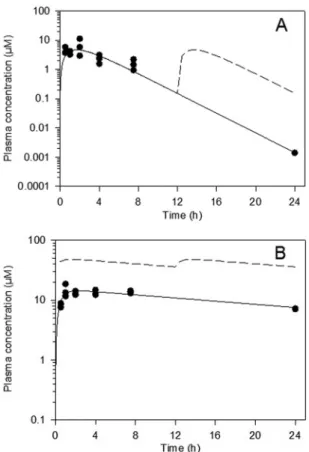
Following a single oral dose of 40 mg/kg,
compound 2 had a moderate rate of absorption (with time to
maximum concentration of drug in serum [
T
max] observed 2 h
postdose), after which concentrations declined gradually over the
24-h postdose period (Fig. 2A). Following a single oral dose of 20
mg/kg posaconazole, maximum plasma concentrations were
ob-served 1 h postdose, but concentrations remained relatively flat
for up to 7.5 h postdose (Fig. 2B). Based on the available data, the
elimination half-lives for compound 2 and posaconazole were
ap-proximately 2 h and 18 h, respectively. The exposure to posaconazole
at 20 mg/kg was higher than that of compound 2 after a single oral
dose of 40 mg/kg based on both
C
maxand AUC
0 –⬁(Table 1).
Con-sistent with the shorter estimated half-life for compound 2, the
sim-ulated repeat (b.i.d.) dose profile suggests that there would be no
significant accumulation after 20 days In contrast, posaconazole
would be expected to exhibit substantial accumulation with repeat
b.i.d. dosing and steady-state exposure parameters would be
approx-imately 3-fold higher than those observed after a single dose.
Comparison of compound 2 (racemic) with posaconazole
and benznidazole in the murine model of
T. cruzi
infection.
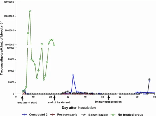
Mice were treated orally with test compounds or vehicle for 20
days beginning 4 days postinfection. Vehicle-treated mice
demon-strated high levels of parasitemia, peaking around day 7
postinfec-tion. All 7 vehicle-treated mice died between days 12 and 24
postinfection. Microscopic parasitemia was dramatically
sup-pressed in mice treated with compound 2 (Fig. 3). However,
low-level reactivation was observed microscopically in 4 mice after
completion of the compound 2 treatments. Five of 5 surviving
mice that were treated with compound 2 had positive PCR tests 30
days after treatment, indicating that they were not
parasitologi-cally cured. In comparison, 2 of 5 mice treated with posaconazole
on March 3, 2017 by UNIVERSIDADE FEDERAL DE OURO PRETO
http://aac.asm.org/
were PCR positive and 2 of 6 mice treated with benznidazole were
PCR positive, indicating that none of these treatments yielded
100% cures with this model (Table 2). Two mice that received
treatment with compound 2 died (days 18 and 34 postinfection).
Each was free of parasitemia at the time of death, so these deaths
were due either to complications from repeated oral gavage
treat-ments or to side effects from the compound. Similarly, two mice
receiving posaconazole died (days 5 and 7 postinfection) and one
mouse receiving benznidazole died (day 5 postinfection). The
av-erage weight gained by mice treated with compound 2 was
below that of untreated controls on the last day of treatment;
however, the weights rebounded to normal by 30 days
post-treatment (Fig. 4).
Separation and testing of enantiomers of compound 2.
The
preclinical drug candidate tipifarnib is an
R
enantiomer, having
⬎
100-fold-greater PFT-inhibitory activity than the corresponding
S
enantiomer (29). The tipifarnib analogs that we previously described
were synthesized and tested as racemic mixtures (11–13). To further
evaluate the activities of compound 2, the two enantiomers were
sep-arated using chiral chromatography. The two enantiomers were
tested for the following biological activities: (i)
T. cruzi
intracellular
amastigote growth inhibition, (ii) rat PFT inhibition, (iii)
T. cruzi
CYP51 binding, (iv) CYP450 inhibition, and (v) stability upon
incu-bation with liver microsomes. As can be seen in
Table 3, the different
enantiomers have significantly different activities. We deduce that the
enantiomer (compound 2.1) with more potent activity on
T. cruzi
amastigotes and the lower
K
d(dissociation constant) on
T. cruzi
CYP51 is the
R
enantiomer since the crystal structure of the closely
related compound 3 (discussed below) demonstrates that the
R
enan-tiomer is bound in the CYP51 active site.
Since the racemic mixture of compound 2 was relatively
inac-tive against rat PFT (IC
50of
⬃
1
M), the separate enantiomers
were not tested for PFT inhibition. The activity of the two
enan-tiomers on human hepatic P450 enzymes demonstrated some
dif-ferences, with lower inhibition fortuitously associated with
com-pound 2.1. In particular, comcom-pound 2.1 inhibited CYP3A4/5 with
an IC
50in the 2.1 to 2.3
M range, which is more than twice as
high as the IC
50for compound 2.2 and
⬎
20 times higher than the
IC
50for posaconazole against this enzyme. Finally, assays of
in
vitro
metabolic stability demonstrated that compound 2.1 was
very stable in human liver microsomes with a
t
1/2of 270 min,
which is even longer than that of the parent drug, tipifarnib. Of
particular interest is that compound 2.1 is metabolized
consider-ably faster in mouse liver microsomes (
t
1/2of 28 min) than in
human liver microsomes.
X-ray crystal structure of CYP51 bound to compound 3.
In
the crystallographic studies, we worked with the
Trypanosoma
brucei
CYP51 (83.4% amino acid sequence identity to the
T. cruzi
CYP51 ortholog). These two trypanosomal enzymes have high
overall structural similarity (15) and only one amino acid
differ-ence within the binding cavity (Fig. 5). Formation of the enzyme/
inhibitor complex does not cause significant structural
rearrange-ments in the CYP51 molecule. The root mean square deviation
between all C
␣
-atoms in the ligand-free structure (PDB ID, 3G1Q
[17]) and compound 3-bound structures is only 0.63 Å, which is
comparable to the deviations between the four compound
3-bound
T. brucei
CYP51 molecules in the asymmetric unit
(0.548
⫾
0.07 Å) and is just slightly exceeded by the deviation
between the compound 3-bound
T. brucei
CYP51 and
posacona-zole-bound
T. cruzi
(PDB ID, 3K1O; 0.857 Å). Although
com-pound 3 was used for cocrystallization as a racemic mixture, only
the
R
enantiomer is present in the structure. In all four molecules
in the asymmetric unit, the inhibitor adopts the same orientation
within the CYP51 active site, no amino acid side chain
reposition-ing affectreposition-ing the heme-protein interactions (15,
17) being
ob-served. The imidazole ring of compound 3 is coordinated with the
heme iron. The length of the N
3-Fe coordination bond (2.08
⫾
0.04 Å) is slightly shorter than the average length observed in the
TABLE 1Pharmacokinetic parameters following oral administration of compound 2 and posaconazole to mice
Parameterc
Compound 2 Posaconazole
Day 1a
Estimated day 20b Day 1a
Estimated day 20b
Dose (mg/kg) 40 40 20 20
Cmax(M) 4.7 4.8 14.3 48.2
dn-Cmax(M/mg/kg) 0.1 0.1 0.7 2.4
AUC0–⬁(M · h) 22.9 23.3 509 1700
dn-AUC0–⬁(M · h/mg/kg) 0.57 0.58 25.5 85.0
Cav,ss(M) 1.9 1.9 42.4 141
aDay 1 data were based on the fit of a single-compartment pharmacokinetic model to
experimental single-oral-dose data.
bDay 20 b.i.d. data (after the second dose on day 20) were estimated using simulated
profiles based on the fitted data after a single dose.
cdn, dose normalized. FIG 2Plasma concentrations of compound 2 (40 mg/kg) (A) and posaconazole
(20 mg/kg) (B) following single oral administration to mice. Filled symbols repre-sent measured concentrations (n⫽3 samples per time point), while the solid line represents profiles fitted with a single-compartment model. The dashed line rep-resents estimated plasma concentrations on day 20 after b.i.d. (q12h) administra-tion.
on March 3, 2017 by UNIVERSIDADE FEDERAL DE OURO PRETO
http://aac.asm.org/
structures of P450-azole complexes (
⬃
2.15 to 2.3 Å) and similar
to that found in
T. brucei
CYP51 complexed with
N
-1-(2,4-di-chlorophenyl)-2-(1
H
-imidazol-1-yl)ethyl-4-(5-phenyl-1,3,4-oxadiazol-2-yl)benzamide (17), which may be indicative of
stronger interaction. The nonligated portion of the inhibitor
mol-ecule is surrounded by 20 protein residues; 17 of them are located
within the van der Waals bond distances (
⬍
4.5 Å). Six residues are
provided by the B
=
-helix/B
=
-C loop (Y103, F105, M106, F110,
A115, and Y116); four are contributed by helix I (A287, F290,
A291, and T295), two are from the
␣
K/

1-4 loop (L356 and
M358), two are from the

4 hairpin (M460 and V461), and one
residue (L127) is from helix C. Unique for this inhibitory scaffold
is the involvement in the interaction of the
␣
F/FF
⬙
loop region
(E205 and L208). This is due to the positioning of the phenyl ring
(attached to the main quinolone group), which occupies an
addi-tional CYP51 active-site cavity bordered by helices F and I and the

4-hairpin. This ring has served as the subject for the hydrogen atom
substitutions in this study (Fig. 1). Altogether, the structural data
imply that the high potency of this inhibitory scaffold is largely due to
the precise hand-in-glove fit between the ligand molecule and the
enzyme active-site geometry, and therefore, minor chemical
modifi-cations that would optimize the scaffold
pharmacokinetic/pharma-codynamic properties may significantly enhance its curative
poten-tial.
DISCUSSION
The anti-
Trypanosoma cruzi
activity of sterol 14
␣
-demethylase
in-hibitors has been recognized since the early 1980s (9). The early
imidazole and triazole antifungal drugs such as ketoconazole,
itra-conazole, and fluconazole appeared to lack sufficient potency
FIG 3Parasitemia of mice infected withTrypanosoma cruziY strain and treated with compound 2, posaconazole, or benznidazole for 20 consecutive days. Arrows indicate the first and the last day of treatment and the first day of cyclophosphamide immunosuppression.
TABLE 2Efficacy experiment with mice infected withTrypanosoma cruziY strain and treated daily with compound 2, posaconazole, or benznidazole for 20 consecutive daysa
Parameter
Treatment group
Compound 2 (40 mg/kg)
Posaconazole (20 mg/kg)
Benznidazole (100 mg/kg) No. of drug doses for clearance of parasitemia (mean⫾SD) 1.71⫾1.11 1.5⫾0.58 1.17⫾0.41 No. of mice with natural parasitemia reactivation after treatment/total no. 4/5 0/5 1/6 No. of mice with parasitemia reactivation after immunosuppression/total no. 3/5 0/5 1/6 No. of mice positive by real-time PCRb(30 days after treatment)/total no. 5/5 2/5 2/6
Total no. of positive mice/total no. 5/5 2/5 2/6
aTreatments were started at the 4th day after infection.
bReal-time PCR was performed in blood of all animals 30 days after treatment.
on March 3, 2017 by UNIVERSIDADE FEDERAL DE OURO PRETO
http://aac.asm.org/
and/or pharmacological properties to cure mice with chronic
T.
cruzi
infection (26). However, newer azole antifungal drugs,
in-cluding D0870 (discontinued in clinical development) and
po-saconazole, have been shown to have curative activity in mouse
models of Chagas disease (20,
28). The outcome of the ongoing
clinical trial with posaconazole will help validate (or invalidate)
CYP51 as a drug target for Chagas disease (4). If CYP51 is
vali-dated, additional CYP51 inhibitors with lower costs of goods may
be necessary to bring treatment to the large numbers of Chagas
disease patients who live in resource-limited settings. The
investi-gations with compound 2 have been pursued with this framework
in mind.
The efficacy study in mice demonstrated that the racemic
mix-ture of compound 2 had strong suppressive activity on
para-sitemia; however, the ability to cure mice was lower than that of
posaconazole. Of 5 surviving mice, all 5 treated with compound 2
remained parasitologically positive by sensitive testing methods,
whereas only 2 of 5 mice treated with posaconazole were positive
after treatment. The finding that 2 of 6 benznidazole-treated mice
remained PCR positive demonstrates the high stringency of the
mouse model with Y strain parasites used in these experiments. In
previous work using a different strain of
T. cruzi
(Tulahuen), we
observed that posaconazole led to negative parasitemia by PCR
(25). In addition to using different strains, we also tested for cure
by performing PCR after inducing immunosuppression, which
further enhances the ability to uncover low-level residual
infec-tions.
The estimated plasma concentrations of compound 2 are
ex-pected to be substantially lower than those for posaconazole after
b.i.d. dosing for 20 days, attributable both to posaconazole’s
higher single-dose exposure and to its longer half-life, leading to
substantial accumulation upon repeat administration. Estimated
C
av,ssvalues on day 20 are approximately 1.9
M for compound 2
and 141
M for posaconazole. The simulated profiles (Fig. 2)
FIG 4Weight gain in grams of animals infected withTrypanosoma cruzistrain and treated with compound 2, posaconazole, or benznidazole in comparison with the noninfected-control groups. The weight gain was calculated by the difference between the weights of animals obtained on days 1 and 20 of the treatment and on days 1 and 30 posttreatment. The weights of animals in-cluded in control groups were obtained at the same times. The statistical dif-ferences among treated and noninfected groups were determined by the non-parametric Tukey multiple comparison test. #, significant difference (P⬍
0.001) in relation to the noninfected group.
TABLE 3Comparisons of tipifarnib, compound 2 racemic mixture, enantiomers of compound 2, and posaconazole
Assay Tipifarnib
Compound 2 racemic
mixture Enantiomer 2.1 Enantiomer 2.2 Posaconazole
T. cruziamastigote; EC50(M) 0.006 0.0005 0.00031 0.0263 0.00016
Rat PFT; IC50(M) 0.0013 0.991 T. cruziCYP51 binding;Kd(M)
a 0.11 0.18 3.8 0.06
Liver microsome stability;t1/2(min)
Human 100 270 ⬎360
Mouse ⬎180 28 ⬎180
CYP450 inhibition (M)
CYP1A2 13.3 ⬎30 ⬎39
CYP2C9 0.29 0.08 9.5
CYP2C19 4.2 3.2 20.9
CYP2D6 6.5 25 ⬎30
CYP3A4/5 midazolam 2.3 0.65 0.09
CYP3A4/5 testosterone 2.1 0.9 0.04
aThe apparentK
ds were calculated by plottingT. cruziCYP51 spectral responses (⌬A420 –390) against free ligand concentration (15).
FIG 5Active site ofTrypanosoma bruceiCYP51 with bound compound 3. The residues located within van der Waals contacts (⬍4.5 Å) with the inhibitor are shown in wire representation and labeled, carbon atoms are colored in gray, and the corresponding secondary structural elements of the protein are pre-sented as ribbons. The single active-site amino acid residue that differs inT. cruziCYP51 (I105) is colored in purple. Coordinated with the heme (orange), compound 3 is colored in magenta. Nitrogens are blue, oxygens are red, chlo-rine is green, and fluochlo-rines are light yellow. A stereoview of the active site in the superimposedT. bruceiandT. cruzi(3K1O) CYP51 structures can be seen as Fig. S2 in the supplemental material.
on March 3, 2017 by UNIVERSIDADE FEDERAL DE OURO PRETO
http://aac.asm.org/
suggest that posaconazole concentrations remained above
ap-proximately 30
M for the full 20-day dosing period, whereas
concentrations of compound 2 were below 10
M, and frequently
below 1
M, throughout the dosing period, which may partly
account for the differences in efficacy. Other factors, such as
plasma protein binding and the volume of distribution (not
avail-able in mice), may also play a role in the different efficacy profiles.
The active enantiomer (compound 2.1) is considerably more
metabolically stable in human microsomes (
t
1/2of 270 min) than
in mouse microsomes (
t
1/2of 28 min) (Table 3). The lower activity
of compound 2 than of posaconazole in mice could therefore be
due to the higher clearance of compound 2, translating to lower
exposure of the parasites to the compound (Fig. 2). It may be
necessary to make additional changes to the tipifarnib scaffold
that further improve metabolic stability and reduce plasma
clear-ance rates. As we know that the pharmacokinetic profile of
tipi-farnib in humans is quite good (5,
30), these results suggest that
the rodent model may underestimate the potential activity of
tipi-farnib analogs in humans.
There were similar numbers of deaths in each group of mice
treated with compound 2, posaconazole, and benznidazole (2, 2,
and 1, respectively). The deaths are best attributed to
complica-tions from repeated oral gavages over the 20-day treatment course
rather than to side effects from the drugs. The average weight of
mice at the end of treatment with compound 2 was lower than that
for untreated mice, but the weight rebounded to normal levels by
30 days posttreatment (Fig. 4). Since the mice fully recover their
weight to levels of the control mice by 30 days posttreatment, this
indicates that the side effects are reversible. It seems unlikely that
the toxicity is due to PFT inhibition, given the low level of activity
of compound 2 on the rodent PFT enzyme (IC
50,
⬃
1
M),
par-ticularly compared to that of tipifarnib (IC
50,
⬃
1 nM), which is
tolerated by rodents. It is also unlikely that compound 2 inhibits
the rodent CYP51 since tipifarnib and several analogs were
previ-ously shown to not inhibit human CYP51 (13). Superposition of
the experimental structures of human CYP51 (24) on the
T. brucei
CYP51 (an excellent proxy for the
T. cruzi
enzyme) complex with
compound 3 reveals several clashes that make it impossible for
compound 3 to bind to the human CYP51. The lack of weight gain
during treatment could be due to inhibition of one of the other
CYP enzymes (19) or to a completely unrelated mechanism that
will require additional investigation to elucidate.
Compound 2 was further characterized by separating the
race-mic mixture into its two enantiomers. Enantiomer 2.1 was
ob-served to be a tighter
T. cruzi
CYP51 ligand and more active
against
T. cruzi
cells than enantiomer 2.2. As discussed above, we
concluded that enantiomer 2.1 corresponds to the
R
enantiomer.
The CYP inhibition studies showed that compound 2.1 had much
less inhibitory activity on CYP3A4/5 than did posaconazole.
Given the important role of CYP3A4 in drug metabolism, the
observation suggests that compound 2.1 has an advantage over
posaconazole with respect to the potential for drug-drug
interac-tions. It is fortuitous that compound 2.1 had lower inhibitory
activity against most of the CYP isoforms than did the 2.2
enan-tiomer. For an imidazole-containing molecule, compound 2.1 has
a good profile against hepatic CYP enzymes, consistent with what
is known about tipifarnib (29).
The crystal structure of the
Trypanosoma brucei
CYP51 bound
to compound 3 reveals a significant difference between the
bind-ing mode of the tipifarnib-related compounds and that predicted
from a structural model of the enzyme that was based on the
mycobacterial CYP51 structure, which is only 30% identical in
sequence (11). This underscores the value of experimentally
de-termining crystal structures rather than depending on molecular
models built on homology. The binding mode of compound 3,
discussed in Results, provides insights for our group to design and
synthesize new analogs to further explore this structural series as
potential antitrypanosomal agents.
In conclusion, the analog of tipifarnib (compound 2) is highly
potent against
T. cruzi
cultures and demonstrated strong
suppres-sive anti-
T. cruzi
activity in the mouse model. It had a lower cure
rate than that of the drug posaconazole, which could possibly be
the result of higher plasma clearance, and hence lower exposure,
of compound 2 than of posaconazole. The data suggest that
fur-ther changes to the tipifarnib scaffold, guided by the recent crystal
structure of CYP51, are needed to optimize the pharmacokinetic
properties while retaining anti-
T. cruzi
activity. The very good
pharmacokinetic attributes of tipifarnib in humans give us
confi-dence that optimized tipifarnib analogs can be achieved.
ACKNOWLEDGMENTS
Funding to F. S. Buckner, C. L. M. J. Verlinde, and M. H. Gelb came from NIH (grants AI070218 and AI054384) and the Drugs for Neglected Dis-eases Initiative. Funding to M. T. Bahia and S. A. Charman came from the Drugs for Neglected Diseases Initiative. Funding to G. I. Lepesheva came from NIH (GM067871).
For the work described in this paper, DNDi received financial support from the following donors: Department for International Development (DFID) (United Kingdom), GiZ on behalf of the Government of the Fed-eral Republic of Germany (Germany), Ministry of Foreign and European Affairs (MAEE) (France), Spanish Agency for International Development Cooperation (AECID) (Spain), Médecins Sans Frontières (Doctors with-out Borders) (international), and a Swiss foundation. The donors had no role in study design, data collection and analysis, decision to publish, or preparation of the manuscript.
REFERENCES
1.Bern C.2011. Antitrypanosomal therapy for chronic Chagas’ disease. N. Engl. J. Med.364:2527–2534.
2.Buckner FS, Verlinde CLMJ, La Flamme AC, Van Voorhis WC.1996. Efficient technique for screening drugs for activity againstTrypanosoma cruziusing parasites expressing-galactosidase. Antimicrob. Agents Che-mother.40:2592–2597.
3.Caldas S, et al.2008. Trypanosoma cruzi: acute and long-term infection in the vertebrate host can modify the response to benznidazole. Exp. Para-sitol.118:315–323.
4.Clayton J.2010. Chagas disease: pushing through the pipeline. Nature
465:S12–S15.
5.Cohen SJ, et al.2003. Phase II and pharmacodynamic study of the farne-syltransferase inhibitor R115777 as initial therapy in patients with meta-static pancreatic adenocarcinoma. J. Clin. Oncol.21:1301–1306. 6.Collaborative Computational Project N4.1994. The CCP4 suite:
pro-grams for protein crystallography. Acta Crystallogr. D Biol. Crystallogr.
50:760 –763.
7.Coura JR, Vinas PA.2010. Chagas disease: a new worldwide challenge. Nature465:S6 –S7.
8.Cummings KL, Tarleton RL.2003. Rapid quantitation of Trypanosoma cruzi in host tissue by real-time PCR. Mol. Biochem. Parasitol.129:53–59. 9.Docampo R.1981. Biochemical and ultrastructural alterations produced by miconazole and econazole inTrypanosoma cruzi. Mol. Biochem. Para-sitol.3:169 –180.
10. Emsley P, Cowtan K.2004. Coot: model-building tools for molecular graphics. Acta Crystallogr. D Biol. Crystallogr.60:2126 –2132.
11. Hucke O, Gelb MH, Verlinde CL, Buckner FS. 2005. The protein farnesyltransferase inhibitor tipifarnib as a new lead for the development of drugs against Chagas disease. J. Med. Chem.48:5415–5418.
on March 3, 2017 by UNIVERSIDADE FEDERAL DE OURO PRETO
http://aac.asm.org/
12. Kraus JM, et al.2010. Second generation analogues of the cancer drug clinical candidate tipifarnib for anti-Chagas disease drug discovery. J. Med. Chem.53:3887–3898.
13. Kraus JM, et al.2009. Rational modification of a candidate cancer drug for use against Chagas disease. J. Med. Chem.52:1639 –1647.
14. LaPlante SR, Edwards PJ, Fader LD, Jakalian A, Hucke O.2011. Revealing atropisomer axial chirality in drug discovery. ChemMedChem6:505–513. 15. Lepesheva GI, et al.2010. Structural insights into inhibition of sterol
14alpha-demethylase in the human pathogen Trypanosoma cruzi. J. Biol. Chem.285:25582–25590.
16. Lepesheva GI, Nes WD, Zhou W, Hill GC, Waterman MR.2004. CYP51 from Trypanosoma brucei is obtusifoliol-specific. Biochemistry 43: 10789 –10799.
17. Lepesheva GI, et al. 2010. Crystal structures of Trypanosoma brucei sterol 14alpha-demethylase and implications for selective treatment of human infections. J. Biol. Chem.285:1773–1780.
18. Leslie M.2011. Infectious diseases. A tropical disease hits the road. Science
333:934.
19. Lewis DF.2004. 57 varieties: the human cytochromes P450. Pharmacog-enomics5:305–318.
20. Molina J, et al.2000. Activities of the triazole derivative SCH 56592 (posaconazole) against drug-resistant strains of the protozoan parasite Trypanosoma (Schizotrypanum) cruzi in immunocompetent and immu-nosuppressed murine hosts. Antimicrob. Agents Chemother.44:150 –155.
21. Moser DR, Kirchhoff LV, Donelson JE.1989. Detection ofTrypanosoma cruziby DNA amplification using the polymerase chain reaction. J. Clin. Microbiol.27:1477–1482.
22. Otwinowski Z, Minor W.1995. The HKL manual. Yale University Press, New Haven, CT.
23. Stordeur P, et al.2002. Cytokine mRNA quantification by real-time PCR. J. Immunol. Methods259:55– 64.
24. Strushkevich N, Usanov SA, Park HW.2010. Structural basis of human CYP51 inhibition by antifungal azoles. J. Mol. Biol.397:1067–1078. 25. Suryadevara PK, et al.2009. Structurally simple inhibitors of lanosterol
14alpha-demethylase are efficacious in a rodent model of acute Chagas disease. J. Med. Chem.52:3703–3715.
26. Urbina JA.2009. Ergosterol biosynthesis and drug development for Cha-gas disease. Mem. Inst. Oswaldo Cruz104(Suppl 1):311–318.
27. Urbina JA.2010. Specific chemotherapy of Chagas disease: relevance, current limitations and new approaches. Acta Trop.115:55– 68. 28. Urbina JA, et al.1996. Cure of short- and long-term experimental
Cha-gas’ disease using D0870. Science273:969 –971.
29. Venet M, End D, Angibaud P.2003. Farnesyl protein transferase inhib-itor ZARNESTRA R115. Curr. Top. Med. Chem.3:1095–1102. 30. Zujewski J, et al.2000. Phase I and pharmacokinetic study of farnesyl
protein transferase inhibitor R115777 in advanced cancer. J. Clin. Oncol.
18:927–941.