Characterization Analysis of
Schistosoma japonicum
Plasma Membrane Repair Relative Gene Myoferlin
Yanian Xiong, Ming Zhang, Yang Hong, Meimei Wei, Dezhou Ai, Peipei Meng, Yanhui Han, Zhiqiang Fu, Yaojun Shi, Jianmei Yang, Jiaojiao Lin*
National Laboratory of Animal Schistosomiasis Control/Key Laboratory of Animal Parasitology, Ministry of Agriculture, Shanghai Veterinary Research Institute, Chinese Academy of Agricultural Sciences, Shanghai, People’s Republic of China
Abstract
Myoferlin is a member of the ferlin family of proteins, which are involved in plasma membrane repair, and has been identified as one of the tegument proteins ofSchistosoma japonicum.The tegument proteins are potential candidates for vaccines and new drug targets. In this study, myoferlin ofS. japonicum(SjMF) was cloned, expressed and characterized, the potential of SjMF recombinant protein (rSjMF) as a vaccine candidate was evaluated, and the effect of praziquantel on SjMF was detected by Real-time PCR. Immunofluorescence showed that this protein was mainly distributed on the surface of worms at different stages. Sequence analysis revealed that the SjMF open reading frame was conserved at all stages of the
S. japonicumlife cycle. And SjMF transcription was upregulated in 42-day-old worms, and was significantly higher in female worms. Western blotting revealed that rSjMF showed strong immunogenicity. The cytokine profile and IgG isotype analysis demonstrated that rSjMF plus ISA206 immunization induced a mixed T helper (Th)1/Th2 response. Purified rSjMF emulsified with ISA206 adjuvant significantly reduced worm burden from 21.8% to 23.21% and liver egg number from42.58% to 28.35%. Besides, SjMF transcription was downregulated when worms were exposed to low-dose praziquantel (PZQ) and upregulated when PZQ was degraded, accompanied by recovery of damaged tegument. When worms were exposed to high-dose PZQ, SjMF transcription was downregulated all the time and the damaged tegument did not recover. These findings indicated that SjMF is a potential vaccine againstS. japonicumand provides the basis for further investigations into the biological function of SjMF.
Citation:Xiong Y, Zhang M, Hong Y, Wei M, Ai D, et al. (2013) Characterization Analysis ofSchistosoma japonicumPlasma Membrane Repair Relative Gene Myoferlin. PLoS ONE 8(6): e66396. doi:10.1371/journal.pone.0066396
Editor:Jason Mulvenna, Queensland Institute of Medical Research, Australia
ReceivedFebruary 4, 2013;AcceptedMay 6, 2013;PublishedJune 18, 2013
Copyright:ß2013 Xiong et al. This is an open-access article distributed under the terms of the Creative Commons Attribution License, which permits unrestricted use, distribution, and reproduction in any medium, provided the original author and source are credited.
Funding:This work was supported by the National Natural Science Foundation of China (no. 31172315), Agro-scientific Research in the Public Interest (200903036), China Postdoctoral Science Foundation (No. 2012M510630), Science Technology and Development Foundation of Shanghai (no. 12140902700) and State-level public welfare scientific research courtyard basic scientific research operation cost (2013JB18). The funders had no role in study design, data collection and analysis, decision to publish, or preparation of the manuscript.
Competing Interests:The authors have declared that no competing interests exist.
* E-mail: [email protected]
Introduction
Schistosomes are parasitic blood helminths that infect millions of people in tropical and subtropical countries [1]. Approximately 779 million people are at risk of being infected in 76 endemic countries and an estimated 280,000 deaths are directly or indirectly attributable to the disease annually [2,3]. Besides humans, .40 types of livestock and wild animals are reservoir hosts for Schistosoma japonicumin China, and cattle are the major source of infection, especially in the lake–marsh endemic area of the Yangtze River. Therefore, schistosomiasis control remains a major challenge in China. Currently, schistosomiasis control strategy is mainly based on treatment of infected individuals with praziquantel (PZQ). PZQ can effectively reduce the morbidity associated with schistosomiasis, but it has been proved not to be sufficient to control disease transmission and prevent reinfection [4,5]. An effective vaccine against schistosomiasis would be essential to the current control strategy, mainly because it would provide long-lasting immunity against infection. In addition, it is suggested that the combined use of chemotherapy and vaccination is the basis for a novel, more versatile method to control schistosomiasis. Therefore, it is important to identify the
appro-priate schistosomal antigens that could induce activity against schistosomal infection or reduce the release of live eggs to limit parasite transmission.
damage [12]. Davis et al. Have found that myoferlin is expressed abundantly in both cardiac and skeletal muscle and is associated with the plasma and nuclear membranes [11]. Doherty et al. have suggested that the interaction of myoferlin with eps15 homology domain protein (EHD2) may facilitate membrane fusion at sites of contact between cells where cytoskeletal rearrangements are needed [13]. Furthermore, Robinson et al. have validated the expression of myoferlin in term placenta and trophoblastic cells, and have speculated that myoferlin also repairs damage to the syncytiotrophoblast apical plasma membrane [14].
In the present study, we described the cloning, expression, and immunolocalization of the myoferlin ofS. japonicum(SjMF) gene, as well as the immunogenicity of recombinant SjMF (rSjMF). We also evaluated the protective immunity induced by rSjMF, and the effect of PZQ on SjMF transcription.
Materials and Methods
Ethics Statement
All animal care and procedures were conducted according to the guidelines for animal use in toxicology (Society of Toxicology USP, 1989). The study protocol was approved by the Animal Care and Use Committee of the Shanghai Veterinary Research Institute, Chinese Academy of Agricultural Sciences.
1. Mice and Parasites
Male BALB/c mice aged 6 weeks were purchased from Shanghai Experimental Animal Centre, Chinese Academy of Sciences. The life cycle ofS. japonicumwas maintained routinely in
Oncomelania hupensissnails and New Zealand rabbits, and cercariae were obtained by exposing infected snails to light for shedding. The number of cercariae and their viability were determined using a light microscope. Parasites at 7, 13, 21, 28, 35, and 42 days were obtained by perfusion of artificially infected New Zealand rabbits.
Partial 42-day-old worms were manually separated into males and females.
2. Molecular Characterization of SjMF
The deduced amino acid sequence of SjMF was analyzed as follows: theoretical isoelectric point (pI) and molecular weight (MW) were calculated at http://www.expasy.org/tools/pi tool.html; prediction of signal peptides with SignalP 3.0 at http://www.cbs.dtu.dk/services/SignalP; search for glycosylpho-sphatidyl anchors in the sequence with NetNGlyc 1.0 at http:// www.cbs.dtu.dk/services/NetNGlyc/; analysis of conserved do-mains using ScanProsite at http://www. ebi.ac.uk/Tools/pfa/ iprscan/; prediction of transmembrane helices using the TMHMM Server v.2.0 at http://www.cbs.dtu.dk/services/ TMHMM-2.0; and protein modeling at http://swissmodel. expasy.org/. The amino acid sequences of the myoferlin protein were obtained from GenBank and were aligned using ClustalX software (http://www.clustal.org/).
3. Cloning and Sequence Analysis of the ORF of SjMF c-DNA
The forward and reverse oligonucleotides, 59- ATAGGATC-CAT GGTAGATTCACAG TGGG-39 and 59 -ATACTC-GAGGGCATCAGTATAGGCAGG-39 (BamHI and XhoI sites are underlined), were used to amplify the complete SjMF open reading frame (ORF; GenBank accession AAW27277.1) from a transcriptome cDNA library of worms at different developmental stages by PCR (the polymerase chain reaction) with Phusion High-Fidelity PCR Master Mix. Amplification was performed in an initial denaturation step at 98uC for 1 min, then 32 cycles at 98uC for 8 s, 52uC for 1 min, and 72uC for 90 s, and a post-PCR step at 72uC for 10 min. dATP was added to the blunt end of the purified PCR product by DNA A-Tailing Kit (TaKaRa). Then, the PCR-generated fragment was cloned into the pMD19-T vector
Figure 1. Clustal X alignment of the derived amino acid sequences of SjMF (AAW27277.1), HsMF (Q9NZM1), MmMF (Q69ZN7) and XtMF (B3DLH6).The regions with high identity and similarity among myoferlin sequences are shown as black and gray columns, according to the Clustal X algorithm. The boxes refer to FerA (central domain A in proteins of the ferlin family), FerB (central domain B in proteins of the ferlin family), and DysfN (Dysferlin domain, N-terminal region) domain respectively.
(TaKaRa) and sequenced. To understand the structural conser-vation of SjMF in worms at different developmental stages, c-DNA encoding the ORF of SjMF was cloned by PCR with Phusion High-Fidelity PCR Master Mix. Then, the PCR-generated fragment was cloned into the pMD19-T vector (TaKaRa) and 40 clones of each stage were sequenced and compared.
4. Real-time Reverse-transcriptase PCR (RT-PCR) Analysis of SjMF
Transcription of SjMF at the mRNA level was evaluated in 7-, 14-, 21-, 28-, 35-, and 42-day-old worms ofS. japonicum, as well as in 42-day-old female and male worms using real-time quantitative RT-PCR. Total RNA was isolated from the different develop-mental stages of worms using TRIzol reagent (Invitrogen) according to the manufacturer’s instructions. RNA samples were treated with RNase-free DNaseI (Takara) and purified with an RNeasy Mini Kit (QIAGEN, USA) following the manufacturer instructions. RNA was quantified by spectrophotometry (Biopho-tometer; Eppendorf, Germany). cDNA was synthesized using PrimeScript RT Reagent Kit with gDNA Eraser (Takara) according to standard protocols. A product of 222 bp was amplified with the primers designed for SjMF (forward primer:
59-GCACAGTTGGCGATTTCT-39; reverse primer: 59 -GGCTTCTCGGTAGGCTTT-39). A negative control which did not include cDNA as a template was set in each PCR run. Real-time PCR was performed in a reaction mixture of 20ml containing 10ml 26SYBR Green PCR Premix Taq (TaKaRa),
6.8ml EASY Dilution Buffer (TaKaRa), 2ml cDNA, 0.8ml primers (10mM), and 0.4ml ROX Reference Dye II (506). The cycling protocol was 95uC for 30 s followed by 40 cycles of 95uC for 5 s, 60uC for 34 s, and a dissociation stage of 95uC for 15 s, 60uC for 1 min, and 95uC for 15 s in a ABI PRISM 7500 Fast Real-Time PCR System. The generation of a specific PCR product was tested using melting curve analysis and sequenced. And the efficiency of the PCR reaction was between 95% and 105%. The independent experiments were repeated three times. Differences in transcription levels were observed by comparison to the housekeeping gene, NADH dehydrogenase with double-standard curves method of relative quantification PCR [15,16].
5. Transcription Level Analysis of SjMF by Treatment with PZQ
Ninety mice were divided randomly into nine groups of 10 Mice were challenged through percutaneous exposure of
Figure 2. Stage and sex differential expression of SjMF inS. japonicum.(A) Expression throughout six stages ofS. japonicum, and (B) expression in male and female adult worms analyzed by real-time PCR. Data were normalized against amplification of an internal housekeeping control gene SjNADH. The data are the means6SD of one representative of three independent experiments.
doi:10.1371/journal.pone.0066396.g002
Figure 3. Effect of praziquantel on the SjMF transcription by real-time RT-PCR.(A) Mice treated with single dose of 200 mg/kg PZQ and sacrificed at various times post-treatment. (B) Mice treated with single dose of 40 mg/kg PZQ and sacrificed at various times post-treatment. Statistically significant compared with the control group denoted by *P,0.05, **P,0.01 or ***P,0.001.
doi:10.1371/journal.pone.0066396.g003
abdominal skin for 15 min in water containing 100 viable cercaria. Thirty-five days after challenge, the infected groups were treated with a single dose of PZQ at 40 or 200 mg/kg in carboxyl methyl cellulose (CMCNa). Infected mice were sacrificed at 30 min, 4 h, 12 h, and 36 h after drug administration. The effect of PZQ on SjMF transcription between the treated and untreated groups was determined by RT-PCR. Scanning electron microscopy (SEM) and transmission electron microscopy (TEM) were used to examine the ultrastructural differences in the tegumental and subtegumental structures between the worms treated with PZQ at different doses. After treatment with PZQ, worms were collected and fixed in 4% formaldehyde (in 0.1 M cacodylate buffer, pH 7.4) overnight at 4uC, and processed according to a standard method [17]. Worms were rinsed three times with ice-cold cacodylate buffer (0.1 M, pH 7.4) and post-fixed in buffered osmium tetroxide, followed by dehydration with decreasing concentrations of alcohol before critical point drying. The specimens were mounted on stubs and sputter-coated with gold, and examined under a JEOL-6380LV scanning electron micro-scope. For TEM, after fixation, post-fixation and dehydration, worms were embedded in spur resin. Ultrathin sections of the schistosomes were stained with uranyl acetate and lead citrate.
The specimens were then examined under a Tecnai G2 Spirit BioTwin transmission electron microscope.
6. Expression and Purification of Recombinant Protein
With overhanging ends generating fromBamHI andXhoI, the cDNA of SjMF was subcloned into the multiple cloning sites present in the pET32a(+) expression vector (Invitrogen) to produce a fusion protein. The recombinant plasmid pET32a(+)-SjMF was transformed intoEscherichia coliBL21(DE3) (Tiangen Biotech Co. Ltd., Beijing, China), and transformants were confirmed by restriction enzyme digestion and sequence analysis. Transformed
E. coli BL21(DE3) cells were grown in 500 ml Luria–Bertani medium plus ampicillin (100mg/ml) at 37uC with shacking until OD600reached 0.6, and overexpressed using 1 mM isopropyl-b -D- thiogalactopyranoside (IPTG) at 37uC for 6 h. The bacteria were harvested by centrifugation at 10,0006g for 15 min.
Following this, the pellet was suspended in 15 ml phosphate buffered saline (PBS, pH 7.4) and extracted using an ultrasonic processor to release the fusion proteins. The lysates were centrifuged at 12,0006g for 15 min to collect inclusion bodies and cellular debris, while leaving other soluble substances. The pellets were resuspended in 5 ml 16binding buffer plus 6 M urea.
After analyzed by SDS-PAGE, rSjMF was expressed mainly in an
Figure 4. Photomicrographs of the dorsal surface of male, adultS. japonicumas observed by SEM.These micrographs provide a visual sample of the comparative damage induced by PZQ in the control group (A), in a mouse at 36 h after treatment with 200 mg/kg PZQ (B), a mouse at 4 h (C) or 36 h (D) after treatment with 40 mg/kg PZQ.
inclusion body form. rSjMF was purified using a Ni-NTA His-Bind Resin (Qiagen GmbH, Hilden, Germany) and dialyzed against PBS (pH 7.4), containing decreasing concentrations of urea (6, 4, 3, 2, and 1 M) and PBS only. The purified recombinant protein was used to inject BALB/c mice emulsified with ISA206 adjuvant three times to produce polyclonal antibodies, which were used for the following immunolocalization test.
7. Immunolocalization of SjMF
To analyze the tissue distribution of SjMF, freshly collected adult worms were embedded in optimal cutting temperature (OCT) compound medium and precooled in a freezing microtome cryostat for 30 min, then cut into 8-mm sections for immunoflu-orescence assay. The sections were fixed with precooled acetone for 30 min and immunolabeled using indirect immunofluores-cence as follows. The sections were blocked with 10% goat serum in PBS containing 0.05% Tween 20 (PBST) for 2 h at 37uC, and incubated with the anti-rSjMF mouse serum diluted 1:200 in blocking buffer for 1 h at 37uC. Serum from nonimmunized mice was used as a negative control. After three washes in PBST, samples were probed with CY3-conjugated goat-anti-mouse IgG
(Rockland, ME, USA), diluted 1:3,000 in blocking buffer for 1 h at 37uC. Sections were washed three times and stained with 10mg/ ml 49,6-diami-dino-2-phenylindole (DAPI) for 8 min at room temperature, and observed by fluorescence microscopy (Nikon, Japan).
8. Western Blot Analysis
The purified rSjMF protein was subjected to 12% SDS-PAGE and transferred electrophoretically onto a 0.45-mm pore nitrocel-lulose membrane (Whatman, Germany) at 130 mA for 1 h at 4uC. The membranes were blocked with PBST plus 5% skimmed milk at 37uC for 2 h, washed three times with PBST, and probed with serum from rabbits immunized against SWAP (soluble adult worms antigen preparation) diluted 1:200 for 1 h at 37uC. After three washes in PBST, membranes were probed with goat-anti-rabbit IgG conjugated to horseradish peroxidase (HRP) diluted 1:3000 in PBST for 1 h. Following a further three washes, the result was visualized using precipitation-type TMB Substrate Solution (Tiangen Biotech).
Figure 5. TEM observation of ultrastructural alterations inS. japonicumcaused by PZQ.These micrographs provide a visual sample of the comparative damage induced by PZQ in the control group (A), 36 h after treatment with 200 mg/kg PZQ (B), 4 h after treatment with 40 mg/kg PZQ (C, D), and 36 h after treatment with 40 mg/kg PZQ (E, F). The outer plasma membrane was smooth and integral and musculature was regular (arrow in A) in the control group. The tegumental matrix and parenchymal tissues lost their definition and became indistinct and there was focal or extensive lysis of muscle bundles and parenchymal tissues, which resulted in vacuole formation (arrow in B). Plates C showed vacuole formation in the matrix ((large arrow in C) and in parenchymal tissues (small arrow in C). The underlying muscle bundles were swollen (small arrow in D). The surface returned to its normal morphological appearance and the large vacuoles were decreased and turned substantial (Plates E), but the muscle bundles were still swollen (arrow in F).
doi:10.1371/journal.pone.0066396.g005
9. Evaluating the Protective Efficacy of rSjMF
Six-week-old male BALB/c mice (10 mice per group) were immunized subcutaneously with 20mg rSjMF emulsified with ISA206 adjuvant on days 0, 15 and 30. In the control groups, adjuvant in PBS or PBS alone was administered using the same immunization protocol. On day 10 after each immunization, blood samples from 10 mice in each experimental group were collected by retro-orbital bleeding and stored at –20uC until use.
Two weeks after the last boost, mice were challenged through percutaneous exposure of abdominal skin for 15 min in water containing 4061 viable cercaria, as described by Smithers and Terry [18]. 42 days post-challenge, adult worms were perfused from the portal system and mesenteric veins. The protection level was calculated by comparing the number of worms recovered from the immunization group with that of the control group, using the formula: protection level = (worms recovered from the control group – worms recovered from the vaccination group)/worms recovered from the control group6100%) [9].
To evaluate the liver egg burden, liver from each mouse in the control and rSjMF immunized groups were collected 42 days post-infection. One gram samples of liver tissue from each mouse were digested in 10 ml 5% NaOH for 30 min at 56uC for 30 min, and mixed thoroughly. An average of three counts per 20ml mixture was taken to estimate the number of eggs, and this count was converted to eggs per gram (EPG). The reduction in liver EPG was calculated using the formula: protection level = (EPG from the control group – EPG from the vaccinated group)/EPG from the control group6100%).
10. Measurement of Specific Antibodies
The measurement of specific anti-SjMF IgG, IgG1 and IgG2a antibodies was performed by ELISA. Maxsorp 96-well microtiter plates were coated with 100ml/well rSjMF(10mg/ml) in carbon-ate–bicarbonate buffer, pH 9.6, for 16 h at 4uC. The plates were then blocked for 2 h at 37uC with 150ml/well PBST plus 1.5% bovine serum albumin. One hundred microliters of each serum sample, diluted 1:200 in PBST, was added per well and incubated for 1 h at 37uC. Plate-bound antibody was detected using goat-anti-rabbit IgG conjugated to horseradish peroxidase (Sigma– Aldrich, St. Louis, MO, USA), goat-anti-rabbit IgG1 conjugated to horseradish peroxidase (AbD Serotech, Kidlington, UK), and goat-anti-rabbit IgG2a conjugated to horseradish peroxidase (AbD Serotech) diluted in PBST to 1:3000. Color reaction was developed by the addition of 100ml/well TMB (Tiangen Biotech) and stopped with 50ml/well 5% sulfuric acid. Absorbance was measured at 450 nm in an ELISA reader.
Figure 6. Immunolocalization of SjMF in the S. japonicum adult worms and lung-stage schistosomes. Secondary antibody CY3-conjugated anti-mouse IgG (red) was used for fluorescence detection of SjMF on adult worms and lung-stage schistosome sections. DAPI (blue) was used to stain parasite nuclei. (A) Section was probed with naı¨ve mouse serum (negative control). (B) Section ofS. japonicumadult worms probed with anti-rSjMF mouse serum.
doi:10.1371/journal.pone.0066396.g006
Figure 7. Western blotting analysis of the immunogenicity of rSjMF.Lane M: molecular mass markers; lane 1: purified rSjMF was probed with serum from rabbit immunized against SWAP.
11. Cytokine Analysis
Cytokine analysis was performed using serum from five mice 7 days after the third immunization with rSjMF emulsified with 206 adjuvant or 206 adjuvant in PBS. The level of stimulated cytokine was analyzed by the Luminex 100 multiplex bead-based immu-noassay system by labeled cytokine capture antibody pairs (Bio-Plex Suspension Array Systembio-plex System; Bio-Rad, Hercules, CA, USA) according to the manufacturer’s procedures. Stimulated levels of interleukin (IL)-12p70, interferon (IFN)-c, IL-4 and IL-10 cytokines were determined using Bio-Plex Manager Software (Bio-Rad). All standards and samples were run in triplicate as recommended.
12. Statistical Analysis
Statistical analyses were performed with paired and unpaired Student’sttest or ANOVA using the software package GraphPad Prism 4.0 (GraphPad Software, San Diego, CA, USA).
Results
1. Molecular Cloning and Sequence Analysis of SjMF
The sequence of the S. japonicum cDNA encoding SjMF was obtained by PCR amplification with specific oligonucleotides. The sequences of 40 cDNA clones encoding the ORF of SjMF from 7-, 14-, 21-, 28-, 35-, and 42-day-old worms were analyzed and compared. All the sequences were conserved in worms from each stage of the parasitic life cycle tested. And there were five substitutions in the deduced amino acid sequence compared with the original sequence deposited in GenBank (AAW27277.1). The coding sequence showed an ORF of 963 bp and encoded a protein of 321 amino acids with a predicted molecular mass of approximately 37,050 Da and a pI of 5.16. Post-translational modifications were assessed in silico, and the results revealed that
SjMF did not contain a signal peptide, N-glycosylation sites, or transmembrane helices. After searching with BlastX, comparison of the amino acid sequences showed that the myoferlin of S. japonicum had 40%, 40% and 41% identity with its orthologs in
Xenopus tropicalis,Mus musculusandHomo sapiens, respectively. The consensus sequence showed that the FerA (central domain A in proteins of the ferlin family), FerB (central domain B in proteins of the ferlin family), and DysfN (Dysferlin domain, N-terminal region) domain was conserved among all of these species (Fig. 1).
2. Transcription Levels of SjMF at Different Stages of the S. japonicum Life Cycle
The results showed that SjMF was transcripted at all developmental stages tested and exhibited a higher transcription level in 42-day-old worms and lower transcription level in 21-day and 14-day schistosomula (Fig. 2). The results also suggested that transcription level in the 42-day female worms were higher than in their male counterparts.
3. Dose Dependent Effect of PZQ on SjMF Transcription and Tegumental Damage
SjMF transcription was significantly inhibited at 36 h after treatment with a single dose of 200 mg/kg PZQ (Fig. 3A). However, when treated with a single dose of 40 mg/kg PZQ, SjMF transcription was significantly inhibited after 30 min, recovered partly after 4 h, and then upregulated significantly after 12 and 36 h (Fig. 3B). The surface of adult worms from untreated mice was composed of grooves and channels arranged regularly and the tegument was distinct and intact (Figs. 4A and 5A), which was similar to that described previously [19]. Severe damage to the tegument was observed at 36 h after treatment with 200 mg/kg PZQ. The typical structural features were almost completely degenerated, and much of the corrugated appearance
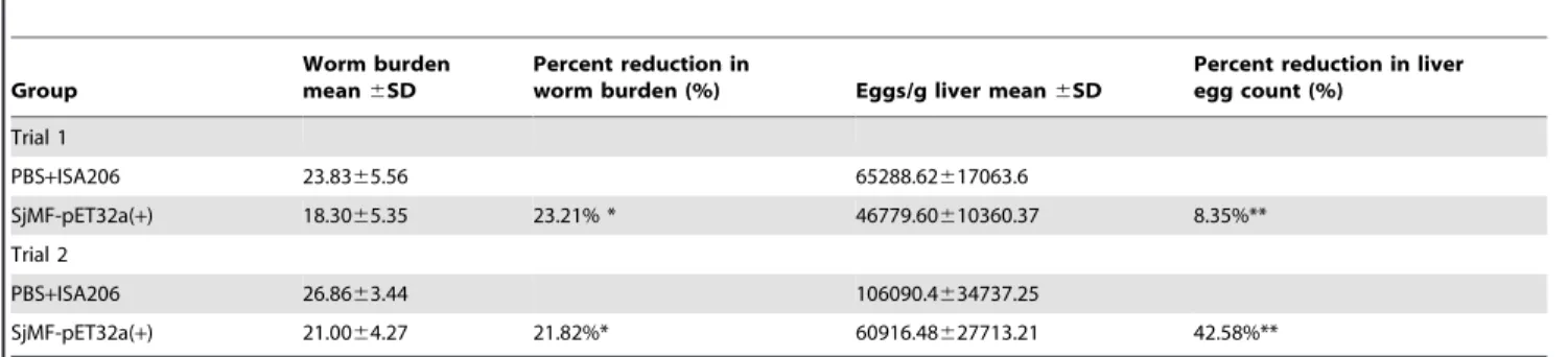
Table 1.Protection induced in mice immunized with rSjMF.
Group
Worm burden mean6SD
Percent reduction in
worm burden (%) Eggs/g liver mean6SD
Percent reduction in liver egg count (%)
Trial 1
PBS+ISA206 23.8365.56 65288.62617063.6
SjMF-pET32a(+) 18.3065.35 23.21% * 46779.60610360.37 8.35%**
Trial 2
PBS+ISA206 26.8663.44 106090.4634737.25
SjMF-pET32a(+) 21.0064.27 21.82%* 60916.48627713.21 42.58%**
Data are expressed as mean6SD and statistically significant compared to control group shown by *P,0.05 or **P,0.01. doi:10.1371/journal.pone.0066396.t001
Table 2.Specific IgG1 and IgG2a antibody level in rSjMF-vaccinated or blank control mice.
IgG1 IgG2a
Days ISA 206 rSjMF-adjuvant ISA 206 rSjMF-adjuvant IgG1/IgG2a ratio
0 0.08360.0075 0.07860.0034 0.07360.0060 0.06860.0031 1.15
10 0.09660.012 0.7560.17** 0.07260.014 0.08660.010 8.72
25 0.08160.0028 0.9960.032*** 0.07060.0057 0.4460.26* 2.25
40 0.1160.020 0.9160.046*** 0.06460.0067 0.6660.25** 1.38
Significantly increased serum antibody titers compared with the ISA 206 adjuvant control is denoted by *P,0.05, **P,0.01 or ***P,0.001. doi:10.1371/journal.pone.0066396.t002
was either swollen or collapsed (Fig. 4B). Meanwhile, the tegumental matrix and parenchymal tissues lost their definition and became indistinct and there was focal or extensive lysis of muscle bundles and parenchymal tissues, which resulted in vacuole
Immunolocalization assay was performed to identify the distribution of the SjMF protein in the S. japonicum with anti-rSjMF and naive mouse serum. Native SjMF was mainly distributed over the tegument and at lower levels in the internal tissues of the parasite, however, no specific staining was observed in sections incubated with naive mouse serum (Fig. 6).
5. Immunogenicity Analysis of rSjMF
The SjMF gene was cloned into the pET32a(+) expression vector, and the recombinant protein was expressed successfully with an expected size of 62 kDa inE. coliBL21 (DE3) cells induced by IPTG. SDS-PAGE showed that the insoluble fractions contained the majority of the recombinant protein, which was mostly soluble by extraction with 8 M urea. rSjMF protein was
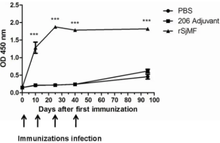
Figure 8. Kinetics of specific anti-rSjMF IgG in mice.Mice were immunized with three doses of a formulation containing rSjMF (20mg/ mouse) or saline in association with ISA 206 adjuvant. Serum samples were obtained from 10 individual mice from each group on days 0,10, 25, 40 and 95 after the first immunization and assayed by ELISA. Arrows indicate the time of each immunization and challenge infection. Results are presented as mean absorbance measured at 450 nm. Significantly increased serum antibody titers compared with the PBS control is denoted by ***P,0.001.
doi:10.1371/journal.pone.0066396.g008
Figure 9. Cytokine profile of mice immunized with rSjMF.Seven days after the last immunization, serum was isolated and assayed for 4, IL-10, IL-12 and IFN-cproduction in response to rSjMF or ISA 206 adjuvant alone, as a control. Significantly increased serum cytokine concentration compared with the ISA 206 adjuvant control is denoted by **P,0.01 or ***P,0.001.
purified by affinity chromatography using His binding columns under denaturing conditions, and was then refolded by dialysis against PBS containing successive decreasing concentrations of urea. rSjMF was identified further by western blot using serum from rabbits immunized against SWAP (soluble adult worms antigen preparation) and naive rabbit serum. A positive band of 62 kDa was observed when probed with rabbit serum specific to SWAP, but not in the naive rabbit serum, which revealed that rSjMF had good immunogenicity (Fig. 7).
6. rSjMF Immunization Elicits Partial Protection against Schistosomal Infection and Reduction in Liver Egg Number
Compared with the adjuvant control group, mice vaccinated with rSjMF had lower worm burdens and liver egg numbers (Table 1). Mice immunized with rSjMF showed 23.21% and 21.82% reductions in worm burden (P,0.05) as well as 28.35% and 50.52% reductions in liver egg numbers (P,0.01) compared to adjuvant control groups in two independent trials.
7. Specific Antibody Responses Induced by rSjMF Immunization
ELISA showed that in the serum of rSjMF-immunized mice, the levels of anti-rSjMF total IgG antibody were significantly higher compared to those in adjuvant control groups after the first immunization, which were maintained until the mice were killed. In the blank and adjuvant control groups, there were no significant differences in specific total IgG levels before or after vaccination (Fig. 8). To evaluate the IgG subtype in the immune response elicited by rSjMF immunization, the levels of IgG1 and IgG2a antibodies specific to rSjMF were also determined by ELISA. No significant changes were observed in the adjuvant control group, and rSjMF immunization induced significant production of specific anti-rSjMF IgG1 and IgG2a antibodies compared to the control group after the first immunization (Table 2).
8. rSjMF plus ISA206 Immunization Induces a Mixed Th1/ Th2 Response
To determine the cytokine profile produced by vaccination with rSjMF, seru from mice after immunization with rSjMF plus 206 adjuvant or 206 adjuvant in PBS were analyzed by the Luminex 100 multiplex bead-based immunoassay system. Mice in the vaccination group had significantly higher concentrations of IL-4, IL-10, IL-12p70 and IFN-c compared with the control group (Fig. 9). IL-12p70 and IFN-care indicative of a Th1-type immune response, and IL-4 are the characteristic cytokines of a Th2 immune response. These results revealed that rSjMF plus ISA206 immunization induces a mixed Th1/Th2 response.
Discussion
Schistosomiasis is one of the most important neglected tropical diseases and continues to be a significant public health problem worldwide. The effective control strategy for this zoonosis is to develop vaccines and improve sanitation. The schistosomal tegumental proteins are not just key molecules for worms to survive in their host, but also targets for the host’s immune attack and drug treatment. Previous studies have demonstrated that some membrane proteins successfully induce protective immune re-sponse against schistosomal infection, such as Sj23, SmTSP-2 and Sm29 [7,9], so it is possible to screen for more vaccine candidates or drug targets by further investigation of schistosomal tegumental proteins. Recently, we have isolated 85 tegumental proteins from
42-day adult worms via S. japonicum tegument surface protein analysis, including myoferlin, dysferlin and some other important schistosomal proteins, which provides the basis for us to identify more effective vaccine candidates or new drug targets for the control of schistosomiasis (data not published).
In the present study, one of the schistosomal tegumental proteins SjMF was characterized, cloned and expressed, and the potential of rSjMF as a vaccine candidate against schistosomal infection in mice was evaluated.
Bioinformatic analysis revealed that SjMF contained the conserved domain of the ferlin family that is involved in vesicle fusion. Therefore, we propose that myoferlin might be a part of the emergency response that mediates resealing-based fusion events in the tegument and muscle ofS. japonicum.
Besides, PZQ could disturb the transcription of myoferlin. We treated mice at 35 days post-infection with high- and low-dose PZQ, and compared the transcript level of SjMF in worms in the treated and untreated groups with RT-PCR. SjMF was signifi-cantly inhibited at 36 h after treatment with a single dose of 200 mg/kg PZQ, accompanied with unrecovered tegumental damage. However, SjMF was upregulated significantly at 12 and 36 h after treatment with a single dose of 40 mg/kg PZQ, accompanied with recovery of tegumental damage. Moreover, many large vacuoles occurred at 4 h after treatment with 40 mg/ kg PZQ, and the vacuoles decreased and turned substantial after 36 h. However, the vacuoles did not change at 36 h after treatment with 200 mg/kg PZQ. In the low-dose PZQ group, the worms were not killed and recovered their normal morpho-logical features. Moreover, Davis et al. have carried out a series of elegant experiments that have shown that myoferlin is associated with the plasma and nuclear membranes [11,20], suggesting that myoferlin is potentially an essential protein for plasma membrane integrity. Therefore, we speculate that SjMF may participate in maintaining the integrity of plasma membrane ofS. japonicum.
Sequence analysis displayed that the ORF of SjMF was conserved in all stages of the S. japonicum life cycle. And real-time PCR analysis showed that SjMF transcripts were expressed in all stages ofS. japonicumtested, with a higher expression level in 42-day worms. Additionally, the expression level in the female worms at 42 days was significantly higher than that in their male counterparts. It is well known that the female worms ofS. japonicum
begin to lay eggs at 24 days [21]. During this process, more materials are needed for the development of the ovaries and vitellaria, which are involved in vesicle trafficking and fusion, while, myoferlin has been proposed to facilitate vesicle trafficking and fusion during membrane repair [22,23], suggesting that SjMF is important for the development of the schistosome.
References
1. Wang L, Utzinger J, Zhou XN (2008) Schistosomiasis control: experiences and lessons from China. Lancet 372(9652): 1793–5.
2. Gryseels B, Polman K, Clerinx J, Kestens L (2006) Human schistosomiasis. Lancet 368 (9541): 1106–18.
3. van der Werf MJ, de Vlas SJ, Brooker S, Looman CW, Nagelkerke NJ, et al. (2003) Quantification of clinical morbidity associated with schistosome infection in sub-Saharan Africa. Acta Trop 86 (2–3): 125–39.
4. Caffrey CR, Secor WE (2011) Schistosomiasis: from drug deployment to drug development. Curr Opin Infect Dis 24 (5): 410–7.
5. Carod AF (2012) Cerebral and Spinal Schistosomiasis. Curr Neurol Neurosci Rep 5 (9): 1420–34.
6. Pearce EJ, MacDonald AS (2002) The immunobiology of schistosomiasis. Nat Rev Immunol 2 (7): 499–511.
7. Tran MH, Pearson MS, Bethony JM, Smyth DJ, Jones MK, et al. (2006) Tetraspanins on the surface of Schistosoma mansoni are protective antigens against schistosomiasis. Nat Med 12 (7): 835–40.
8. Da’Dara AA, Skelly PJ, Wang MM, Harn DA (2001) Immunization with plasmid DNA encoding the integral membrane protein, Sm23, elicits a protective immune response against schistosome infection in mice. Vaccine 20 (3–4): 359–69.
9. Cardoso FC, Macedo GC, Gava E, Kitten GT, Mati VL, et al. (2008) Schistosoma mansoni tegument protein Sm29 is able to induce a Th1-type of immune response and protection against parasite infection. PLoS Negl Trop Dis 2 (10): e308.
10. McManus DP, Loukas A (2008) Current status of vaccines for schistosomiasis. Clin Microbiol Rev 21 (1): 225–42.
11. Davis DB, Delmonte AJ, Ly CT, McNally EM (2000) Myoferlin, a candidate gene and potential modifier of muscular dystrophy. Hum Mol Genet 9 (2): 217– 26.
12. Demonbreun AR, Lapidos KA, Heretis K, Levin S, Dale R, et al. (2010) Myoferlin regulation by NFAT in muscle injury, regeneration and repair. J Cell Sci 123 (Pt 14): 2413–22.
13. Doherty KR, Demonbreun AR, Wallace GQ, Cave A, Posey AD, et al. (2008) The endocytic recycling protein EHD2 interacts with myoferlin to regulate myoblast fusion. J Biol Chem 283 (29): 20252–60.
14. Robinson JM, Ackerman WT, Behrendt NJ, Vandre DD (2009) While dysferlin and myoferlin are coexpressed in the human placenta, only dysferlin expression is responsive to trophoblast fusion in model systems. Biol Reprod 81 (1): 33–9. 15. Gobert GN, Moertel L, Brindley PJ, McManus DP (2009) Developmental gene expression profiles of the human pathogen Schistosoma japonicum. BMC Genomics 10: 128.
16. Hong Y, Han H, Peng J, Li Y, Shi Y, et al. (2010) Schistosoma japonicum: cloning, expression and characterization of a gene encoding the alpha5-subunit of the proteasome. Exp Parasitol 126 (4): 517–25.
17. Thomas JM, Zewail AH (2010) 4D electron microscopyimaging in space and time. London: Imperial College Press. 341 p.
18. Smithers SR, Terry RJ (1965) The infection of laboratory hosts with cercariae of Schistosoma mansoni and the recovery of the adult worms. Parasitology 55 (4): 695–700.
19. Xiao SH, Shen BG, Horner J, Catto BA (1996) Tegument changes of Schistosoma japonicum and Schistosoma mansoni in mice treated with artemether. Zhongguo Yao Li Xue Bao 17 (6): 535–7.
20. Davis DB, Doherty KR, Delmonte AJ, McNally EM (2002) Calcium-sensitive phospholipid binding properties of normal and mutant ferlin C2 domains. J Biol Chem 277 (25): 22883–8.
21. He YX, Yang HZ (1980) Physiological studies on the post-cercarial development of Schistosoma japonicum. Acta Zoologica Sinica;26: 32–39.
22. Mellgren RL, Zhang W, Miyake K, McNeil PL (2007) Calpain is required for the rapid, calcium-dependent repair of wounded plasma membrane. J Biol Chem 282 (4): 2567–75.
23. Mellgren RL, Miyake K, Kramerova I, Spencer MJ, Bourg N, et al. (2009) Calcium-dependent plasma membrane repair requires m- or mu-calpain, but not calpain-3, the proteasome, or caspases. Biochim Biophys Acta 1793 (12): 1886–93.
24. Finkelman FD, Holmes J, Katona IM, Urban JJ, Beckmann MP, et al. (1990) Lymphokine control of in vivo immunoglobulin isotype selection. Annu Rev Immunol 8: 303–33.