Entrapment of H1N1 Influenza Virus Derived
Conserved Peptides in PLGA Nanoparticles
Enhances T Cell Response and Vaccine
Efficacy in Pigs
Jagadish Hiremath1, Kyung-il Kang1, Ming Xia3, Mohamed Elaish1,
Basavaraj Binjawadagi1, Kang Ouyang1, Santosh Dhakal1, Jesus Arcos2, Jordi B. Torrelles2, X. Jiang3, Chang Won Lee1*, Gourapura J. Renukaradhya1*
1Food Animal Health Research Program, Ohio Agricultural Research and Development Center, 1680 Madison Avenue, Wooster, Ohio, 44691, United States of America, and Department of Veterinary Preventive Medicine, College of Veterinary Medicine, The Ohio State University, Columbus, Ohio, 43210, United States of America,2Department of Microbial Infection and Immunity, The Ohio State University, Columbus, Ohio, United States of America,3Division of Infectious Diseases, Cincinnati Children's Hospital Medical Center, University of Cincinnati College of Medicine, Cincinnati, Ohio, United States of America
*[email protected](GJR);[email protected](CWL)
Abstract
Pigs are believed to be one of the important sources of emerging human and swine influ-enza viruses (SwIV). Influinflu-enza virus conserved peptides have the potential to elicit cross-protective immune response, but without the help of potent adjuvant and delivery system they are poorly immunogenic. Biodegradable polylactic-co-glycolic acid (PLGA) nanoparti-cle (PLGA-NP) based vaccine delivery system enhances cross-presentation of antigens by the professional antigen presenting cells. In this study, Norovirus P particle containing SwIV M2e (extracellular domain of the matrix protein 2) chimera and highly conserved two each of H1N1 peptides of pandemic 2009 and classical human influenza viruses were entrapped in PLGA-NPs. Influenza antibody-free pigs were vaccinated with PLGA-NPs peptides cock-tail vaccine twice with or without an adjuvant,Mycobacterium vaccaewhole cell lysate, intranasally as mist. Vaccinated pigs were challenged with a virulent heterologous zoonotic SwIV H1N1, and one week later euthanized and the lung samples were analyzed for the specific immune response and viral load. Clinically, pigs vaccinated with PLGA-NP peptides vaccine had no fever and flu symptoms, and the replicating challenged SwIV was undetect-able in the bronchoalveolar lavage fluid. Immunologically, PLGA-NP peptides vaccination (without adjuvant) significantly increased the frequency of antigen-specific IFNγsecreting
CD4 and CD8 T cells response in the lung lymphocytes, despite not boosting the antibody response both at pre- and post-challenge. In summary, our data indicated that nanoparticle-mediated delivery of conserved H1N1 influenza peptides induced the virus specific T cell response in the lungs and reduced the challenged heterologous virus load in the airways of pigs.
a11111
OPEN ACCESS
Citation:Hiremath J, Kang K-i, Xia M, Elaish M, Binjawadagi B, Ouyang K, et al. (2016) Entrapment of H1N1 Influenza Virus Derived Conserved Peptides in PLGA Nanoparticles Enhances T Cell Response and Vaccine Efficacy in Pigs. PLoS ONE 11(4): e0151922. doi:10.1371/journal.pone.0151922
Editor:Balaji Manicassamy, The University of Chicago, UNITED STATES
Received:October 26, 2015
Accepted:March 7, 2016
Published:April 19, 2016
Copyright:© 2016 Hiremath et al. This is an open access article distributed under the terms of the Creative Commons Attribution License, which permits unrestricted use, distribution, and reproduction in any medium, provided the original author and source are credited.
Data Availability Statement:All relevant data are within the paper
Funding:This project was supported by Agriculture and Food Research Initiative Competitive Grant no. 2013-67015-20476 from the USDA National Institute of Food and Agriculture. The first author of this paper was supported by International Fellowship (2010-11), by Indian Council for Agricultural Research, India.
Introduction
Swine influenza is a highly contagious acute respiratory viral disease of pigs, caused by H1N1, H1N2 and H3N2 subtypes of Influenza A virus (IAV). The disease is responsible for significant economic loss to the swine industry [1]. Pigs also play a critical role in the emergence of new strains of influenza viruses by acting as a“mixing vessel”[2]. Current swine flu vaccines are strain specific and they have been failed to induce cross-protection against genetically variant flu viruses [3]. Moreover, intramuscularly delivered flu vaccine induces poor mucosal IgA anti-body and T cell responses [4].
The highly conserved influenza viral proteins across IAV subtypes are matrix (M1 and M2), nucleocapsid (NP) and stalk domain of hemagglutinin (HA). Promising new generation flu vac-cine platforms include use of highly conserved peptides in a vacvac-cine formulation; because, recent developments in biotechnology tools have made the large scale production of antigenic peptides highly feasible at low cost. Attempts were made to develop IAV peptide vaccine by coexpressing conserved peptides of M protein 2 ectodomain (M2e) with Hepatitis B capsid protein [5], and also using cocktail of conserved T and B cells peptides [6]. But due to lack of identified effective vaccine delivery and potent adjuvant system, peptides based vaccine candidates have been unsuc-cessful to induce robust response in pigs. Moreover, in intramuscularly vaccinated animals mucosal immune system is weakly activated. Recently, chimeric construct that express M2e on the surface loop of norovirus P particle (M2e-PP) was shown to produce high levels of antibody response and protect mice from a lethal challenge [7]. In pigs, M2e-PP also induced specific immune response, but failed to provide protection from disease (unpublished data).
Potent vaccine delivery and adjuvant systems are essential to enhance immunogenicity of peptides vaccine [8]. One of the endeavors of 21stcentury is delivery of vaccines and drugs through biocompatible and biodegradable polymer based nano or microparticles. PLGA (poly lactic-co-glycolic acid) is a nontoxic, FDA and European Medicines Agency approved polymer, and widely used as a vehicle for drug and vaccine delivery [9,10]. The properties of PLGA made it ideal to entrap even soluble vaccine Ags in nanoparticles (NP) (200–600 nm) to elicit strong immune response [11]. PLGA-NP entrapped peptide Ags are being protected from enzymatic or ionic degradationin vivountil they are uptaken by antigen presenting cells (APCs) [12]. Particulate antigens are readily taken up by mucosal M cells and APCs in the nasal-associated lymphoid tissues in intranasally vaccinated animals [13], which enhances anti-gen specific IFNγsecreting T cell response and production of high-affinity neutralizing anti-bodies in pigs [14,15]. Benefits of PLGA based intranasal vaccine delivery system with the inactivated porcine reproductive and respiratory syndrome virus (PRRSV) in pigs and Hepati-tis B Ags in rodents has been demonstrated [14–17].
In this study, a cocktail of conserved two each of T and B cell peptides of human H1N1 IAV and M2e-PP of SwIV H1N1 were entrapped in PLGA-NPs and characterized their vaccine proper-tiesin vitro. Further, efficacy of the candidate vaccine was evaluated against a virulent heterologous zoonotic SwIV H1N1 challenge in intranasally vaccinated pigs, coadministered with or without an adjuvantM.vaccaeWCL. Our results indicated induction of peptide specific T cell response, reduc-tion in the lung viral load and clinical flu symptoms, but the specific antibody response was not boosted both in the pre- and post-challenged NP based H1N1 peptides vaccinated pigs.
Materials and Methods
Cells, SwIV and reagents
Dulbecco’s minimum essential medium (DMEM, Lonza) supplemented with 10% fetal bovine serum (Atlanta Biologicals) at 37°C with 5% CO2. For virus infection DMEM containing
TPCK (L-1-tosylamido-2-phenylethyl chloromethyl ketone) trypsin (1μg/ml) and without fetal bovine serum was used. For preparation of nanoparticles, polylactic-co-glycolic acid (PLGA) 75:25 (MW 40,000–75,000) (Lakeshore Biomaterials, AL), poly-vinyl alcohol (MW 30,000–70,000) (Sigma–Aldrich) and dicholoro methane (Acros Organics) were used. Micro BCA (bicinchoninic acid) protein assay kit (Pierce) was used for estimation of the peptides concentration.
Peptide antigen
Cytotoxic T lymphocyte specific peptides specific to SLA-1 0401, NP (A1): NSDTVGWSW; and PA (A2): ATEYIMKGVY of the pandemic 2009 H1N1 virus were used [20]. The SLA-1 0401 and HLA-I-A 0101 present the same antigenic peptides to CD8 T cells [20] and it is com-monly expressed in five swine breeds [21], indicating that it is a valuable SLA I allele that has survived long-term evolutionary selection [20]. Other two peptides used in the study were A7: NSENGTCYPGDFIDYEELREQLSSVSSFEKFEIF of the HA1 domain of human A/South
Carolina/1/18 (H1N1) strain [GenBank AF117241] [6]; and A8: NPENGTCYPGYFADYEEL
REQLSSVSSFERFEIF of NP protein chosen from human influenza H1N1 strain (A/New Cale-donia/20/99) [6,22]. Of these four peptides, A7 and A8 were evaluated as vaccine candidates against a SwIV challenge in pigs [6].
M2e-P particle (M2e-PP)
M2e peptide, SLLTEVETPTRSEWECRCSDSSD, was from a SwIV H1N1 consensus M2e sequence expressed as M2e-PP chimera, constructed using the P-particle expression vector [pGEX-4T-1 containing P-domain sequence of norovirus VA387, genogroup II, cluster 4 (GII.4)] as described previously [7]. M2e-PP chimera was studied earlier in mice [7].
Preparation of PLGA-NPs entrapped conserved IAV peptides
PLGA-NPs entrapped with four conserved H1N1 peptides of HA, NP and PA, and M2e-PP were prepared using double emulsion method (water/oil/water) as described previously [23]. Briefly, 1.2 mg each of the four peptides (total 4.8 mg) were suspended in 500μl PBS, and mixed with 2% PVA (w/v), 2% (w/v) sucrose and 2% (w/v) Mg (OH)2and emulsified in 200
mg of PLGA dissolved in 5 mL dichloromethane using the probe sonicator (6 mm) for 30 sec at 30% power on ice. To the resulting water-in-oil (w/o) primary emulsion 23 mL of 2.5% w/v polyvinyl alcohol (PVA) containing 1% polaxmer was added and divided equally into two tubes, and the emulsion was sonicated for 60 sec at 30% power on ice. The secondary emulsion was stirred overnight at 4°C to allow evaporation of the organic solvents. Resulting PLGA-NP were washed thrice with cold sterile milli Q water and centrifuged at 10,000 rpm for 30 min. Finally, the PLGA-NP was resuspended in 5% sucrose in milli Q water and lyophilized. Simi-larly, 5 mg of M2e-PP chimera suspended in 500μl of PBS was entrapped in PLGA-NP.
Characterization of PLGA-NPs
supernatant was collected at 0 (2 hr after reconstitution), 1, 3, 5, 10, 15, 20, 25 and 30 days and stored at -20°C. On the last day, NP-Peptide was hydrolyzed using the buffer containing 5% SDS in 0.1 N NaOH and recovered the remaining entrapped peptides; and the peptide concen-tration in all the samples were estimated using the micro BCA protein assay kit [23]. The pep-tides released until day 30 and the amount released after hydrolysis were together considered to estimate the total peptide amount and the cumulative release was expressed in percentage. The amount of entrapped peptides in PLGA-NPs was determined as described previously [23,25]. Briefly, freeze-dried NPs (10 mg) were dissolved in 1 ml of freshly prepared buffer (5% SDS in 0.1 N NaOH) and incubated for 2 hr at 37°C with constant stirring. The mixture was centrifuged at 11,400 ×gfor 10 min, and the supernatant was collected and total protein con-centration was quantified using the micro BCA protein assay kit [23]. Shape and surface mor-phology of PLGA-NP was determined by scanning electron microscopy (Hitachi S-3500 N) as described previously [15]. Briefly, freeze-dried particles were dispersed in PBS at concentration of 1 mg/ml and was spread on to an adhesive stub, dried and then coated with gold/palladium under vacuum using an ion coater. The coated specimen was examined under the microscope at 10 kV. Size distribution of the PLGA-NPs was determined using NICOMP 370 particle sizer (Particle Sizing Systems, FL); and the zeta potential of the PLGA-NPs was determined by Zeta PALS (Brookhaven Instruments Corp, Holtsville, NY, USA) as described previously [15].
Production of
M
.
vaccae
WCL
M.vaccae(ATCC#23027) was grown in endotoxin free 7H9 medium at 37°C in accordance to ATCC instructions, and whole cell lysate (WCL) was prepared as previously described [26]. Briefly, liveM.vaccaeculture was harvested, washed twice in sterile endotoxin free PBS (pH 7.4), and suspended in PBS containing 8 mM EDTA, proteinase inhibitors, DNase and RNase. Bacteria were disrupted under endotoxin free condition using the Bead Beater until approxi-mately 90% breakage was observed. Lysates were centrifuged at 30,000 xgfor 20 min to pellet the unbroken cells and insoluble cell wall components. The collected supernatant (WCL con-taining soluble components) was filter-sterilized using a 0.2μm filter. Protein content and endotoxin levels in theM.vaccaeWCL was quantified by the BCA protein assay kit [23,26], and lyophilized aliquots (5 mg protein) were stored at -80°C until used in the study.
Vaccination
Influenza specific antibodies are commonly found in majority of commercial pigs; hence, cae-sarean delivered colostrum-deprived 4 week old (n = 32) conventional Large White-Duroc crossbred pigs were used. Pigs were randomly divided into five groups (n = 6 or 7 pigs per group). Group 1: mock (MOK); Group 2: mock-challenge (MKC); Group 3: peptides (PEP); Group 4: entrapped peptides with adjuvant (NPA) and; Group 5: nanoparticle-entrapped peptides (NPP) (Table 1). In each vaccine dose of PEP, NPP, and NPA, 50μg each four peptides and 200μg of M2e-PP chimera were included, and the vaccine was inoculated twice at 14 days interval as mist using a multidose aerosol device developed exclusively to use in pigs by Prima Tech (Neogen Corporation, USA). The adjuvantM.vaccaeWCL (5 mg per pig) was mixed with PLGA-NP entrapped peptide just before inoculation. Pig groups 2, 3, 4 and 5 were challenged 14 days after the booster with the SwIV H1N1 (Sw/OH/24366/07) (6 x10⁶TCID50in 3ml), 50% volume delivered by intranasal and 50% by intratracheal route [19].
Clinical signs and gross examination of lungs
SwIV H1N1 challenged pigs were observed daily for clinical signs of influenza at DPC 1 to 7 and the rectal temperature was recorded daily beginning 1 day before challenge. During nec-ropsy the lungs were examined for gross pathological changes and scored for virus induced consolidation as described previously [27].
Quantification of lung viral load
SwIV titer in BAL fluid collected at DPC 7 from euthanized pigs was quantified by qRT-PCR as previously described [28]. Briefly, total RNA was extracted from 50μl of BAL fluid using MagMAX™-96 RNA Multi-Sample Kit (Ambion) according to the protocol provided by the manufacturer. The automated MagMAX™Express Magnetic Particle Processor (Life Technol-ogy) was used for sample processing, and qPCR was performed to estimate viral RNA load in samples. SwIV RNA load in test samples were calculated using the standard curve with Y-axis being virus concentration in TCID50and X-axis was being the Ct value.
The replicating challenged SwIV in the BAL fluid was quantified by the indirect immunoflu-orescence assay (IFA) as described previously [15]. Briefly, MDCK cell monolayer cultured in 96-well plate was treated with ten-fold serially diluted BAL fluid (100μl) diluted in serum free DMEM and supplemented with TPCK trypsin (1μg/mL), and after 24 hr cells were washed and fixed in 80% acetone in water. The air dried plates were immunostained using the influ-enza nucleoprotein specific mAb (M058, CalBioreagents, CA) followed by goat anti-mouse IgG Alexa 488 conjugated secondary antibody (Invitrogen). The plates were washed and treated with PBS/Glycerol (60:40) and the immunofluorescence was evaluated using an inverted fluorescent microscope (Olympus IX51, Center Valley, PA).
Quantification of virus specific IgA, IgG, hemagglutination inhibition and
virus neutralizing antibody titers
SwIV specific IgA and IgG isotype antibodies in the BAL and plasma samples were analyzed as described previously [29,30]. Briefly, 96-well plates were coated with pretitrated quantity of the challenge SwIV (1x10³TCID50per well) in carbonate-bicarbonate buffer (pH 9.6) overnight at
4°C, and plates were washed and treated with blocking buffer (1% BSA and 0.1% Tween 20 in PBS) for 2 hr at RT. BAL fluid samples (1:25 diluted) were added to first wells and subsequently two-fold serially diluted and incubated for 2 hr at RT. Bound virus specific isotype antibodies were detected using affinity purified Goat anti-pig IgA secondary antibody conjugated with HRP (Bethyl Laboratories Inc. TX). Finally, plates were developed using chromogen TMB
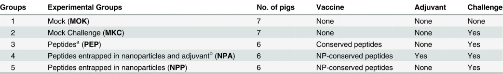
Table 1. Experimental Design forin vivoexperiment.
Groups Experimental Groups No. of pigs Vaccine Adjuvant Challenge
1 Mock (MOK) 7 None None None
2 Mock Challenge (MKC) 7 None None Yes
3 Peptidesa(PEP) 6 Conserved peptides None Yes
4 Peptides entrapped in nanoparticles and adjuvantb(NPA) 6 NP-conserved peptides Yes Yes
5 Peptides entrapped in nanoparticles (NPP) 6 NP-conserved peptides None Yes
Note:Pigs were vaccinated twice at two weeks interval, intranasally, challenged at two weeks booster vaccination and euthanized on day 7 post-challenge.
aPeptides: M2e-PP and four conserved SwIV peptides (A1, A2, A7, and A8) bAdjuvant:Mycobacterium vaccaewhole cell lysate
(3,30,5,50-Tetramethylbenzidine) (KPL) and the reaction was stopped using 1 M phosphoric
acid. Plates were read using SPECTRA max microplate reader at 450 nm. For BAL fluid, the end point titer was determined based on the highest dilution of the sample in which the mea-sured OD value was greater than the mean OD value of seven SwIV negative pig BAL fluid samples plus two standard deviations at the respective dilutions was used. Similarly, SwIV spe-cific IgG antibody end point titer from both BAL fluid and plasma were determined by ELISA. Peptide specific IgA level was also determined by coating peptides at the concentration of 2μg/ ml and OD values were compared between the experimental groups.
SwIV specific hemagglutination inhibition (HI) titer in pig BAL fluid and plasma samples was determined in accordance with the recommendation of World Organization for Animal Health (OIE) [31] (World Organization for Animal Health, 2014). SwIV specific neutralizing antibody and hemagglutination inhibition titers in plasma and BAL fluid were analyzed as described previously [32]. For determining the virus neutralizing (VN) antibody titers, plasma and BAL fluid samples were heat inactivated (56°C for 30 min), two-fold diluted and incubated with an equal volume of 50 TCID50of the SwIV per well for 2 hr at 37°C. The suspension was
transferred into microtiter plates containing confluent monolayer of MDCK cells and incu-bated for 24 hr at 37°C, followed by incubation with anti-SwIV Nucleoprotein specific mAb (M058, Cal Bioreagents, CA), Alexa-488 conjugated mouse IgG (H+L) secondary anti-body and mounted with glycerol in PBS (6:4 ratio). VN titer is the reciprocal of the highest dilution of a test sample which inhibited greater than 95% of the virus infectivity.
Peptide-specific lymphocytes response by flow cytometry analysis
The peptide specific recall lymphocyte response was assayed as described previously [14,33] with few modifications. Briefly, lung mononuclear cells (LMNCs) of all the pigs at DPC 7 were isolated by enzymatic digestion of lung tissues as described previously [14]. Five million LMNCs were restimulated with individual peptides (M2e, A1, A2, A7 and A8) (2μg/ml), SwIV (multiplicity of infection [MOI] 1) or Phytohaemagglutinin (10μg/ml) for 72 hr at 37°C in enriched RPMI 1640 (E-RPMI) (10% FBS, gentamicin [100μg/ml], ampicillin [20μg/ml], 20 mM HEPES, 2 mM L-glutamine, 0.1 mM nonessential amino acids, 1 mM sodium pyruvate and 50 nM 2-ME) in Bre-feldin A (B7651, SIGMA) were added during the last 6 hr of incubation of LMNCs treated with or without the indicated stimulants. The surface immunostained cells were fixed with 1% para-formaldehyde and permeabilized with the cell-permeabilization buffer (85.9% deionized water, 11% PBS without Ca2+or Mg2+, 3% formaldehyde solution and 0.1% saponin) overnight at 4°C. The cells were washed and stained with fluorochrome-conjugated anti-pig IFNγ(clone P2G10) or its isotype control mAb (BD Biosciences) in 0.1% saponin containing fluorescence-activated cell-sorting (FACS) buffer. Immunostained cells were acquired using the FACS Aria II (BD Bio-sciences) flow cytometer and analyzed using FlowJo (Tree Star, Ashland, OR, USA) software. All specific cell population frequencies were presented as the percent of total CD3+lymphocytes.
IFN
γ
ELISA
The culture supernatant of stimulated LMNCs with SwIV as described above was harvested after centrifuging the 48 well LMNCs culture plates for 2 min at 3,500 rpm and subjected to IFNγELISA as described previously [30]. Background cytokine levels from each pig groups were subtracted from the specific stimulation.
Ethics Statement
Council’s Guide for the Care and Use of Laboratory Animals and the Federation of Animal Sci-ence Societies’Guide for the Care and Use of Agricultural Animals in Agricultural Research and Teaching, and all relevant institutional, state and federal regulations and policies regarding animal care and use at The Ohio State University. The protocol was approved by the Commit-tee 48 well tissue culture plates. Stimulated LMNCs were immunostained and analyzed by flow cytometry as previously described [14,30]. Briefly, LMNCs were first surface-labeled with pig lymphocyte specific purified, fluorochrome or biotin conjugated mAbs [CD3έ(clone PPT3), CD4α(clone 74–12–4) and CD8α(clone 76-2-11)] (SouthernBiotech) followed by treatment with fluorochrome labeled streptavidin or anti-mouse isotype specific antibodies. For intracel-lular IFNγstaining, GolgiPlug™(BD Biosciences, San Jose, CA, USA) and on the Ethics of Ani-mal Experiments of The Ohio State University (Protocol Number: 2014A00000099). All the pigs were maintained, samples collected and euthanized, and all efforts were made to minimize the suffering of pigs.
Statistical analysis
All the data were expressed as the mean of 6 or 7 pigs ± standard error of the mean (SEM). Sta-tistical analyses were performed using one way analysis of variance (ANOVA) followed by post-hoc Tukey’s test using GraphPad InStat Prism (software version 5.0) to establish varia-tions among MKC, PEP, NPP and NPA pig groups. Statistical significance was assessed at P<0.05 (), P<0.01 () and P<0.001 ().
Results
Characterization of H1N1 influenza virus conserved peptides entrapped
PLGA-NP
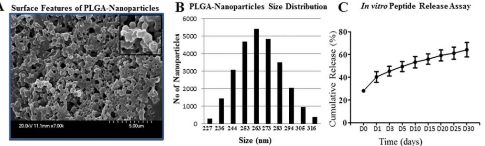
In the PLGA particulate vaccine delivery system, optimum antigen loading efficiency, charge, stability and size of the NP are important [34]. We entrapped M2e-PP and pooled four pep-tides separately in PLGA-NP using the similar procedure.In vitrocharacterization of both pep-tides and M2e-PP entrapped NPs showed comparable results and the data of M2e-PP is shown here. Entrapment efficiency of M2e-PP in NP was 50–54%. Scanning Electron Microscopy (SEM) imaging of NP revealed them as spherical particles (Fig 1A). Size distribution profile of the NP was determined by dynamic light scattering technique and it was 227–316 nm with the majority of NPs around 260 nm (Fig 1B). Surface charge of the particles was -21.93±2.93 mV (an average value of ten runs) as measured by zeta potential analysis.In vitroprotein release profile of M2e-PP from NP-entrapped M2e-PP suspended in PBS overtime was quantified under physiological conditions. PLGA-NP containing the vaccine cargo carry a small quantity of surface anchored Ags and they rapidly release upon reconstitution in PBS (10 min) called the burst release; while the entrapped Ags in NP release over a period of 4–6 weeks [15]. We observed a burst release of 14.3%, cumulative release of 25% (including the burse release) after 2 hr (day 0) and the total release of 64% over a period of 4 weeks (Fig 1C). Thus, our results indicated that PLGA-NP release entrapped M2e-PP gradually, supporting the principle of depot-effect provided by PLGA-NP underin vivoconditions.
SwIV antigen specific activated lymphocyte subsets in the lungs of pigs
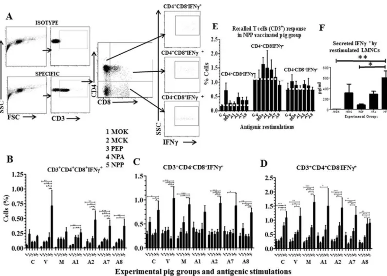
representative graph depicting the gating pattern of restimulated porcine lymphocyte subsets secreting IFNγbelongs to SwIV infected pigs is shown (Fig 2A). Porcine immune system has a unique abundant population of CD4 and CD8 double positive T cell subset, which have com-bined T-helper, memory and cytotoxic T cell properties [35,36]. Our results detected both the virus and peptides (A1, A2, A7 and A8) specific increased frequency of CD4+CD8α+IFNγ+T cells in LMNCs of NPP vaccinated pigs compared to all the other vaccine formulations received animals (Fig 2B). This data indicated influenza Ags specific activation of CD4 and CD8 double positive T cell subset in the lungs of NPP vaccinated pigs.
In pigs, naïve T-helper cells are identified based on the expression of combination of pheno-typic markers, CD3+CD4+CD8α-. Similarly, CD3+CD4-CD8α+markers bearing cells are either cytotoxic T cells (CTLs) orγδT cells; and CD3+CD4-CD8αβ+expressing cells are exclusively CTLs [37]. Frequency of IFNγ+T-helper cells and CTLs/γδT cells in LMNCs were significantly higher in NPP vaccinated pigs, irrespective of the cells unstimulated or stimulated with the viral Ags or most of the peptides (Fig 2C and 2D). This data indicated the presence of activated other T cell subsets in NPP vaccinated pigs, which continued to secrete IFNγ+when culturedex vivo.
Further, to determine influenza Ags specific recalled T cell response exclusively in NPP vacci-nated pigs, all the three T cell subsets which were IFNγ+in NPP pig group were plotted separately (Fig 2E). A white dotted line showed in the figure in each T cell subset separates the specific recalled T cell response over the control (C) unstimulated cells (Fig 2E). Our results detected the presence of increased recalled CD4+CD8α+IFNγ+T cell subset in both SwIV and peptides A2, A7 and A8 stimulated LMNCs (Fig 2E, 1stpanel). Similarly, increased recalled T-helper cell pop-ulation specific to peptides M2e, A1 and A2 (Fig 2E, 2ndpanel), and recalled specific CD3 +-CD4-CD8α+cell response against the virus, M2e and A2 peptides (Fig 2E, 3rdpanel) in NPP vaccinated pigs were detected. This data analysis revealed that nanoparticle entrapped conserved influenza peptides induced the peptide specific T cell response in pigs. In addition, supernatants harvested from the cultured LMNCs restimulated with the SwIV had significantly higher levels of secreted IFNγin NPP compared to peptides- and mock-vaccinated pig groups (Fig 2F).
Humoral immune response in vaccinated and SwIV challenged pigs
To quantify humoral immune response in the vaccinated pigs, we performed IgG antibody analysis in plasma samples collected at day pre-challenge 0 (DPC 0) using pretitrated amounts of H1N1 SwIV and SwIV M2e protein by ELISA. Our results showed very low levels of specific
Fig 1. Characterization of influenza A virus specific M2e-PP entrapped PLGA-NP.(A) The surface morphology of M2e-PP entrapped PLGA-NP (x7K) showing spherical and uniform sized particles; (B) Size distribution of M2e-PP entrapped PLGA-NP; (C)In vitroprotein release profile of M2e-PP entrapped PLGA-NP.
antibody response, and the data was not statistically significant among the vaccinated pig groups (Fig 3A and 3B). Even in post-challenge plasma and BAL fluid samples of vaccinated pigs collected at DPC 7, the virus specific IgA and IgG antibody, hemagglutination inhibition (HI) and virus neutralizing (VN) antibody titers were low and the data was not significantly different among the experimental pig groups (Fig 3C–3H). Thus, our study indicated that anti-body response in NPP and NPA vaccinated pigs at pre-challenge was weak or absent, and the detected antibody response at post-challenge was induced by the challenge virus. Our results suggested the need of using additional B cell antigenic peptides or the inactivated virus
Fig 2. Immunophenotyping of activated lymphocyte subsets in LMNCs of vaccinated and SwIV challenged pigs.(A) A representative flow cytometry plots depicting porcine IFNγpositive T lymphocytes subpopulations. Gating pattern of lymphocytes of a pig infected with SwIV H1N1 and restimulatedex vivo with the virus for 3 days and immunostained using fluorochrome conjugated pig specific CD3έ, CD4αand CD8αmarkers and intracellular IFNγto identify the
frequency of CD3+CD4+CD8α+IFNγ+, CD3+CD4-CD8α+IFNγ+and CD3+CD4+CD8α-IFNγ+cells is shown. Lung MNCs(LMNCs) isolated on the day of
necropsy from all the experimental pigs were either unstimulated (Control, C) or stimulated with the SwIV (V), conserved peptides (A1, A2, A7 and A8 peptides) or M2e (M) for 3 days and immunostained cells were gated for T cell subsets as described above for: (B) CD3+CD4+CD8
α+IFNγ+; (C)
CD3+CD4-CD8
α+IFNγ+; and (D) CD3+CD4+CD8α-IFNγ+cells. (E) Delineating the SwIV specific T cell response in NPP vaccinated pigs. Three activated
(IFNγ+) T lymphocyte subsets in the LMNCs of only NPP vaccinated pigs either unstimulated (Control, C) or stimulated with the SwIV or indicated peptides
are shown. A white dotted line drawn across each T cell subset separates the Ags specific recalled T cell response over the control unstimulated cells of NPP vaccinated pigs. (F) Secreted IFNγin the culture supernatant of virus restimulated LMNCs of vaccinated and SwIV challenged pigs was analyzed by ELISA.
Background cytokine levels from each pig groups were subtracted from the specific stimulation. Each bar indicates the average frequency of indicated T cell subset or cytokine from 6 or 7 pigs±SEM. Asterisk denotes statistically significant difference at P<0.05 (*), P<0.01 (**) and P<0.001 (***) between the
indicated two pig groups determined by One way ANOVA followed by Tukey Post-hoc test for the significance between the indicated pig groups.
entrapped in NP vaccine, and coadministering with a potent adjuvant to boost the specific anti-body response in pigs.
Clinical signs and lung virus load in the SwIV challenged pigs
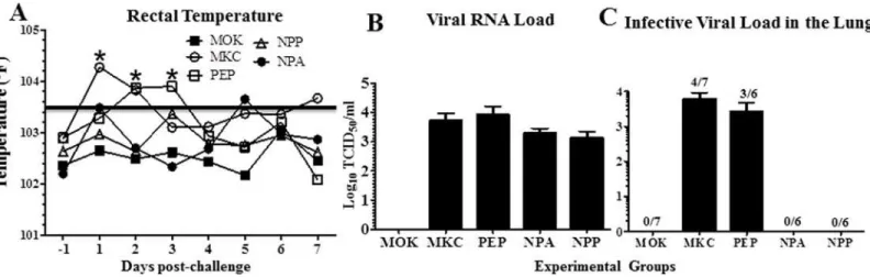
In the mock and unentrapped peptides vaccinated and virulent SwIV H1N1 challenged pigs, the rectal temperature was 1.5 to 2 °F higher at DPC 1 to 3 compared to mock animals (Fig 4A). The pigs which had fever were anorexic and lethargic during those initial days of viral challenge. Similarly challenged, but NPP and NPA vaccinated pig groups did not suffer from hyperthermia (Fig 4A). To associate the body temperature to viral load, we measured the viral RNA copies in BAL fluid and observed comparable RNA load among all the virus-challenged pig groups (Fig 4B). But the replicating SwIV was detected in50% of MKC and PEP pig groups, and it was undetectable in the NPP and NPA vaccinated animals (Fig 4C). The assay was repeated three times to confirm the live SwIV titer in the BAL fluid samples. Nasal swabs collected at DPC 4 and 7 did not show any detectable replicating virus in any of the virus chal-lenged pig groups (data not shown). In summary, clinical signs of flu and replicating detectable lung viral load were detected only in SwIV challenged control (MCK and PEP), but not in PLGA-NP peptides (NPP and NPA) vaccinated pig groups.
Discussion
Control of influenza in pigs and humans is a challenge due to continuous genetic changes in the viral surface proteins. Therefore, enhancing influenza virus specific immune response to
Fig 3. Humoral immune response in vaccinated and SwIV challenged pigs.In pre-challenge pigs, the levels of specific IgG in plasma against (A) H1N1 SwIV and (B) SwIV M2e protein at DPC 0. In post-challenge pigs at DPC 7, the levels of SwIV specific IgA and IgG antibody titers (C and D);
hemagglutination inhibition (HI) titers (E and F); and virus neutralization (VN) titers (G and H) in the BAL fluid and plasma samples, respectively, are shown. Each bar represents the average titer of 6 or 7 pigs±SEM. The data of antibody titers shown in the graph are corrected values after subtracting from the background OD values.
highly conserved antigenic epitopes is important to achieve increased breadth of protective immunity. Hence, to elicit strong specific response to influenza, we entrapped highly conserved H1N1 peptides in PLGA-NPs and inoculated intranasally in the form of mist to pigs to effi-ciently reach the mucosal inductive sites in the respiratory tract. Our results demonstrated a significantly reduced detectable infectious challenged virus in the airways of NPP and NPA vaccinated pigs, associated with augmented antigen-specific T cell response in the lung lym-phocytes of NPP inoculated animals. Amounts of each of the four peptides entrapped in NP-Peptides vaccine is assumed to be the same, because in another independent study individ-ual peptides entrapped in PLGA NPs had comparable levels of entrapment efficiency. In addi-tion, results ofex vivorestimulation of LMNCs with all the four individual peptides elicited comparable peptide specific T cell response in NPP vaccinated pigs.
Studies in mice demonstrated that intranasal administration of M2e-PP chimera induces high levels of M2e specific antibodies [7]. Conserved A7 and A8 peptides elicit antibody response in pigs, but failed to reduce the flu symptoms [5,6]. Soluble antigens and peptides are poorly immunogenic, but when entrapped in NPs elicit strong immune response [38,39]. Pul-monary immune response varies with the vaccine particle size and surface charge, because par-ticles of500 nm are readily phagocytized by dendritic cells and M cells at mucosal sites [40,41]. Consistent with that the size of our candidate NP-Peptide vaccine was approximately 300 nm. PLGA-NP entrapped Ags are stable for 4–6 weeks and facilitate activation and matu-ration of APCs [40,42]. Mechanism of induction of increased breadth of protective response by NPs based vaccine delivery system is mediated through efficient internalization of particulate Ags, followed by processing and cross-presentation of entrapped Ags by dendritic cells and macrophages to naïve CD8 T cells [43–45]. This has been shown specifically using PLGA NPs delivery system mediated by their ability to disrupt phagosomes and release of cargo into cyto-sol for MHC class I loading [44,45]. Based on our data with heightened peptide-specific CD4 CD8 double positive T cell response in LMNCs of NPP pig group, it is expected that both den-dritic cells and macrophages in the lungs of pigs would have uptaken and efficiently cross-pre-sented the PLGA-NP delivered peptide Ags to naïve T cells. Pigs vaccinated with M2eHBc fusion proteins without NP delivery system has failed to protect animals from a challenge virus induced flu symptoms [5].
Fig 4. Clinical outcome and virus load in the lungs of vaccinated and SwIV challenged pigs.(A) Rectal temperature was recorded everyday post-challenge (day -1 to 7). SwIV titer in BAL fluid at DPC 7 was detected by (B) viral RNA load by qRT-PCR, and (C) replicating infectious virus load using MDCK cells by immunofluorescence assay. The ratio indicated above each bar represents the number of pigs positive for virus and contributed to the data out of 6 or 7 animals. The viral titer is expressed in tissue culture infective dose 50 (TCID50) in each ml of the BAL fluid. Each data point or bar represents the average value of 6 to 7 pigs±SEM. Asterisk denotes statistically significant difference at P<0.05 (*) between MKC and NPP or NPA pig groups determined by One
way ANOVA followed by Tukey Post-hoc test.
In pigs, a significant upregulation of activated CD4 CD8 double positive T cells has been associated with protective response against classical swine fever, Aujeszky’s disease and PRRS viruses [14,15,46,47]. In particular in our study, NPP but not NPA vaccinated pigs elicited anti-gen-specific increased activated IFNγsecreting CD4 CD8 double positive T cell response in the lungs. Interestingly, complete clearance of detectable infectious challenge virus was observed in the airways of both NPP and NPA vaccinated animals, despite the comparable low levels of humoral response. This data suggested the critical role played by NPs entrapped peptides deliv-ery system in pigs, as well as the role of adjuvantM.vaccaeWCL in possibly skewing the response of NPP towards the Th2 or Th1-Th2 balanced state, which needs further investigation.
Earlier, we have shown strong virus specific antibody response in pigs vaccinated intrana-sally using PLGA-NPs entrapped PRRSV with a potent adjuvantM.tbWCL, which elicited a strong Th1-Th2 balanced response [14,15]. In place of the adjuvantM.tbWCL, we used a non-pathogenic mycobacterium,M.vaccaederived WCL; because,M.vaccaehas comparable bio-chemical and cellular components ofM.tb[48], and also it was shown to possess potent adjuvant effects in rodents [49,50]. Further,M.vaccaeWCL was not entrapped in PLGA-NP, because greater adjuvant effects by soluble compared to NP entrappedM.tb WCLin intrana-sally vaccinated pigs was observed [14,15]. Overall, our study suggested thatM.vaccaeWCL is not a potent adjuvant to NPP vaccine in pigs, indicating the need of including additional highly antigenic B cell peptides or inactivated SwIV entrapped in NPs and coadministered with a potent adjuvant to boost the specific antibody response in pigs.
In conclusion, our study in pigs for the first time demonstrated that influenza H1N1 con-served peptides cocktail entrapped in biodegradable nanoparticles and delivered intranasally as mist induced epitope specific T cell response, despite not boosting the antibody response. Therefore, our future studies are focused on including additional highly antigenic influenza virus B cell peptides and potent adjuvant/s to enhance specific mucosal and systemic antibody responses to nanoparticle flu vaccine candidate. This is important because, biodegradable nanoparticle vaccine delivery platform has translational value to improve cross-protective immunity against genetically variant influenza viruses.
Acknowledgments
The authors would like to thank Dr. Juliet Hanson and Megan Strother for assistance with ani-mal studies.
Author Contributions
Conceived and designed the experiments: JH CWL GJR. Performed the experiments: JH KK ME BB KO SD GJR. Analyzed the data: JH GJR. Contributed reagents/materials/analysis tools: MX XJ JA JBT. Wrote the paper: JH GJR.
References
1. Chen Q, Madson D, Miller CL, Harris DL (2012) Vaccine development for protecting swine against influ-enza virus. Anim Health Res Rev 13: 181–195. doi:10.1017/S1466252312000175PMID:23253165 2. Myers KP, Olsen CW, Gray GC (2007) Cases of swine influenza in humans: a review of the literature.
Clin Infect Dis 44: 1084–1088. PMID:17366454
3. Vincent AL, Ma W, Lager KM, Janke BH, Richt JA (2008) Swine influenza viruses a North American perspective. Adv Virus Res 72: 127–154. doi:10.1016/S0065-3527(08)00403-XPMID:19081490 4. Heath WR, Carbone FR (2001) Cross-presentation in viral immunity and self-tolerance. Nat Rev
5. Heinen PP, Rijsewijk FA, de Boer-Luijtze EA, Bianchi AT (2002) Vaccination of pigs with a DNA con-struct expressing an influenza virus M2-nucleoprotein fusion protein exacerbates disease after chal-lenge with influenza A virus. J Gen Virol 83: 1851–1859. PMID:12124449
6. Vergara-Alert J, Argilaguet JM, Busquets N, Ballester M, Martin-Valls GE, Rivas R, et al. (2012) Con-served synthetic peptides from the hemagglutinin of influenza viruses induce broad humoral and T-cell responses in a pig model. PLoS One 7: e40524. doi:10.1371/journal.pone.0040524PMID:22815759 7. Xia M, Tan M, Wei C, Zhong W, Wang L, McNeal M, et al. (2011) A candidate dual vaccine against
influ-enza and noroviruses. Vaccine 29: 7670–7677. doi:10.1016/j.vaccine.2011.07.139PMID:21839795 8. Purcell AW, McCluskey J, Rossjohn J (2007) More than one reason to rethink the use of peptides in
vaccine design. Nat Rev Drug Discov 6: 404–414. PMID:17473845
9. Lu JM, Wang X, Marin-Muller C, Wang H, Lin PH, Yao Q, et al. (2009) Current advances in research and clinical applications of PLGA-based nanotechnology. Expert Rev Mol Diagn 9: 325–341. doi:10. 1586/erm.09.15PMID:19435455
10. Aguado MT, Lambert PH (1992) Controlled-release vaccines—biodegradable polylactide/polyglycolide (PL/PG) microspheres as antigen vehicles. Immunobiology 184: 113–125. PMID:1587538
11. McNeil SE (2005) Nanotechnology for the biologist. J Leukoc Biol 78: 585–594. PMID:15923216 12. Panyam J, Labhasetwar V (2003) Biodegradable nanoparticles for drug and gene delivery to cells and
tissue. Adv Drug Deliv Rev 55: 329–347. PMID:12628320
13. Zhang L, Tian X, Zhou F (2007) Intranasal administration of CpG oligonucleotides induces mucosal and systemic Type 1 immune responses and adjuvant activity to porcine reproductive and respiratory syndrome killed virus vaccine in piglets in vivo. Int Immunopharmacol 7: 1732–1740. PMID:17996683 14. Binjawadagi B, Dwivedi V, Manickam C, Ouyang K, Torrelles JB, Renukaradhya GJ (2014) An
innova-tive approach to induce cross-protecinnova-tive immunity against porcine reproducinnova-tive and respiratory syn-drome virus in the lungs of pigs through adjuvanted nanotechnology-based vaccination. Int J Nanomedicine 9: 1519–1535. doi:10.2147/IJN.S59924PMID:24711701
15. Binjawadagi B, Dwivedi V, Manickam C, Ouyang K, Wu Y, Lee LJ, et al. (2014) Adjuvanted poly(lactic-co-glycolic) acid nanoparticle-entrapped inactivated porcine reproductive and respiratory syndrome virus vaccine elicits cross-protective immune response in pigs. Int J Nanomedicine 9: 679–694. doi: 10.2147/IJN.S56127PMID:24493925
16. Dwivedi V, Manickam C, Binjawadagi B, Renukaradhya GJ (2013) PLGA nanoparticle entrapped killed porcine reproductive and respiratory syndrome virus vaccine helps in viral clearance in pigs. Vet Micro-biol 166: 47–58. doi:10.1016/j.vetmic.2013.04.029PMID:23764272
17. Thomas C, Rawat A, Hope-Weeks L, Ahsan F (2011) Aerosolized PLA and PLGA Nanoparticles Enhance Humoral, Mucosal and Cytokine Responses to Hepatitis B Vaccine. Mol Pharm 8: 405–415. doi:10.1021/mp100255cPMID:21189035
18. Hossain MJ, Perez S, Guo Z, Chen LM, Donis RO (2010) Establishment and characterization of a Madin-Darby canine kidney reporter cell line for influenza A virus assays. J Clin Microbiol 48: 2515– 2523. doi:10.1128/JCM.02286-09PMID:20504984
19. Yassine HM, Khatri M, Zhang YJ, Lee CW, Byrum BA, O'Quin J, et al. (2009) Characterization of triple reassortant H1N1 influenza A viruses from swine in Ohio. Vet Microbiol 139: 132–139. doi:10.1016/j. vetmic.2009.04.028PMID:19477087
20. Zhang N, Qi J, Feng S, Gao F, Liu J, Pan X, et al. (2011) Crystal structure of swine major histocompati-bility complex class I SLA-1 0401 and identification of 2009 pandemic swine-origin influenza A H1N1 virus cytotoxic T lymphocyte epitope peptides. J Virol 85: 11709–11724. doi:10.1128/JVI.05040-11 PMID:21900158
21. Ho CS, Lunney JK, Ando A, Rogel-Gaillard C, Lee JH, Schook LB, et al. (2009) Nomenclature for fac-tors of the SLA system, update 2008. Tissue Antigens 73: 307–315. doi:10.1111/j.1399-0039.2009. 01213.xPMID:19317739
22. Daum LT, Canas LC, Smith CB, Klimov A, Huff W, Barnes W, et al. (2002) Genetic and antigenic analy-sis of the first A/New Caledonia/20/99-like H1N1 influenza isolates reported in the Americas. Emerg Infect Dis 8: 408–412. PMID:11971776
23. Dwivedi V, Manickam C, Binjawadagi B, Joyappa D, Renukaradhya GJ (2012) Biodegradable Nano-particle-Entrapped Vaccine Induces Cross-Protective Immune Response against a Virulent Heterolo-gous Respiratory Viral Infection in Pigs. PLoS One 7: e51794. doi:10.1371/journal.pone.0051794 PMID:23240064
25. Rajapaksa TE, Bennett KM, Hamer M, Lytle C, Rodgers VG, Lo DD (2010) Intranasal M cell uptake of nanoparticles is independently influenced by targeting ligands and buffer ionic strength. J Biol Chem 285: 23739–23746. doi:10.1074/jbc.M110.126359PMID:20511224
26. Shi L, Torrelles JB, Chatterjee D (2008) Lipoglycans of Mycobacterium tuberculosis: Isolation, Purifica-tion,and Characterization. In: Parish T, Brown CA, eds Mycobacteria Protocols 2nd Edition ed Totowa, NJ: Humana Press: 23–45.
27. Jung K, Alekseev KP, Zhang X, Cheon DS, Vlasova AN, Saif LJ (2007) Altered pathogenesis of porcine respiratory coronavirus in pigs due to immunosuppressive effects of dexamethasone: implications for corticosteroid use in treatment of severe acute respiratory syndrome coronavirus. J Virol 81: 13681– 13693. PMID:17942563
28. Lee CW, Suarez DL (2004) Application of real-time RT-PCR for the quantitation and competitive repli-cation study of H5 and H7 subtype avian influenza virus. J Virol Methods 119: 151–158. PMID: 15158597
29. Guo YJ, Sun SH, Zhang Y, Chen ZH, Wang KY, Huang L, et al. (2004) Protection of pigs against Taenia solium cysticercosis using recombinant antigen or in combination with DNA vaccine. Vaccine 22: 3841–3847. PMID:15364430
30. Dwivedi V, Manickam C, Patterson R, Dodson K, Murtaugh M, Torrelles JB, et al. (2011) Cross-protec-tive immunity to porcine reproducCross-protec-tive and respiratory syndrome virus by intranasal delivery of a live virus vaccine with a potent adjuvant. Vaccine 29: 4058–4066. doi:10.1016/j.vaccine.2011.03.006 PMID:21419162
31. Sa e Silva M, Swayne DE (2012) Serum and egg yolk antibody detection in chickens infected with low pathogenicity avian influenza virus. Avian Dis 56: 601–604. PMID:23050482
32. Khatri M, Dwivedi V, Krakowka S, Manickam C, Ali A, Wang L, et al. (2010) Swine influenza H1N1 virus induces acute inflammatory immune responses in pig lungs: a potential animal model for human H1N1 influenza virus. J Virol 84: 11210–11218. doi:10.1128/JVI.01211-10PMID:20719941
33. Kimman TG, De Bruin TM, Voermans JJ, Peeters BP, Bianchi AT (1995) Development and antigen specificity of the lymphoproliferation responses of pigs to pseudorabies virus: dichotomy between sec-ondary B- and T-cell responses. Immunology 86: 372–378. PMID:8550073
34. Ito F, Fujimori H, Makino K (2008) Factors affecting the loading efficiency of water-soluble drugs in PLGA microspheres. Colloids Surf B Biointerfaces 61: 25–29. PMID:17719753
35. Zuckermann FA, Husmann RJ (1996) Functional and phenotypic analysis of porcine peripheral blood CD4/CD8 double-positive T cells. Immunology 87: 500–512. PMID:8778040
36. Denyer MS, Wileman TE, Stirling CM, Zuber B, Takamatsu HH (2006) Perforin expression can define CD8 positive lymphocyte subsets in pigs allowing phenotypic and functional analysis of natural killer, cytotoxic T, natural killer T and MHC un-restricted cytotoxic T-cells. Vet Immunol Immunopathol 110: 279–292. PMID:16325923
37. Talker SC, Kaser T, Reutner K, Sedlak C, Mair KH, Koinig H, et al. (2013) Phenotypic maturation of por-cine NK- and T-cell subsets. Dev Comp Immunol 40: 51–68. doi:10.1016/j.dci.2013.01.003PMID: 23352625
38. Bacon A, Makin J, Sizer PJ, Jabbal-Gill I, Hinchcliffe M, Illum L, et al. (2000) Carbohydrate biopolymers enhance antibody responses to mucosally delivered vaccine antigens. Infect Immun 68: 5764–5770. PMID:10992483
39. Bertram U, Bernard MC, Haensler J, Maincent P, Bodmeier R (2010) In situ gelling nasal inserts for influenza vaccine delivery. Drug Dev Ind Pharm 36: 581–593. doi:10.3109/03639040903382673 PMID:19954407
40. Blank F, Stumbles PA, Seydoux E, Holt PG, Fink A, Rothen-Rutishauser B, et al. (2013) Size-depen-dent uptake of particles by pulmonary antigen-presenting cell populations and trafficking to regional lymph nodes. Am J Respir Cell Mol Biol 49: 67–77. doi:10.1165/rcmb.2012-0387OCPMID:23492193 41. Kim B, Bowersock T, Griebel P, Kidane A, Babiuk LA, Sanchez M, et al. (2002) Mucosal immune
responses following oral immunization with rotavirus antigens encapsulated in alginate microspheres. J Control Release 85: 191–202. PMID:12480324
42. Yoshida M, Babensee JE (2006) Differential effects of agarose and poly(lactic-co-glycolic acid) on den-dritic cell maturation. J Biomed Mater Res A 79: 393–408. PMID:16886225
43. Woodrow KA, Bennett KM, Lo DD (2012) Mucosal vaccine design and delivery. Annu Rev Biomed Eng 14: 17–46. doi:10.1146/annurev-bioeng-071811-150054PMID:22524387
45. Schliehe C, Redaelli C, Engelhardt S, Fehlings M, Mueller M, van Rooijen N, et al. (2011) CD8- den-dritic cells and macrophages cross-present poly(D,L-lactate-co-glycolate) acid microsphere-encapsu-lated antigen in vivo. J Immunol 187: 2112–2121. doi:10.4049/jimmunol.1002084PMID:21795597 46. De Bruin TG, Van Rooij EM, De Visser YE, Bianchi AT (2000) Cytolytic function for pseudorabies
virus-stimulated porcine CD4+ CD8dull+ lymphocytes. Viral Immunol 13: 511–520. PMID:11192298 47. Saalmuller A, Werner T, Fachinger V (2002) T-helper cells from naive to committed. Vet Immunol
Immunopathol 87: 137–145. PMID:12072228
48. Lederer E, Adam A, Ciorbaru R, Petit JF, Wietzerbin J (1975) Cell walls of Mycobacteria and related organisms; chemistry and immunostimulant properties. Mol Cell Biochem 7: 87–104. PMID:1095912 49. Hernandez-Pando R, Aguilar D, Orozco H, Cortez Y, Brunet LR, Rook GA (2008) Orally administered Mycobacterium vaccae modulates expression of immunoregulatory molecules in BALB/c mice with pul-monary tuberculosis. Clin Vaccine Immunol 15: 1730–1736. doi:10.1128/CVI.00286-08PMID: 18827195