online | memorias.ioc.fiocruz.br
Since the identification of human immunodeficiency virus (HIV)-1 as the aetiological agent of acquired im-mune deficiency syndrome (AIDS), several studies have associated different viral molecular characteristics, es-pecially the diversity of the envelope gene, with disease progression (Potts et al. 1993, Santoro-Lopes et al. 2000). Most of these studies are focused on the envelope third variable region, or V3 loop, which is involved in HIV-1 binding to the chemokine co-receptors 5 (R5) and/or CX-CR4 (X4). This step is necessary for viral fusion to the host cell (Shioda et al. 1991). A change in viral tropism has been associated with different phases of HIV-1 infec-tion, with a predominance of R5 tropic virus observed at the initial stages of infection. X4-tropic viruses emerge at advanced disease stages in about half of all cases and this emergence is correlated with clinical progression and CD4 T cell depletion (Richman & Bozzette 2004, Goetz et al. 2009). The pathogenic potential of X4 virus is highlighted by its capacity to induce syncytia forma-tion (Richman & Bozzette 2004). Several bioinformat-ics tools have been used to determine viral genotypic
tropism, such as the website geno2pheno and the 11/25 rule. Additionally, amino acid signatures at the V3 loop tip tetramer, which commonly harbour a proline at codon 16 (15GPGR18), show a substitution to tryptophan (W) at this position in some Brazilian clade B envelopes and are referred to as B’ or Brazilian-B (Shioda et al. 1991, Potts et al. 1993, Pinto et al. 2008). The GWGR signature has a prevalence of 20-50% in Brazil and has been associ-ated with a slower progression to AIDS (Santoro-Lopes et al. 2000, Casseb et al. 2002, 2004, de Brito et al. 2006). However, the actual relevance of this molecular diversity on treatment response is not yet clear. To expand the un-derstanding of the potential role of genotypic viral tro-pism, false-positive rate (FPR) values obtained at geno2-pheno and the presence of the GWGR motif in patients on antiretrovirals (ARV), the correlations of these charac-teristics with viral loads (VLs) and CD4+ T lymphocyte counts were assessed in patients during 48 weeks of first-line highly active antiretroviral therapy (HAART).
PATIENTS, MATERIALS AND METHODS
Study population - HIV-1-infected individuals not
exposed to antiretroviral therapy (ART), but with indi-cations for treatment according to the Brazilian Clinical Guideline, were invited and those eligible were randomly assigned in a 1:1 ratio to receive either lopinavir or efa-virenz with azithothymidine (AZT)/3TC backbone. Clini- cal follow-up was conducted at the HIV Clinic, Medicine School Hospital, Ribeirão Preto, São Paulo University to evaluate the virological and immunological responses of the patients to HAART during the 48 weeks of follow-Financial support: FAPESP (2009/8215-0, 2006/61311-0), CNPq
(144076/2010-1)
+ Corresponding author: lubrigido@gmail.com Received 15 June 2011
Accepted 24 December 2011
HIV-1 tropism and CD4 T lymphocyte recovery in a prospective
cohort of patients initiating HAART in Ribeirão Preto, Brazil
Andre Minhoto Lanca1, Jeova Keny Baima Collares2, João Leandro de Paula Ferreira1,
Danielle Malta Lima2, Luis Fernando de Macedo Brigido1/+,
Rosangela Rodrigues1, Benedito Antonio Lopes da Fonseca3
1Laboratório de Retrovirus, Centro de Virologia, Instituto Adolfo Lutz, São Paulo, SP, Brasil 2Universidade de Fortaleza, Fortaleza, CE, Brasil 3Centro de Pesquisa em Virologia, Faculdade de Medicina, Universidade de São Paulo, Ribeirão Preto, SP, Brasil
While human immunodeficiency virus (HIV)-1 chemokine co-receptors 5 tropism and the GWGR motif in the envelope third variable region (V3 loop) have been associated with a slower disease progression, their influence on antiretroviral response remains unclear. The impact of baseline V3 characteristics on treatment response was evalu-ated in a randomised, double blind, prospective cohort study with patients initiating highly active antiretroviral ther-apy with lopinavir or efavirenz plus azithothymidine/3TC (1:1) over 48 weeks. Similar virological and immunological responses were observed for both treatment regimens. The 43 individuals had a mean baseline CD4 T cell count of
119 cells/mm³ [standard deviation (SD) = 99] and a mean viral load of 5.09 log10 copies/mL (SD = 0.49). The GWGR
motif was not associated with a CD4 T cell response, but predicted R5 tropism by the geno2pheno[clinical20%] algorithm
correlated with higher CD4 T cell levels at all monitoring points (p < 0.05). Moreover, higher false-positive rates (FPR) values from this analysis revealed a strong correlation with CD4 T cell recovery (p < 0.0001). Transmitted drug resistance mutations, documented in 3/41 (7.3%) cases, were unrelated to the assigned antiretroviral regimen and had no impact on patient outcomes. In conclusion, naïve HIV-1 R5 infected patients exhibited higher CD4 T cell
counts at baseline; this difference was sustained throughout therapy. The geno2pheno[clinical] option FPR positively
correlated with CD4 T cell gain and may be useful in predicting CD4 T cell recovery.
up. Data from patients who died before completing the follow-up were included in the baseline evaluation and the individuals who did not complete all visits were in-cluded in an intention to treat (ITT) analysis. A written informed consent was obtained from all patients and both Institutional Review Boards approved the study.
Laboratory and molecular analysis - CD4 T cell
counts and percentages (flow cytometry, BD, USA) and VLs (Branched DNA, Siemens, USA) were determined at baseline and at weeks 6, 12, 24, 36 and 48. CD4 T cell gain was calculated as the absolute cell count from each week of observation minus the baseline CD4 T cell count. HIV-1 RNA and genomic DNA were extracted using the QIAamp® RNA/DNA extraction kit (Qiagen, Germany) according to manufacturer instructions and were stored at -70ºC until used. Synthesis of comple-mentary DNA (cDNA) was performed by reverse tran-scription with Superscript III (Invitrogen, USA). Poly-merase chain reaction amplification was performed on cDNA (pol) and proviral DNA (env) using a nested protocol as previously described (Ferreira et al. 2008). Amplicons were sequenced directly using BigDye and were resolved with an ABI 3100 Genetic Analyzer (Ap-plied Biosystems, USA). Sequences of partial env and
pol genes were manually edited with Sequencher 4.14 (GeneCodes, USA) software. Multiple sequence align-ments were performed using the CLUSTALW algorithm with a reference set obtained from the Los Alamos Se-quence Database. HIV subtyping was screened using NCBI Genotyping and was confirmed by phylogenet-ic methods using the software PAUP* version 4.0b10 (Sinauer Associates, USA). Transmitted drug resistance mutations (tDRM) were defined according to Calibrat-ed Population Resistance v.6 (HIV Stanford Database) and the International AIDS Society (IAS) 2010 muta-tion list. Viral tropism was evaluated using the web-site geno2pheno[co-receptor] bioinformatics tool (Sing et al. 2007) and both clonal and clinical options were used to obtain FPR values. Cut-offs of 5.75% (MOTIVATE op-tion) and 20% (Vanderkerckhove et al. 2011) were em-ployed to predict X4 tropism using clonal and clinical data. To obtain the FPR using the clinical option, base-line (absolute and percentage) CD4 T cells counts and VLs were added to the model. These predictions were analysed both as categorical variables (X4 or R5) and as continuous variables. V3 loop amino acid composi-tion was determined by analysis of alignments with the HXB2 reference sequence from the Los Alamos Se-quence Database (accession K03455).
Statistical analysis - Categorical variables were test-ed using Fischer’s exact test or the Yates correcttest-ed test (two-tailed), as appropriate. Continuous variables were tested using the Mann-Whitney test, two-way analysis of variance (ANOVA) (non-parametric), the Spearman correlation and linear regression. Epinfo6 (Centers for Disease Control and Prevention)and GraphPadPrism 5.0 software were used in the statistical analysis. Viro-logical and CD4 T cell outcomes were evaluated as ITT, missing equals failure (ITT, M = F) and as treated (AT), missing equals exclusion (AT, M = E).
RESULTS
The 43 patients enrolled from 2004-2006 were most-ly male (70%) and had a mean age of 41 years [standard deviation (SD) 15 years], a mean baseline CD4 T cell count of 119 cells/mm3 (SD = 99 cells/mm3) and a mean VL of 5.09 log10 c/mL (SD = 0.49). CD4 T cell counts and VLs are depicted in Table. Three patients died and two abandoned therapy before their first return at week 6 of the follow-up. One patient discontinued treatment be-fore 24 weeks and three left between 24-36 weeks of the follow-up. All three deaths occurred before concluding the clinical follow-up and these patients had baseline CD4 T lymphocyte counts below 50 cells/mm3.
Patients were randomly assigned in a 1:1 ratio to re-ceive either lopinavir (48.8%) or efavirenz (51.2%) with AZT/3TC backbone. The tDRM frequency at baseline (available after treatment initiation) was 7.3%, all mu-tations were observed in the protease gene (mumu-tations V82L, I85V and G73S) and the predicted loss of ART susceptibility was not relevant to the assigned ARV regimen. V82L and I85V substitutions were observed in patients assigned to receive lopinavir; these individuals exhibited a similar response to those without detectable mutations. The G73S mutation was observed in a patient receiving efavirenz who died two weeks after enrolment without an evaluation of the ARV response.
Virological and immunological outcomes after HAART on both regimen arms were comparable, with no differences between lopinavir and efavirenz response except for a higher occurrence of hypertriglyceridemia in the lopinavir group (data not shown). Viral suppres-sion was achieved in most patients by week 24, when 89% of individuals had viremia below detection limits. After 48 weeks of HAART, only two individuals (6%) had a detectable VL (> 50 copies/mL), with one on each regimen arm (AT, M = E). Further genotyping test anal-ysis of one of these cases, RP16 on lopinavir + AZT/3TC therapy with several adherence issues, revealed the emergence of the mutation M184V, which is predicted to reduce 3TC susceptibility.
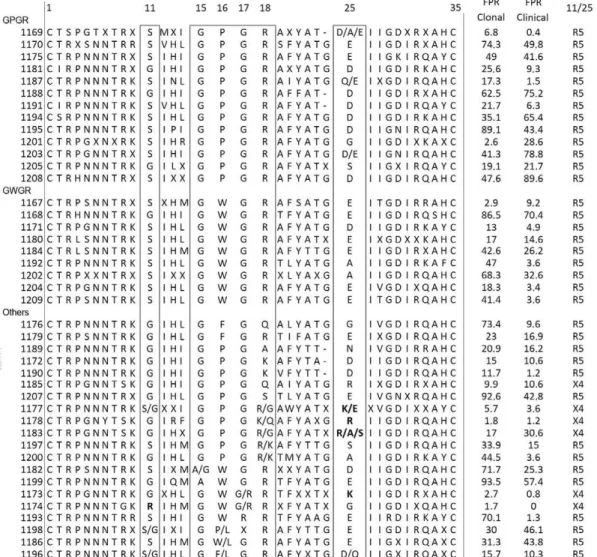
a 30% FPR value when we used the clinical algorithm, despite presenting an arginine (R) at position 25. The V3 loop alignment and tropism prediction using clonal data, clinical data and the 11/25 rule are shown in Fig. 1.
CD4 T cell counts at each interval were lower for pa-tients with X4-tropic viruses and were significantly dif-ferent when tropism was determined by geno2pheno[clinical
20%] (Fig. 2A, B). CD4 T cell counts and VLs according to different tropism prediction criteria are described in Table. CD4 T lymphocyte cell counts throughout the 48 weeks (Fig. 2C) and VLs (Fig. 2D) were comparable for patients infected with either GWGR or GPGR variants.
Taking into consideration the FPR values gener-ated using geno2pheno[clinica20%l] for each isolate from all cases (ITT analysis), the CD4 T cell gain showed a positive, significant correlation with FPR values at all observation weeks. This was observed by both linear regression (p < 0.0001) (Fig. 3) and the Spearman linear correlation (p < 0.01). A similar trend was also observed when only those completing week 48 of follow-up were evaluated (“as treated”, data not shown). Baseline CD4 T cell counts and geno2pheno[clonal20%] FPR values were not significantly correlated with CD4 T cell gain (p = 0.3 and 0.2, respectively).
DISCUSSION
This study evaluated the potential role of the molecu-lar characteristics of the HIV-1 envelope on CD4 T lym-phocyte gains in patients initiating their first HAART regimen from a structured cohort study performed to compare two first-line regimens. Viral tropism was de-termined by bioinformatics tools, using the geno2pheno algorithm with different criteria and inspecting the ami-no acid composition of the V3 loop of env (11/25 rule). The geno2pheno clinical option incorporated VL, CD4 T lymphocyte cell counts and percentages to generate an FPR value in which the predicted tropism was weighted by these parameters. Sequences were also evaluated us-ing the MOTIVATE option, which is the most specific criterion for detection of X4-tropic viruses, as it uses a more stringent FPR cut-off. It is of note that both the MO-TIVATE study and the European consensus recommend lower cut-offs of 5.75% and 10%, respectively, when only three or more sequences of each individual are available. Therefore, a cut-off of 20% was used for all analyses, as recommended by the European consensus for single sequences. The results exhibited some discordance in tropism interpretation when different algorithms were TABLE
CD4 T cell counts and viral load (VL) according to highly active antiretroviral therapy (HAART) allocation group, V3 motif and predicted viral tropism
n (%)
Baseline VL mean (SD)
Baseline CD4 T cell mean (SD)
CD4 T cells gain at 48 week mean (SD)
Randomization
Lopinavir + zidovudina /3TC 21 (49) 5.09 (0.12) 117 (22) 223 (22)
Efavirenz + zidovudina /3TC 22 (51) 5.09 (0.09) 122 (21) 212 (29)
V3 Motif
GPGR 12 (29.3) 5.12 (0.12) 104 (23) 235 (43)
GWGR 9 (21.9) 5.22 (0.17) 134 (34) 187 (38)
Other 20 (48.8) 4.99 (0.12) 117 (24) 219 (23)
Tropism 11/25 rule
X4 6 (14.3) 4.98 (0.27) 99 (48) 180 (41)
R5 36 (85.7) 5.11 (0.08) 125 (17) 223 (21)
MOTIVATE (5.7%)
X4 6 (14.3) 5.13 (0.19) 70 (31) 183 (53)
R5 36 (85.7) 5.09 (0.08) 130 (17) 222 (20)
geno2pheno[clonall20%]
X4 17 (40.5) 5.07 (0.15) 90 (23) 187 (19)
R5 25 (59.5) 5.11 (0.08) 142 (20) 237 (28)
geno2pheno[clinical20%]
X4 24 (57.1) 5.16 (0.11) 84 (20)a 192 (18)
R5 18 (42.9) 5.00 (0.10) 170 (20)a 245 (34)
a: p < 0.01. CD4 T (cells/mm3) and VL (log
used. Interestingly, most patients had advanced disease at the beginning of treatment, with a mean CD4 T cell count of 119 cells/mm3; this could explain the tropism disparities when clinical data were incorporated.
To assess the impact of viral tropism on clinical evolu-tion, both VL suppression and CD4 T cell recovery were followed-up for one year. Treatment, tDRM, V3 loop motif and viral tropism did not impact VL suppression. Lower numbers of CD4 T cells were observed at both the baseline and the follow-up weeks in patients infected with X4 virus using both clinical and clonal algorithms. Fur-thermore, CD4 T cell numbers were significantly lower in patients harbouring X4 variants when prediction used the geno2pheno[clinical20%] option (Fig. 2A). Theuse of baseline CD4 T cell counts on the clinical algorithm could be re-sponsible for this stronger association. However, baseline CD4 T cell counts did not show a correlation with CD4 T cell gain (p = 0.3). Although CD4 T cell counts of R5-infected patients remained higher at most determinations, CD4 T cell recovery was similar for both groups. In other
words, despite the higher CD4 T cell counts in R5-infect-ed patients, the response to HAART was comparable for both R5 and X4-infected individuals.
Values of FPR as continuous variables were also eval-uated to assess its association to CD4 T cell gain. Using the geno2pheno[clinical] option, envelope sequences FPR values exhibited a strong correlation with CD4 T cell gain (p < 0.0001) (Fig. 3). Again, although CD4 T cell count is used in this model to determine geno2pheno[clinical] FPR, neither CD4 T cell baseline counts (p = 0.3) nor the clon-al FPR (p = 0.2) per se revealed a significant correlation with CD4 T cell gain. This suggests that FPR generated by the geno2phenoclinical algorithm may constitute a potential predictor for CD4 T cell gain after therapy. The usefulness of this option has also been proposed in pre-vious studies (Prosperi et al 2010).
prevalence of X4 variants in GWGR viruses was ob-served using both the geno2pheno[clonal20%] (44%) and the geno2pheno[clinical20%] (67%) algorithms, contrary to previous studies, which suggested that GWGR viruses are rarely X4 (da Silva 2006, Leal et al. 2008). This difference could be due to the algorithm used, as most studies use the 11/25 rule as a prediction criterion. To clarify this matter, the presence of basic amino acids at positions 11 and 25 was evaluated in our sequences; none of the GWGR or GPGR isolates had basic amino acids at these codons. Interestingly, three V3 tip se-quences of non-GWGR tryptophan-harbouring virus-es, classified here as “other motifs”, were predicted as
X4. These viruses had a G→R polymorphism in the V3
loop tip at position 17 (15GWRR18), two had basic amino
acids at codons 11 or 25 and one had a G→R substi
-tution at position 28, a highly conserved codon (Leal et al. 2008, Franca et al. 2011). GWRR seems to be a very rare motif previously documented in the state of Rio de Janeiro, Brazil and Paraguay and its biological relevance is yet to be understood (Cabello et al. 1995, Tanuri et al. 1999)
The small number of samples in this study limits the strength of these observations and it is conceivable that these outcomes may be different if a larger cohort was sampled and a longer follow-up period was employed. Further studies may provide support for the use of geno2pheno[clinical] analysis of V3 envelope sequences as a surrogate marker for CD4 T cell gain after ART.
ACKNOwLEDgEMENTS
To the volunteers and the staff of the STI clinic involved in the study, and to Gabriela Ribeiro Santos, for the English review.
REFERENCES
Cabello A, Cabral M, Vera ME, Kiefer R, Azorero RM, Eberle J 1995. Analysis of the V3 loop sequences from 10 HIV type 1-in-fected AIDS patients from Paraguay. AIDS Res Hum Retrovi-ruses11: 1135-1137.
Casseb J, Komninakis SCV, Abdalla L, Brigido LFM, Rodrigues R, Araujo F 2002. HIV disease progression: is the Brazilian vari-HIV disease progression: is the Brazilian vari-ant subtype B’ (GWGR motif) less pathogenic than US/European subtype B (GPGR)? Inter J Infect Dis6: 64-69.
Casseb J, Montanheiro P, Komninakis S, Brito A, Duarte AJS 2004. Human immunodeficiency virus type 1 Brazilian subtype B variant showed an increasing avidity of the anti-V3 antibodies over time compared to the subtype B US/European strain in São Paulo, Brazil. Mem Inst Oswaldo Cruz99: 69-71.
da Silva J 2006. Site-specific amino acid frequency, fitness and the mutational landscape model of adaptation in human immunode-ficiency virus type 1. Genetics174: 1689-1694.
de Brito A, Komninakis SCV, Novoa P, de Oliveira RM, Fonseca LAM, Duarte AJS 2006. Women infected with HIV type 1 Bra-Women infected with HIV type 1 Bra-zilian variant, subtype B (B’-GWGR motif ) have slower progres-sion to AIDS compared with patients infected with subtype B (B-GPGR motif ). Clin Infect Dis43: 1476-1481.
Ferreira JLP, Thomaz M, Rodrigues R, Harrad D, Oliveira CM, Ol-iveira CAF, Batista JPGB, Ito TS, Brigido LFMB 2008. Molecu- Molecu-lar characterizations of newly identified HIV-1 infections in Cu-ritiba, Brazil: preponderance of clade C among males with recent infections. Mem Inst Oswaldo Cruz103: 800-808.
Franca RFO, Castro-Jorge LA, Neto RJP, Jorge DMM, Lima DM, Co-lares JKB 2011. Genotypic characteristics of HIV type 1 based on gp120 hypervariable region 3 of isolates from southern Brazil.
Aids Res Hum Retroviruses27: 903-909.
Goetz MB, Leduc R, Kostman JR, Labriola AM, Lie Y, Weidler J 2009. Relationship between HIV coreceptor tropism and disease progression in persons with untreated chronic HIV infection.
J Acquir Immune Defic Syndr50: 259-266.
Leal E, Silva WP, Sucupira MC, Janini LM, Diaz RS 2008. Mo- Mo-lecular and structural characterization of HIV-1 subtype B Bra-zilian isolates with GWGR tetramer at the tip of the V3-loop.
Virology381: 222-229. Fig. 3: linear regression of CD4 T cell gain plotted according to
geno2pheno[clinical] option false-positive rate (FPR) obtained from base-line samples (intention to treat, p < 0.0001). Replicates represent CD4 T cell gain at each observation week.
Pinto ME, Schrago CG, MirandaAB, Russo CAM 2008. A molecular study on the evolution of a subtype B variant frequently found in Brazil. Genet Mol Res7: 1031-1044.
Potts KE, Kalish ML, Lott T, Orloff G, Luo C, Bernard M, the Brazil-ian Collaborative AIDS Research Group 1993. Genetic hetero-geneity of the V3 region of the HIV-1 envelope glycoprotein in Brazil. AIDS7: 1191-1197.
Prosperi M, Bracciale L, Fabbiani M 2010. Comparative determina-tion of HIV-1 co-receptor tropism by enhanced sensitivity tro-file, gp120 V3-loop RNA and DNA genotyping. Retrovirology 7: 56.
Richman DD, Bozzette SA 2004. The impact of the syncytium in-ducing phenotype of human immunodeficiency virus on disease progression. J Infect Dis169: 968-974.
Santoro-Lopes G, Harrison LH, Tavares MD, Xexéo A, dos Santos ACE, Schechter M 2000. HIV disease progression and V3 sero-HIV disease progression and V3
sero-types in Brazil: is B different from B-Br? AIDS Res Hum Retro-viruses16: 953-958.
Shioda T, Levy JA, Cheng-Mayer C 1991. Macrophage and T cell-line tropisms of HIV-1 are determined by specific regions of the envelope gp120 gene. Nature349: 167-169.
Sing T, Low AJ, Beerenwinkel N, Sander O, Cheung PK, Domingues F, Büch J, Däumer M, Kaiser R, Lengauer T, Harrigan PR 2007. Predicting HIV co-receptor usage based on genetic and clinical covariates. Antivir Ther12: 1097-1106.
Tanuri A, Swanson P, Devare S, Berro OJ, Savedra A, Costa LJ 1999. HIV-1 subtypes among blood donors from Rio de Janeiro, Brazil.
J Acquir Immune Defic Syndr Hum Retrovirol20: 60-66.
![Fig. 3: linear regression of CD4 T cell gain plotted according to geno2pheno [clinical] option false-positive rate (FPR) obtained from base-line samples (intention to treat, p < 0.0001)](https://thumb-eu.123doks.com/thumbv2/123dok_br/15785293.644956/5.892.84.424.102.443/regression-plotted-according-clinical-positive-obtained-samples-intention.webp)
