Prevalence of
a
-thalassemia 3.7 kb deletion in the adult population
of Rio Grande do Norte, Brazil
Gustavo Henrique de Medeiros Alcoforado
1, Christiane Medeiros Bezerra
2,
Telma Maria Araújo Moura Lemos
1, Denise Madureira de Oliveira
3, Elza Miyuki Kimura
3,
Fernando Ferreira Costa
4, Maria de Fátima Sonati
3and Tereza Maria Dantas de Medeiros
11
Departamento de Análises Clínicas e Toxicológicas, Universidade Federal do Rio Grande do Norte,
Natal, RN, Brazil.
2
Departamento de Microbiologia e Parasitologia, Universidade Federal do Rio Grande do Norte,
Natal, RN, Brazil.
3
Departamento de Patologia Clínica, Faculdade de Ciências Médicas,
Universidade Estadual de Campinas, Campinas, SP, Brazil.
4
Hemocentro, Universidade Estadual de Campinas, Campinas, SP, Brazil.
Abstract
a-Thalassemia, arising from a defect ina-globin chain synthesis, is often caused by deletions involving one or both of thea-genes on the same allele. With the aim of investigating the prevalence ofa-thalassemia 3.7 kb deletion in the adult population of Rio Grande do Norte, 713 unrelated individuals, between 18 and 59 years-of-age, were analyzed. Red blood cell indices were electronically determined, and A2and F hemoglobins evaluated by HPLC. PCR was ap-plied to the molecular investigation ofa-thalassemia 3.7 kb deletion. Eighty (11.2%) of the 713 individuals investi-gated presenteda-thalassemia, of which 79 (11.1%) were heterozygous (-a3.7
/aa) deletions and 1 (0.1%) homozy-gous (-a3.7
/-a3.7
). Ethnically, heterozygous deletions were higher (24.8%) in Afro-Brazilians. Comparison of hemato-logical parameters between individuals with normal genotype and those with heterozygousa+-thalassemia showed a statistically significant difference in the number of erythrocytes (p < 0.001), MCV (p < 0.001), MCH (p < 0.001) and Hb A2(p = 0.007). This study is one of the first dedicated to investigatinga-thalassemia 3.7 kb deletion in the population of the State Rio Grande do Norte state. Results obtained demonstrate the importance of investigating this condition in order to elucidate the causes of microcytosis and hypochromia.
Key words:alpha-thalassemia, -a3.7 kb deletion, Brazilian population. Received: October 21, 2011; Accepted: April 3, 2012.
Alpha (a)-thalassemia results from a defect ina -glo-bin chain synthesis, often caused by deletions involving one (-ahaplotype,a+thalassemia) or both genes (- - haplo-type,a0thalassemia) ofa-globin (HBA1andHBA2), lo-cated in theacluster on chromosome 16 (16p13.3). Less frequently, it can also be caused by point mutations or oligonucleotide insertions and deletions involving the ca-nonical sequences that control gene expression, also de-nominated non-deletion variants (Higgs, 1993).
Differenta-thalassemic haplotypes can be combined, thereby forming various genotypes, whose clinical pheno-types range from minimal or non-hematological alterations (-a/aagenotype), to slight microcytosis and hypochromia
(-a/-aor - -/aagenotypes), or even to hematological alter-ations with 5 to 30% Hb H in adulthood (- -/-agenotype). Homozygous a0 thalassemia (- -/- -) corresponds to Hb Bart’s hydrops fetalis, which leads to intrauterine demise or death a few hours after birth (Harteveld and Higgs, 2010). The most commona+-thalassemia deletions are -a3.7 and -a4.2 resulting from homologous recombination be-tween misaligned chromosomes. Incorrect pairing of the
HBA2andHBA1genes ina3.7 deletion leads to the pro-duction of theHBA2/HBA1hybrid gene and a 3.7 kb dele-tion, whereas ina4.2 deletion, the recombination removes 4.2 kb from the intervening sequence (Borget al., 2009).
The overall distribution ofa-thalassemias is similar to that ofb-thalassemias, extending from sub-Saharan Af-rica, throughout the Mediterranean region and Middle East, to the Indian sub-continent and East and Southeast Asia (Higgs and Weatherall, 2009). Frequency of a+ -thalas-Send correspondence to Tereza M. Dantas de Medeiros.
Depar-tamento de Análises Clínicas e Toxicológicas, Universidade Fed-eral do Rio Grande do Norte, Rua GenFed-eral Cordeiro de Farias s/n, 1° andar, 59010-180 Natal, RN, Brazil. E-mail: tdantas@ufrnet.br.
semia ranges from 10 to 20% in some regions of Africa, 40% or more in some Middle Eastern countries and indige-nous populations, and up to 80% in the north of Papua New Guinea and isolated groups in northeast India (Weatherall and Clegg, 2001).
Despite the high prevalence ofa-thalassemia in Bra-zil, few studies have been dedicated to examining its preva-lence in the general population. On investigatinga+ -thalas-semia in blood donors of African ancestry, in Campinas, southeast Brazil, 21.3% were found to be heterozygous and 2.1% homozygous for deletiona3.7(Sonatiet al., 1991). In the same region, Borgeset al.(2001) studied 339 individu-als with microcytosis and hypochromia without anemia, documenting a prevalence of 49.9% witha-thalassemia. In the remaining individuals witha-thalassemia, 1.5% were non-deletional heterozygous. In another study in northeast-ern Brazil, heterozygosity fora-thalassemia (-a3.7/aa) was 21.7% and 19.7%, respectively, in the study-population (Coutoet al., 2003; Adornoet al., 2005), whereas in a re-cent research in the south of Brazil , involving 191 African and 201 European descendants, the frequencies of the -a3.7 deletion were 23.1% and 4.5%, respectively (Wagneret al., 2010). The remaining molecular forms ofa-thalassemia in-vestigated in the same study (-a4.2; -a20.5; - -SEA; - -MED) were not detected. In the state of Rio Grande do Norte, Bezerra and Meissner (2010) observed the presence of the a+-thalassemia -a3.7 deletion in 319 individuals with microcytosis and/or hypochromia, 29.1% of which hetero-zygous and 3.8% homohetero-zygous.
The aim in the present study was to establish the prev-alence of the a-thalassemia 3.7 kb deletion in the adult population of Rio Grande do Norte State, irrespective of microcytic or hypochromic conditions.
An analysis was undertaken of blood samples from 713 unrelated individuals, comprising 407 females and 306 males, between 18 and 59 years-of-age. Self-declared eth-nic groups were classified as follows: 333 Caucasians, 308 mulattoes and 72 Afro-Brazilians. Sample size was calcu-lated through stratified random sampling (SRS) based on the 2007 census carried out by the Brazilian Institute of Ge-ography and Statistics (IBGE). The population of Rio Grande do Norte was considered within the age-range es-tablished for the study and 17 counties were selected for sample collection, according to population representative-ness and ease of access to health services. Participants were randomly recruited from among individuals undergoing routine examination, in connivance with those in charge of the basic-health units of the county. The study was ap-proved by the Research Ethics Committee of the Federal University of Rio Grande do Norte (protocol no. 243/08). All gave written informed consent prior to participation.
Blood samples were collected in vacuum tubes con-taining EDTA as anticoagulant, and hematological data ob-tained in an automated cell counter (ABX Micros 60,
Horiba Group, Japan). All the samples were submitted to hemoglobin electrophoresis on cellulose acetate, at pH 8.5 (Dacie and Lewis, 1995). Concentrations of hemoglobins A2 and F were quantified by cation-exchange
high-per-formance liquid chromatography (HPLC; VariantTM Bio-Rad Laboratories, USA). Genomic DNA isolation from peripheral blood leukocytes was with the Blood Geno-micPrep Mini-Spin kit (GE Healthcare, Little Chalfont, Buckinghamshire, UK). Molecular investigation of a -thalassemia 3.7 kb deletion was done by PCR with a GeneAmp 9700 thermocycler (Applied Biosystem, Foster City, CA, USA), in accordance with the described method-ology (Dodéet al., 1992). PCR amplification products were visualized after 0.8% agarose gel electrophoresis and ethi-dium bromide staining. A sample with heterozygous dele-tion was applied to each gel as positive control and l DNA/HindIII fragments (Invitrogen) were used as molecu-lar-size standards. Serum ferritin levels were determined by an automated chemoluminescent immunoenzimatic me-thod (Immulite, Diagnostic Products Co., Los Angeles, CA, USA) for all a-thalassemic individuals. Statistical analysis of the data was by descriptive statistics (mean and standard deviation), and the Studentt-test for relative risk and its confidence interval. “Statistica” 7.0 software was employed, and a significance level of 5% (p < 0.05) estab-lished for all the tests.
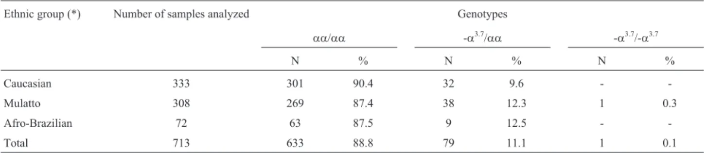
Eighty (11.2%) of the 713 individuals investigated presenteda+-thalassemia, 79 (11.1%) of which heterozy-gous (-a3.7/aa) and 1 (0.1%) homozygous (-a3.7/-a3.7) for the deletion. Genotype distribution among the studied par-ticipants remained within Hardy-Weinberg equilibrium (p = 0.365 with 1 degree of freedom), while -a3.7allele fre-quency was 0.06 (81 in 1430 chromosomes).
As to the prevalence ofa-thalassemia 3.7 kb deletion, according to ethnic group, among the 380 Afro-Brazilians and mulattoes, 47 (24.8%) were heterozygous fora+ -tha-lassemia (-a3.7/aa), in comparison to 32 (9.6%) among the 333 Caucasians (Table 1).
Comparison of hematological parameters between normal and heterozygousa-thalassemia 3.7 kb deletion in-dividuals showed a statistically significant difference (p < 0.05), as to the number of erythrocytes, mean corpus-cular volume (MCV), mean corpuscorpus-cular hemoglobin (MCH), and Hb A2for males and females, as well as
hemo-globin concentration in females (Table 2). Although indi-viduals heterozygous for deletion -a3.7 presented lower MCV, MCH, and Hb A2values than those with the normal
without microcytosis and/or hypochromia with anemia; and IV) those without any hematological alteration (Table 3). Significant relative risks toa-thalassemia were noted, on comparing individuals with microcytosis and/or hypochromia (groups I and II) to individuals without microcytosis and/or hypochromia (groups III and IV). As no significant risks were found on comparing group I to group II, neither group III to group IV (Table 3),a -thalas-semia can be considered as strongly associated with the presence of microcytosis and/or hypochromia. Serum ferritin levels determined for cases ofa-thalassemia were
all above the lower normal limits (10 ng/mL for women and 28 ng/mL for men). The exception was 4 individuals with microcytosis and/or hypochromia presenting lower levels of ferritin, a characteristic of iron deficiency associated with thalassemia.
Alpha+thalassemia with 3.7 kb deletion has been ob-served worldwide, with higher frequencies in some African and Mediterranean populations (Higgs and Weatherall, 2009). In studies, such as those by Pereset al. (1995) in Portugal, and Mouéléet al., (2000) in Africa, the -a3.7allele was detected in 7% and 59.7% of newborns, respectively. Table 2- Comparison of hematological parameters (mean±standard deviation) between individuals with normal genotype and those heterozygous for
deletion -a3.7.
Genotype Gender Hematological parameters
Hemoglobin (g/dL) Erythrocytes (x1012/L) MCV (fL) MCH (pg) Hb A2(%) Hb F (%)
M F M F M F
aa/aa(n = 623*) 265 358 14.6±1.1 12.8±1.0 5.05±0.41 4.46±0.36 86.4±4.8 28.8±1.9 2.8±0.5 0.4±0.3
-a3.7/aa(n = 77*) 38 39 14.4±1.0 12.3±0.8 5.46±0.50 4.79±0.33 80.5±4.5 26.18±1.7 2.6±0.6 0.5±0.3
p-value(**) p = 0.211 p = 0.003 p < 0.001 p < 0.001 p < 0.001 p < 0.001 p = 0.020 p = 0.750
Reference values†
15.0±2.0 13.5±1.5 5.00±0.5 4.30±0.5 92±0.6 29.5±2.5 2.4±1.4 < 1.0
M: male; F: female; MCV = mean corpuscular volume; MCH = mean corpuscular hemoglobin; Hb = hemoglobin. *10 samples with normal genotype and 2 witha+-thalassemia were excluded due to hemolysis and/or coagulation. ** p-value of the Student’s t-test for independent samples.
†Dacie and Lewis (1995).
Table 1- Prevalence ofa+-thalassemia 3.7 kb deletion among the 713 individuals, according to ethnic group.
Ethnic group (*) Number of samples analyzed Genotypes
aa/aa -a3.7/aa -a3.7/-a3.7
N % N % N %
Caucasian 333 301 90.4 32 9.6 -
-Mulatto 308 269 87.4 38 12.3 1 0.3
Afro-Brazilian 72 63 87.5 9 12.5 -
-Total 713 633 88.8 79 11.1 1 0.1
(*)Ethnicity was defined by self-declaration.
Table 3- Distribution of the 701 individuals§according to the presence of microcytosis, hypochromia and/or anemia.
Group N Genotype
aa/aa a3.7/aaor -a3.7/-a3.7
(I) With microcytosis and/or hypochromia with anemia 33 20 (60.6%) 13 (39.4%)
(II) With microcytosis and/or hypochromia without anemia 101 56 (55.4%) 45 (44.6%)
(III) Without microcytosis and/or hypochromia with anemia 49 48 (98.0%) 1 (2.0%)
(IV) Without hematological alterations 518 499 (96.3%) 19 (3.7%)
§10 samples with normal genotype and 2 witha+-thalassemia were excluded due to hemolysis and/or coagulation.
In the few studies undertaken in Brazilian populations, -a3.7 deletion was found to be the most common. Sonatiet al.
(1991) founda-thalassemia 3.7 kb deletion in 23.4% of Afro-Brazilian blood donors from Campinas, São Paulo State. In the Northeast,a-thalassemia 3.7 kb deletion was investigated by Adornoet al.(2005) in 514 newborns from Salvador, Bahia, whereupon it was observed that 114 (22.2%) presenteda3.7thalassemia, of which 101 (19.7%) were heterozygous and 13 (2.5%) homozygous. Borgeset al.(2001), on studying 339 adult outpatients with micro-cytosis and hypochromia without anemia at the Unicamp University Hospital, found 169 (49.9%) with a -thalas-semia, of which 145 (42.8%) were heterozygous (-a3.7/aa) and 18 (5.3%) homozygous (-a3.7/-a3.7). In a recent study in a north Brazilian population, the frequency of a -thalas-semia 3.7 kb deletion among 103 patients with microcytic hypochromic anemia was 19.4% (Souzaet al., 2009).
The present study detecteda-thalassemia 3.7 kb dele-tion in 11.2% (80/713) of an adult populadele-tion from the state of Rio Grande do Norte. Few studies have been dedicated to investigating a+-thalassemia in the general population. These usually involve defined population groups with a greater likelihood of encountering deletion, such as indi-viduals with microcytosis and hypochromia, or African de-scendants (Sonatiet al., 1991; Borgeset al., 2001; Souzaet al., 2009; Bezerra and Meissner, 2010).
Data from the Brazilian Institute of Geography and Statistics (IBGE) (2010), in which inclusion into a distinct ethnic group was based on self-declaration, showed that the population of Rio Grande do Norte was composed of 41.15% Caucasians, 52.48% mulattoes, 5.24% Afro-Bazilians, 1.04% yellow-skinned, 0.08% indigenous peo-ple, and 0.01 individuals with no declared color or race, to a total of 3,168,027 inhabitants.
According to ethnic distribution, prevalence of a+-thalassemia was greater among African descendants (24.8%), while among Caucasians this was 9.6%. Since in Brazil the degree of ethnic miscegenation is high, it appears that even in the non-African population, prevalence is also high.
African influence in the ethnic composition of the state of Rio Grande do Norte was not so high as that in other states, since fewer Africans were sent there as slaves during the colonization period, mainly due to the lack of adequate ports (Cascudo, 1984).
In spite of the different ancestral influence between the populations of Santarem (PA) and Rio Grande do Norte, the prevalence of heterogygotes (12.7%) was found to be similar to that of our study (11.1%) (Souzaet al., 2009), thereby revealing the elevated frequency and wide distribu-tion of -a3.7deletion in our environment.
In southern Brazil, Wagneret al., (2010) studied the prevalence ofa-thalassemia in various ethnic groups. On dividing the studied population into African and European
descendants, they observed greater prevalence in the for-mer (23.1%) than in the latter (4.5%), thus corroborating the strong correlation between thalassemia and ethnic group.
a-Thalassemia is characterized by slight micro-cytosis and hypochromia, reduced levels of hemoglobin, and an increased number of erythrocytes, when compared to parameters of normal individuals. These modifications are more marked in homozygousa-thalassemia individuals than in their heterozygous counterparts (Harteveld and Higgs, 2010).
As observed in studies by Adornoet al., (2005) and Souzaet al., (2009), there was a statistically significant dif-ference (p < 0.05) in the number of erythrocytes, MCV, MCH and HbA2ina-thalassemia carriers (-a3.7/aa),
com-pared to individuals with normala-genes (aa/aa). This was to be expected, since the unbalanced globin synthesis ina-thalassemia gives rise to deficiency in the amount of Hb (accounts for 30%-35% of the red cell content) per cell, thereby leading to the production of hypochromic and microcytic cells (Higgs, 2009). The lower level of HbA2
can be explained by the preferential formation of HbA in relation to HbA2. Under normal conditions of a-globin
chain sufficiency or slight excess, ab dimers assemble more readily thanaddimers. When supply of thea-globin chain is limited, as ina-thalassemia, there is a compatible increase in dimer formation, withbchains competing more effectively thandchains for scarcea-globin (Steinberg and Nagel, 2009).
The high prevalence of -a3.7deletion in individuals with microcytosis and/or hypochromia has already been observed (Borgeset al., 2001; Sankaret al., 2006; Rahim, 2009; Bezerra and Meissner, 2010; Wagneret al., 2010). In the present study, 44.6% of those with microcytosis and/or hypochromia without anemia had a+-thalassemia, thus confirming similar findings in a southeastern Brazilian population, in which 48.1% of individuals under the same conditions were homozygous or heterozygous for the -a3.7 deletion (Borges et al., 2001). Elevated percentages of a+-thalassemia in individuals with this disorder give em-phasis to the importance of -a3.7deletion as a possible cause of hematological alterations.
Notwithstanding, alterations in the hematological pa-rameters evident ina+heterozygotes, through being mini-mal, just below the lower limits, or sometimes even normini-mal, can hinder the detection of -a3.7 deletion carriers during molecular screening (Harteveld and Higgs, 2010). In the present study, and corroborating findings in the literature, and according to Souzaet al.(2009), the -a3.7deletion was detected in 3.7% of the individuals with hematological in-dices within reference limits.
im-portant to recognize the condition in order to elucidate the etiology of microcytosis and/or hypochromia, often con-founded with and treated as iron deficiency anemia.
Acknowledgments
This study was supported by the Conselho Nacional de Desenvolvimento Científico e Tecnológico (CNPq, grant n. 475855/2006-0), the Fundação de Apoio à Pes-quisa do Estado do Rio Grande do Norte (FAPERN/PPSUS III grant n. 011/2009) and the Fundação de Amparo à Pesquisa do Estado de São Paulo (FAPESP, grant n. 2008/57441-0).
References
Adorno EV, Couto FD, Moura Neto JP, Menezes JF, Rêgo M, Reis MG and Gonçalves MS (2005) Hemoglobinopathies in newborns from Salvador, Bahia, Northeast Brazil. Cad Saú-de Pública 21:292-298.
Bezerra CM and Meissner RV (2010) Diagnóstico molecular da talassemia alfa+(deleção -a3.7) em indivíduos com micro-citose e/ou hipocromia atendidos no Hemocentro Dalton Barbosa Cunha em Natal, Rio Grande do Norte. Rev Bras Hematol Hemoter 32:90-91 (Abstract in English).
Borg J, Georgitsi M, Aleporou-Marinou V, Kollia P and Patrinos GP (2009) Genetic recombination as a major cause of muta-genesis in the human globin gene clusters. Clin Biochem 42:1839-1850.
Borges E, Wenning MRSC, Kimura EM, Gervásio SA, Costa FF and Sonati MF (2001) High prevalence of alpha-thalassemia among individuals with microcytosis and hypochromia without anemia. Braz J Med Biol Res 34:759-762. Cascudo LC (1984) História do Rio Grande do Norte. 2 edition.
Fundação José Augusto, Natal, 524 pp.
Couto FD, Albuquerque ABL, Adorno EV, Moura Neto JP, Frei-tas AL, Oliveira JLB, Reis MG and Gonçalves MS (2003) Alpha-thalassemia-2, 3.7 kb deletion and hemoglobin AC heterozygosity in pregnancy: A molecular and hematologi-cal analysis. Clin Lab Haematol 25:29-34.
Dacie JV and Lewis SM (1995) Practical Haematology.Churchill Livingstone, Edinburgh, 608 pp.
Dodé C, Krishnamoorthy R, Lamb J and Rochette J (1992) Rapid analysis of -a3.7thalassaemia andaaaanti3.7triplication by enzymatic amplification analysis. Br J Haematol 83:105-111.
Harteveld LC and Higgs DR (2010)a-thalassaemia. Orphanet J Rare Dis 5:1-21.
Higgs DR (1993) a-Thalassaemia. Baillière’s Clin Haematol 6:117-150.
Higgs DR (2009) The pathopysiology and clinical features ofa
thalassemia. In: Steinberg MH, Forget BG, Higgs DR and Weatherall DJ (eds) Disorders of Hemoglobin. 2ndedition. Cambridge University Press, New York, pp 266-295. Higgs DR and Weatherall DJ (2009) The alpha thalassaemias.
Cell Mol Life Sci 66:1154-1162.
Mouélé R, Pambou O, Feingold J and Galactéros F (2000)a -tha-lassemia in Bantu population from Congo-Brazzaville: Its interaction with sickle cell anemia. Hum Hered 50:118-125. Peres MJ, Romão L, Carreiro H, Picanço I, Batalha L, Magalhães
HA, Martins MC and Lavinha J (1995) Molecular basis of
a-thalassemia in Portugal. Hemoglobin 19:343-352. Rahim F (2009) Microcytic hypochromic anemia patients with
thalassemia: Genotyping approach. J Med 63:101-108. Sankar VH, Arya V, Tewari D, Gupta UR and Pradhan M (2006)
Genotyping of alpha-thalassemia in microcytic hypo-chromic anemia patients from North India. Indian J Med Res 47:391-395.
Sonati MF, Farah SB, Ramalho AS and Costa FF (1991) High prevalence of alpha-thalassemia in a black population of Brazil. Hemoglobin 15:309-311.
Souza AES, Takanashi SYL, Cardoso G and Guerreiro JF (2009)
a-thalassemia (3.7 kb deletion) in a population from the Brazilian Amazon region: Santarém, Pará State. Genet Mol Res 8:477-481.
Steinberg MH and Nagel RL (2009) Hemoglobins of the embryo, fetus and adult. In: Steinberg MH, Forget BG, Higgs DR and Weatherall DJ (eds) Disorders of Hemoglobin. 2ndedition.
Cambridge University Press, New York, pp 119-135. Wagner SC, Castro SM, Gonzalez TP, Santin AP, Filippon L,
Zaleski CF, Azevedo LA, Amorin B, Callegari-Jacques SM and Hutz M (2010) Prevalence of commona-thalassemia determinants in south Brazil: Importance for the diagnosis of microcytic anemia. Genet Mol Biol 33:641-645. Weatherall DJ and Clegg JB (2001) Inherited haemoglobin
disor-ders: An increasing global health problem. Bull World Health Organ 79:704-712.
Internet Resources
Instituto Brasileiro de Geografia e Estatística (IBGE). Censo demográfico 2010. Resultados preliminares da amostra. http://www.sidra.ibge.gov.br (September 5, 2011).
Associate Editor: Mara Hutz