Tiago P. Carvalho
1,2, Roberto E. Reis
3and John P. Friel
4A new miniature species of banjo catfish of the genus Hoplomyzon is described from the Lake Maracaibo Basin in Venezuela. The new species is distinguished from all its congeners by the straight anterior margin of the mesethmoid (vs. a medial notch); a smooth and straight ventral surface of the premaxilla (vs. presence of bony knobs on the ventral surface of premaxilla); absence of teeth on dentary (vs. teeth present on dentary); configuration of ventral vertebral processes anterior
to anal fin, which are composed of single processes anterior to anal-fin pterygiophore (vs. paired process); presence of
several filamentous barbel-like structures on the ventral surface of head of adults (vs. small papillous structures in the
ventral surface of head of adults); and 8 anal-fin rays (vs. 6 or 7). An extensive osteological description is made of the
holotype using high-resolution x-ray computed microtomography (HRXCT).
Keywords: Endemism, Ernstichthys intonsus, Miniaturization, Synapomorphy, Taxonomy.
Se describe una nueva especie miniatura de pez banjo perteneciente al género Hoplomyzon, proveniente de tributarios del Lago Maracaibo en Venezuela. La nueva especie se diferencia de sus congéneres por presentar el margen anterior del mesetmoide recto (vs. con una hendidura central); superficie ventral de la premaxila lisa y recta (vs. superficie ventral de la premaxila con protuberancias óseas); ausencia de dientes en el dentario (vs. dientes presentes); configuración de los procesos anteriores ventrales de la aleta anal, los que están compuestos de procesos simples anteriores al pterigióforo de la aleta anal (vs. procesos dobles); presencia de varias estructuras en forma de barbillas en la superficie ventral de la cabeza (vs. pequeñas estructuras en forma de papilas en la superficie ventral de la cabeza); y 8 radios en la aleta anal (vs. 6 o 7).
Una extensiva descripción osteológica del holotipo fue realizada utilizando microtomografía computarizada de rayos X de alta resolución (HRCXCT).
Palabras Clave: Endemismo, Ernstichthys intonsus, Miniaturización, Sinapomorfia, Taxonomía.
1Laboratório de Ictiologia, Departamento de Zoologia, Universidade Federal do Rio Grande do Sul. Av. Bento Gonçalves, 9500,
91501-970 Porto Alegre, RS, Brazil. [email protected] (corresponding author)
2Department of Ichthyology, The Academy of Natural Sciences of Drexel University, 1900 Benjamin Franklin Parkway, 19103-1195
Philadelphia, PA, USA.
3Laboratório de Sistemática de Vertebrados, Faculdade de Biociências, PUCRS. Caixa Postal 1429, 90619-900 Porto Alegre, RS,
Brazil. [email protected]
4Alabama Museum of Natural History, The University of Alabama, Box 870340, 35487-0340 Tuscaloosa, AL, USA. [email protected] Introduction
Hoplomyzon Myers, 1942 is a member of the aspredinid
tribe Hoplomyzontini, a small and peculiar group of armored banjo catfishes of small body sizes and distinctive, specialized morphologies (Stewart, 1985). Hoplomyzontins,
composed of genera Hoplomyzon, Dupouyichthys Schultz, 1944, Ernstichthys, Fernández-Yépez, 1953 and Micromyzon Friel & Lundberg, 1996, are generally collected in the deep channels of medium to large sized rivers and as a consequence they are rare in museum collections
Stewart (1985), in a review of Hoplomyzontini, based on the relatively small number of specimens available at that time, diagnosed the genus Hoplomyzon by possessing
four or five stout, fleshy papillae on upper lip, 11 pre-anal-fin plates (corresponding two anterior unpaired elements, four sets of paired elements, and the anal-fin pterygiophore
exposed ventrally), short cleithral and scapulocoracoid
processes (10.1-12.5 and 8.9-10.8% SL, respectively), and short pectoral-fin spine (less than 25% SL). Friel (1994),
in a phylogenetic analysis of the family Aspredinidae, diagnosed Hoplomyzon from other aspredinid genera by having two bony knobs on ventral surface of premaxilla,
superficially covered by fleshy papillae, dorsal and ventral
plates not overlapping adjacent ones, two or three sets of
paired pre-anal-fin plates, a maxillary barbel adnate to head by membrane, and a short pectoral-fin spine (less than 25%
SL). Of these characters, only the presence of premaxillary knobs on ventral surface of the premaxilla was recovered as an unambiguous synapomorphy (Friel, 1994), other
diagnostic features ambiguous within Hoplomyzontini. Contrastingly, the tribe Hoplomyzontini is perhaps the most
well supported group of Aspredinidae with 27 unambiguous morphological synapomorphies (see Friel, 1994: 91)
reflecting its specialized and unique morphology.
The Maracaibo Basin has 127 species of freshwater
fishes and more than half of those are endemic (Albert et al., 2011). Here we describe a new species of Hoplomyzon endemic to the Maracaibo Basin, and present osteological
descriptions of this miniature fish based on high-resolution x-ray computed microtomography (HRXCT).
Material and Methods
Measurements were taken point to point with digital calipers. Relative measurements are expressed as percent of the standard length (SL), except subunits of head, expressed
as percent of the head length (HL). The measurements
follow those proposed by Friel (1995) and Cardoso (2010), except for cleithral process length, which was taken from the anterior margin of the cleithrum along its lateral portion to the posterior tip of the cleithral process. Vertebral counts
include all preural vertebrae, including the five vertebrae modified into the Weberian Apparatus, plus the PU1+U1
and U2 elements on the caudal skeleton counted as a single element according to Lundberg, Baskin, (1969) and de Pinna, Ng (2004). Comparative cleared and stained specimens (cs) were prepared according to the method described by Taylor, Van Dyke (1985). Osteological descriptions focused on aspects and characters used in previous phylogenetic studies of the family Aspredinidae (Friel, 1994; de Pinna, 1996; Cardoso, 2008). Anatomical terminology generally follows the Teleost Anatomical Ontology (TAO; Dahdul et al., 2010) an integral part of the Uberon Ontology covering anatomical structures in animals (Mungall et al., 2012; http://uberon.github.io/). The dorsal and ventral vertebral processes are, respectively, dorsolateral and ventrolateral
distally-flattened struts off the neural and hemal arches
that form the dorsal and ventral body armor on the body
surface (Taphorn, Marrero, 1990; fig. 4a; Friel, Lundberg,
1996; Carvalho et al., 2016). Institutional abbreviations follow Sabaj (2016). Photographs were taken of specimens
mounted in a photo-tank following the techniques described
by Sabaj Pérez (2009) with a Nikon D90 and a Nikon D7100 digital SLR. Results Hoplomyzon cardosoi urn:lsid:zoobank.org:act:8331CD18-B92C-4F13-A403-CB9718586E3F Figs. 1-11
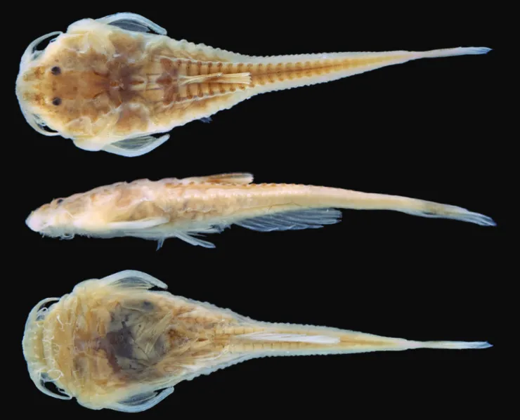
Holotype. MCNG 375, 18.5 mm SL, Venezuela, Zulia,
Colon, Caño La Raya tributary to Río Escalante, Hacienda San Jose, approx. 08°50′N 71°45′W, 8 Sep 1977, D. Taphorn
and others.
Paratypes. MCNG 11560, 1, 15.7 mm SL, Venezuela, Zulia, Río Ariquaisa [Aricuaisa] bridge via Machiches, approx.
09°24′N 72°36′W,14 Mar 1981, D. Taphorn. MCNG 26955,
1, 15.1 mm SL, Venezuela, Zulia, Río Santa Ana, approx.
09°23′N 72°36′W, 10 Dec 1982, F. Mago Leccia.
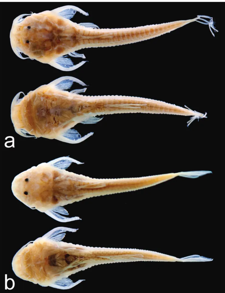
Diagnosis. Hoplomyzon cardosoi is distinguished from all congeners by the straight anterior margin of the mesethmoid (vs. with medial notch); a smooth and straight ventral surface of the premaxilla (vs. presence of bony humps on the ventral surface of premaxilla); absence of teeth on dentary (vs. teeth present on dentary); vertebrae anterior
to anal-fin pterygiophore with simple ventral processes
(vs. bifurcated processes); presence of several filamentous
barbel-like structures on the ventral surface of head (vs. small papillous structures on the ventral surface of head);
and anal-fin rays 8 (vs. 6 or 7). Additionally, Hoplomyzon cardosoi differs from remaining congeners by the following
morphometric features: greater cleithral width 29.8-33.5%
SL, mean 31.1 (vs. 23.5-28.7% SL); shorter scapulocoracoid
process 3.2-3.8% SL, mean: 3.4 (vs. 5.9-6.9% SL); longer
cleithral process 14.1-20.5% SL, mean 16.4 (vs. 9.6-13.4%
SL); narrower interorbital width 26.8-27.5% HL, mean 27.1
(vs. 28.6-36.4% HL); lesser distance between anterior nares
14.6-20.0% HL, mean 17.8 (vs. 25.0-28.6% HL); and lesser
distance between posterior nares 22.0-29.2% HL, mean:
25.4 (vs. 31.7-36.8% HL).
Description. Morphometric data summarized in Tab. 1.
Head and body depressed. Dorsal profile rising gently from snout tip to dorsal-fin origin with several small humps
in between, then descending gradually to end of caudal
margin of eye. Rostrum overhanging ventrally displaced mouth; gape small. Upper lip with pair of lateral main papillae and median unpaired one, additional small papillae displaced anterior to these ones.
Three main pairs of barbels, all simple. Maxillary barbel
short, almost reaching pectoral-fin origin. Three rictal
barbels within ventral portion of axillary region of maxillary barbel, longest at origin of maxillary barbel and two shorter
ones posteriorly displaced about half-length of longest one.
openings separated by distance approximating length of one aperture.
Skin roughened with small rounded unculiferous tubercles. Largest tubercles on dorsal portion of head and body, smaller tubercles on ventral surface of body. Small,
oblique, slit-like pore at pectoral-fin axilla. No apparent
sexual dimorphism. Largest specimen (holotype, 18.5 mm SL) female, with several large eggs visible in ventral and lateral portions of belly (Fig. 1).
Neurocranium. External surfaces of skull roof bones with pitted texture and somewhat interdigitating sutures between bones. Dorsal surface of neurocranium with pronounced bony knobs at posterior dorsal surface of mesethmoid, anterior portion of frontals at orbit margin,
frontal-sphenotic joint, and posterior tip of
parietal-supraoccipital. Mesethmoid long and deep; its middle portion constricted in dorsal view. Anterior margin of mesethmoid straight to slightly convex in dorsal view; no medial notch and weakly projected cornua laterally
(Figs. 3-4). Mesethmoid-frontal joint flat, without
dorsally projecting processes. Lateral ethmoid elevating dorsoposteriorly to contact frontal, not contributing to dorsal surface of neurocranium. Lateral process of lateral ethmoid extended laterally, articulating with mesial face of autopalatine at its midlength. Frontal moderately compact (length about twice its width), deeply constricted at its middle. Lateral margin of frontal forming concave orbital
margin. Frontal-parietal-supraoccipital joint somewhat interdigitating; frontal-sphenotic joint elevating from
main cranial surface. Anterior cranial fontanel ovoid, short and wide (its length about half frontal length), its anterior margin surrounded by mesethmoid, remainder by frontals. Posterior cranial fontanel similarly short and wide (slightly broader than anterior cranial fontanel),
diamond shaped, enclosed equally by frontal and
parietal-supraoccipital (Fig. 3a). Epiphyseal bar with broad median suture (suture length slightly shorter than length of either cranial fontanel).
Parietal-supraoccipital compact, its greatest length about
same as greatest width, somewhat round dorsal view with
deep anterior notch Anterior arms of parietal-supraoccipital
short, contacting frontal medially and sphenotic laterally.
Posterior process of parietal-supraoccipital short, broad
and gently rounded, direct contacting anterodorsal edge of
dorsal lamina of Weberian apparatus.
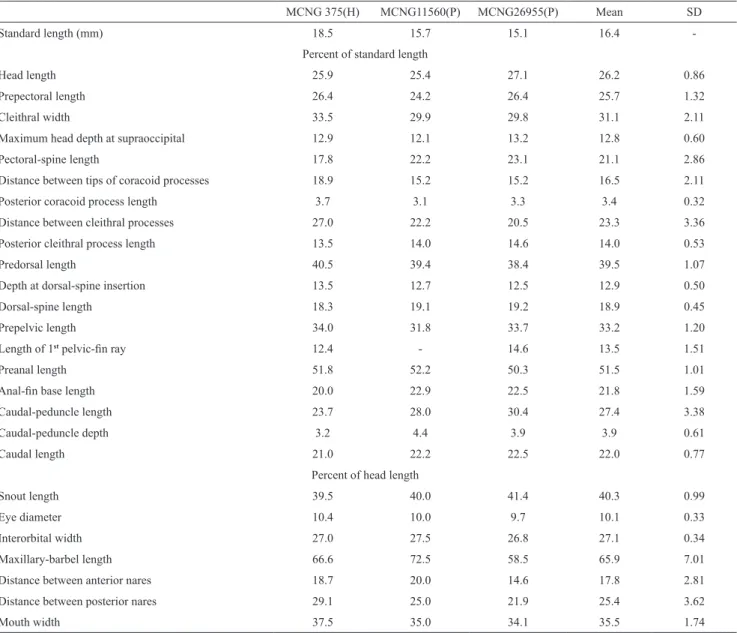
Tab. 1. Morphometric data of holotype (H) and two paratypes (P) of Hoplomyzon cardosoi. SD=standard deviation.
MCNG 375(H) MCNG11560(P) MCNG26955(P) Mean SD
Standard length (mm) 18.5 15.7 15.1 16.4
-Percent of standard length
Head length 25.9 25.4 27.1 26.2 0.86
Prepectoral length 26.4 24.2 26.4 25.7 1.32
Cleithral width 33.5 29.9 29.8 31.1 2.11
Maximum head depth at supraoccipital 12.9 12.1 13.2 12.8 0.60
Pectoral-spine length 17.8 22.2 23.1 21.1 2.86
Distance between tips of coracoid processes 18.9 15.2 15.2 16.5 2.11
Posterior coracoid process length 3.7 3.1 3.3 3.4 0.32
Distance between cleithral processes 27.0 22.2 20.5 23.3 3.36
Posterior cleithral process length 13.5 14.0 14.6 14.0 0.53
Predorsal length 40.5 39.4 38.4 39.5 1.07
Depth at dorsal-spine insertion 13.5 12.7 12.5 12.9 0.50
Dorsal-spine length 18.3 19.1 19.2 18.9 0.45
Prepelvic length 34.0 31.8 33.7 33.2 1.20
Length of 1st pelvic-fin ray 12.4 - 14.6 13.5 1.51
Preanal length 51.8 52.2 50.3 51.5 1.01
Anal-fin base length 20.0 22.9 22.5 21.8 1.59
Caudal-peduncle length 23.7 28.0 30.4 27.4 3.38
Caudal-peduncle depth 3.2 4.4 3.9 3.9 0.61
Caudal length 21.0 22.2 22.5 22.0 0.77
Percent of head length
Snout length 39.5 40.0 41.4 40.3 0.99
Eye diameter 10.4 10.0 9.7 10.1 0.33
Interorbital width 27.0 27.5 26.8 27.1 0.34
Maxillary-barbel length 66.6 72.5 58.5 65.9 7.01
Distance between anterior nares 18.7 20.0 14.6 17.8 2.81
Distance between posterior nares 29.1 25.0 21.9 25.4 3.62
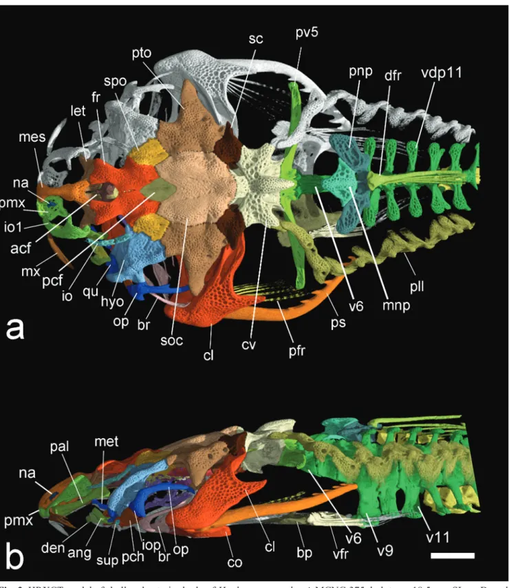
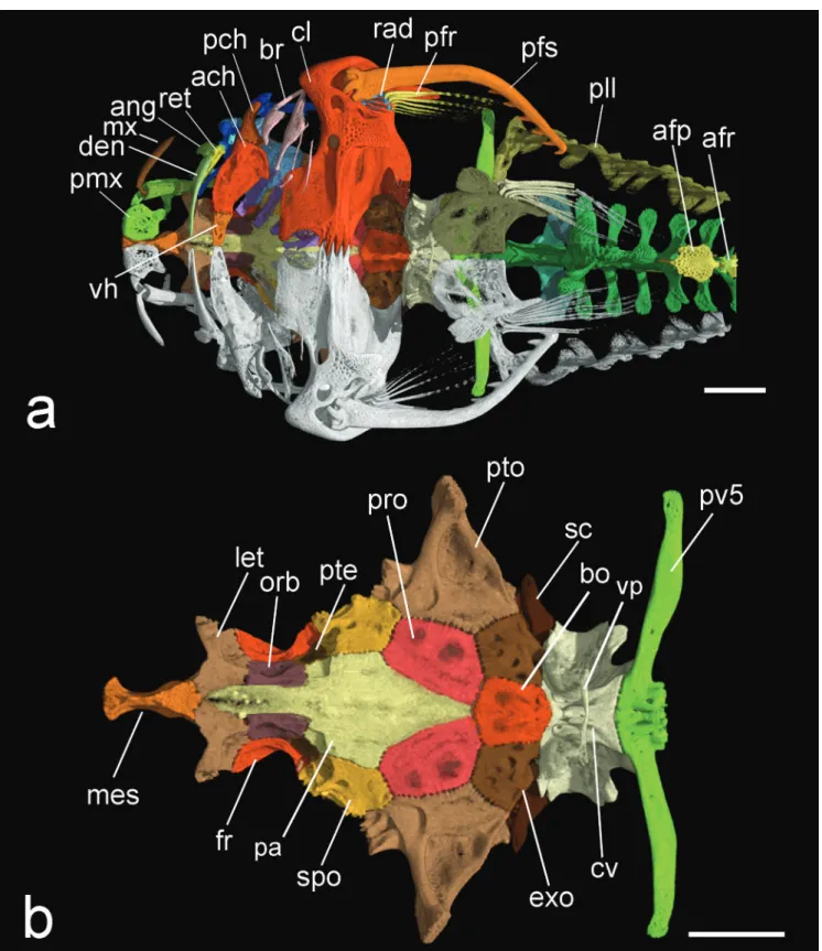
Fig. 3. HRXCT model of skull and anterior body of Hoplomyzon cardosoi, MCNG 375, holotype, 18.5 mm SL. a. Dorsal view. b. Lateral view of left side. acf: anterior cranial fontanel; ang: anguloarticular; bp: basipterygium; br: branchiostegal
rays; cl: cleithrum; cv: complex vertebrae; den: dentary; dfr: dorsal-fin rays; fr: frontal; hyo: hyomandibula; iop:
interopercle; let: lateral ethmoid; mes: mesethmoid; met: metapterygoid; mnp: middle nuchal plate; mx: maxilla; na: nasal;
pal: autopalatine; pcf: posterior cranial fontanel; pch: posterior ceratohyal; pfr: pectoral fin rays; pll: posterior lateral line plates; pmx: premaxilla; ps: pectoral-fin spine; pto: pterotic; qu: quadrate; sc: postemporo-supracleithrum; soc:
parietal-supraoccipital; spo: sphenotic; io1: infraorbitals; iop: interopercle; infraorbital 1; sup: subpreopercle; pv5: parapophysis of
vertebra 5; v6: vertebra 6; v9: vertebra 9; v11: vertebra 11; vdp11: vertebral dorsal process of vertebra 11; vfr: pelvic-fin
Fig. 4. HRXCT model of skull and anterior body of Hoplomyzon cardosoi, MCNG 375, holotype, 18.5 mm SL. a. Ventral view of skull and anterior body. b. Ventral view of neurocranium and first vertebrae. ang: anguloarticular; ach: anterior ceratohyal; afp: anal-fin pterygiophore; afr: anal-fin ray; bo: basioccipital; br: branchiostegal rays; cl: cleithrum; co: scapulocoracoid;
cv: complex vertebrae; den: dentary; exo: exoccipital; fr: frontal; let: lateral ethmoid; mes: mesethmoid; met: metapterygoid;
mx: maxilla; orb: orbitosphenoid; pa: parasphenoid; pch: posterior ceratohyal; pfr: pectoral-fin rays; pmx: premaxilla; po: preopercle; pro: prootic; ps: pectoral-fin spine; pte: pterosphenoid; pto: pterotic; pv5: parapophyses of vertebra 5; rad: pectoral-fin radial; ret: retroarticular; sc: postemporo-supracleithrum; soc: parietal-supraoccipital; spo: sphenotic; tr: tripus;
Sphenotic somewhat diamond shaped in dorsal view, its posterior margin contacting pterotic laterally; and extensively articulating with hyomandibula anterolaterally. Pterotic somewhat expanded anterolaterally, contacting sphenotic and hyomandibula; anterolateral margin with strongly concave margin. Pterotic wing greatly expanded laterally, tip acute. Supratemporal fossa closed; concavity at its typical position between pterotic and
parietal-supraoccipital. Extrascapula absent. Postemporo-supracleithrum plate-like with pitted surface, contributing to dorsal aspect of skull; contacting pterotic and
parietal-supraoccipital anteriorly and parapophysis of complex vertebrae posteriorly; bordering dorsal fenestra medially;
encompassed also by of the Weberian apparatus (posteriorly) and parietal-supraoccipital (anteriorly).
Postemporo-supracleithrum strongly sutured to surrounding bones, with laterally pointed process overlying dorsal process of cleithrum.
In ventral view of neurocranium (Fig. 4) vomer absent and parasphenoid relatively short, its anterior margin not surpassing lateral ethmoid; its posterior margin not surpassing prootic; broadest at about its middle portion (Fig. 4). Orbitosphenoid and pterosphenoid lateroventrally displaced; former four times larger than latter. Prootic somewhat hexagonal in shape, anteriorly contacting
sphenotic; having two unossified regions displaced laterally;
anterior foramen smaller than posterior one (likely optic and
trigemino-facial nerve foramina, respectively). Basioccipital
hexagonal in shape, contacting prootic and posterior tip
of parasphenoid anteriorly, and Weberian compound
vertebra posteriorly. Epiotic apparently absent or fused to exoccipital. Exoccipital not contributing to dorsal surface of neurocranium, and extensively contacting basioccipital.
Suspensorium and oral jaws. Premaxilla (Fig. 4a)
broad and plate-like with anterior and posterior margins
somewhat curved, its ventral surface rugose and not presenting bony knobs; teeth absent. Anterior margin of premaxilla with dorsally expanded process aside mesethmoid cornua; posterior margin with rounded process displaced laterally. Premaxilla displaced lateroventrally to ventral margin of mesethmoid, not contacting counterpart medially and articulating laterally with infraorbital 1 (Fig.
3a). Maxilla (Figs. 3-4) long and slender, distally bifurcate for about two-thirds its length with its ventral arm longer
than dorsal. Anterior portion of maxilla with two rounded articular processes, contacting anterior face of autopalatine. Dentary slender, not contacting counterpart medially; teeth absent (Fig. 5). Anguloarticular with dorsally expanded process. Anguloarticular strongly fused with retroarticular (boundaries tentatively reconstructed in Fig. 5); both forming socket for joint with quadrate. Coronomeckelian bone absent.
Hyomandibula with well-developed anterodorsal
process contacting ventral surface of contact between
frontal and sphenotic. Hyomandibula extensively contacting
quadrate ventrally by interdigitating suture; fused to
preopercle laterally (Fig. 5). Hyomandibula with elongate
posteroventrally directed process and condyle on posterior margin articulating with opercle. Preopercle strongly fused
to lateral margin of hyomandibula. Preopercle with
well-developed, laterally directed expansion, bearing canal, this exiting dorsally and passing through posterolateral portion of expanded hyomandibula before entering pterotic. Suprapreopercle absent. Quadrate subtriangular.
Subpreopercle ossified as tubular canal ventral to quadrate.
Entopterygoid absent or perhaps fused with anterior portion of metapterygoid. Metapterygoid elongate, constricted at midlength. Anterior margin of metapterygoid reaching lateral process of lateral ethmoid, posterior margin not contacting quadrate or hyomandibula posteriorly. Autopalatine elongate, posterior end rounded, laterally compressed and not bifurcate (Fig. 3b). Autopalatine articulating with lateral ethmoid at about its posterior third length.
Opercle (Fig. 5), boomerang shaped with posterior arm about two times longer than ventral arm. Interopercle
present, wedge shaped with dorsoposterior margin firmly
attached to anterior margin of ventral arm of opercle.
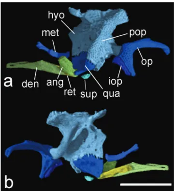
Fig. 5. HRXCT model of left side suspensorium and lower jaw of Hoplomyzon cardosoi, MCNG 375, holotype, 18.5 mm SL. a. Lateral view. b. Medial view. ang: anguloarticular; den: dentary; hyo: hyomandibula; iop: interopercle; met: metapterygoid; op: opercle; qu: quadrate; ret: retroarticular; sup: subpreopercle. Scale bar = 1 mm.
(Fig. 7); fifth ceratobranchial with one or two irregular rows
of acicular teeth on dorsal surface along mesial margin. Four
epibranchials, first and second slender and third and fourth
broader; third epibranchial not bearing typical uncinate
process (Fig. 7). Pharyngobranchials 1-2 and 4 absent.
Pharyngobranchial 3 slender; positioned anterolateral to expanded circular tooth plate (Fig. 7b). Dorsal tooth plate bearing elongate acicular teeth.
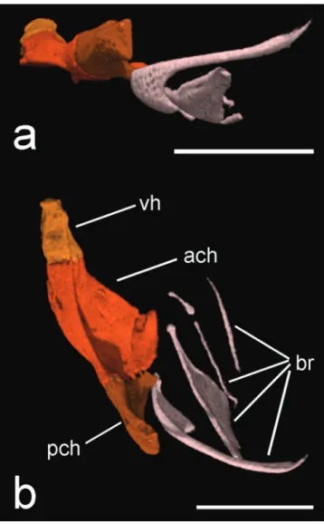
Fig. 6. HRXCT model of hyoid arch of Hoplomyzon cardosoi, MCNG 375, holotype, 18.5 mm SL. a. Left side in lateral view. b. dorsal view (anterior towards left). ach: anterior ceratohyal; br: branquiostegal rays; vh: ventral hypohyal. Scale bars = 1 mm.
Fig. 7. HRXCT model of left branchial arches of Hoplomyzon cardosoi, MCNG 375, holotype, 18.5 mm SL. a. Dorsal view (anterior toward top). b. Ventral view. cb1: ceratobranchial 1; eb1: epibranchial 1; pb3: pharyngobranchial 3; tp: tooth plate. Margin of epibranchials and pharyngobranchial 3 highlighted in white. Scale bar = 1 mm.
Remaining infraorbitals reduced to three small, disjointed
ossified tubules; infraorbital canal entering anterolateral
portion of sphenotic. Sphenotic receiving both infraorbital
and supraorbital canals anteriorly. Preopercle-mandibular
canal incomplete represented by single short tubular
ossification (subpreopercle) anterior to canal entering preopercle; mandibular portion of preopercle-mandibular canal absent. Posterior portion of preopercle-mandibular
canal associated with dorsal portion of hyomandibula and entering pterotic posteriorly. Postotic canal exiting from posterior portion of pterotic and entering anterior portion of
postemporo-supracleithrum. Pterotic branch and first branch
of main lateral line exiting from posteroventral portion of pterotic. Posterior lateral line associated with parapophyses
of complex vertebrae of Weberian apparatus, exiting
posterolateral portion of that bone and entering posterior
lateral-line canal tubules. Posterior lateral line complete
reaching hypural region posteriorly. Posterior lateral line
canal follow zig-zag course associated with intercalate dorsally and ventrally expanded plate-like structures (lateral plates). Lateral line pores in body, posterior to dorsal-fin origin, associated with elevated posteriorly oriented
hook-shaped structures of lateral plates (Fig. 3).
Axial skeleton. Dorsal crest of complex vertebrae of
Weberian apparatus reaching dorsal surface of body, its
dorsal margin strongly concave anteriorly and rising to elevated hump at its posterior tip (Fig. 3). Parapophysis of complex vertebrae short, its posterior end narrowed at contact with parapophyses of vertebra 5. Ventral portion
of complex vertebrae of Weberian apparatus with ventral process forming ring-like arch over aortic canal and
additional laterally directed arm; this arm with expanded thin bony blade surrounding gas bladder anteriorly (Fig. 4b). Parapophysis of vertebra 5 long, extending beyond lateral limits of parapophysis of vertebra 4 and reaching body wall. Parapophysis of vertebra 5 directed slightly posteriorly with distal margin not distinctly expanded. Anterior nuchal plate absent. Middle nuchal plate smaller than posterior one, somewhat triangular in shape (dorsal view) and contacting posterior nuchal plate posterolaterally and extensively. Middle nuchal plate distant from dorsal crest of complex vertebrae, ventrally contacting vertebrae 6 and 7. Posterior nuchal plate expanded anterolaterally forming elongate wing with rounded lateral end. Posterior nuchal plate lateral wing posteriorly contacting and overlapping dorsolateral process of vertebra 9 (Fig. 9). Vertebra 9 to
last (or penultimate vertebra ventrally) bearing dorso- and ventro-lateral projecting processes, these not overlapping
successive ones. Ventrally directed processes of vertebra 9 expanded laterally and bifurcated distally, processes from vertebrae 10 and 11 simple; from 12 and 13 displaced
laterally to anal-fin pterygiophore and bifurcated (Fig. 9). Dorsally and ventrally directed vertebral processes bifid next to dorsal and anal fins; fused medially after last vertebrae associated with pterygiophores of those fins. Two entrances
for hemal canal; on anteroventral portions of vertebrae 6
and 9. Hemal and neural spines short not elongate except
for hemal spines associated with last two vertebrae; hemal
spines associated with anal-fin pterygiophores slightly bifid distally. Consecutive vertebrae with interdigitating
articulations between their neural and hemal arches. Ribs
absent. Hypurals fused; ventral lobe longer than dorsal one, diastema greatly pronounced. Hypurapophysis elongate
and greatly expanded laterally to contact lateral plates. Fins and girdles. Dorsal-fin I,4. Dorsal-fin spinelet absent. First ray simple (not pungent or rigid), slender but
thicker than following branched fin rays. Last dorsal-fin ray adnate to dorsum by membrane. Adipose dorsal-fin absent. Anal fin iv,4; last ray not adnate. Caudal fin with 9
principal rays, 5 in dorsal lobe and 4 in ventral lobe; distal margin obliquely truncate, with lower lobe slightly
longer than upper lobe. Outermost principal caudal-fin rays unbranched and expanded proximally. Procurrent caudal-fin rays small; difficult to visualize in X-ray.
Pectoral fin I,6. First ray spine-like, not pungent; other fin rays branched. Pectoral-fin spine curved with strong deflection inwards at about its midlength (Fig. 8a); conspicuously longer than pectoral-fin branched rays. Anterior margin of pectoral-fin spine smooth; posterior
margin with 6 or 7 hooks of similar size except two proximal
most ones smaller. Three ossified proximal radials associated with pectoral-fin rays; first radial somewhat triangular in shape; positioned within pectoral-spine proximal cleft;
remaining two radials elongate and rod shaped. Dorsal portion of cleithrum with two processes: dorsal process long and narrow with lateral cleft; posterior process articulating
to ventral portions of postemporo-supracleithrum and parapophysis of complex vertebrae of Weberian apparatus;
posterior process somewhat elongate and triangular, pointed distally (Fig. 8). Cleithrum and scapulocoracoid partly fused, sutures almost indistinguishable. In ventral view, anterior margin of cleithrum transversely straight at and near midline, then deeply concave before prominent joint
with pectoral-fin spine (cleithral bulge). Symphysis between
cleithrae contacting anteriorly followed by large, circular
fenestra on midline, followed posteriorly by
heavily-interdigitating symphyseal suture of scapulocoracoids (Fig. 4a). Cleithrum bearing two small foramina of similar size on its ventral blade: anteromedial one positioned parallel to anterior concavity of cleithrum (Fig. 8: f1) and lateral one
positioned near articulation with pectoral-fin spine (Fig. 8:
f2). Scapulocoracoid bearing small foramina posterolaterally displaced; positioned lateral to scapulocoracoid posterior process (Fig. 8: f3). Scapulocoracoid bearing short and posteriorly directed process with somewhat squared distal margin.
Pelvic fin i,5; second ray longest, just reaching anal-fin origin. Lateral and medial anterior processes of
Fig. 8. HRXCT model of left pectoral skeleton of Hoplomyzon cardosoi, MCNG 375, holotype, 18.5 mm SL. a. ventral view (anterior towards left). b. medial view (anterior towards right). c. lateral view (anterior towards left). d. Dorsal view (anterior towards bottom).
1-3 pr: pectoral-fin proximal radials; cl/co: fused cleithrum-scapulocoracoid; f1: Cleithral arrector dorsalis foramen; f2: Cleithral arrector superficialis foramen; f3: Scapulocoracoidal abdutor superficialis foramen; pfr:
pectoral-fin rays; pfs: pectoral-fin spine. Scale bars = 1 mm.
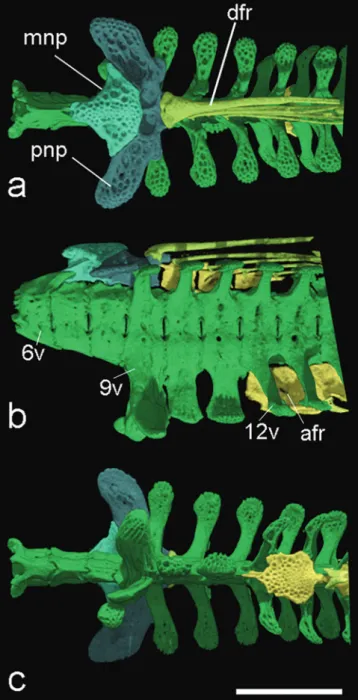
Fig. 9. HRXCT model of anterior vertebrae and associated dorsal and anal fin structures of Hoplomyzon cardosoi, MCNG 375, holotype, 18.5 mm SL. a. Dorsal view; b. lateral view, and c. ventral view (anterior portion towards left). 6v: vertebrae 6; 9v: vertebrae 9; 12v: vertebrae 12;
afr: anal-fin radials; dfr: dorsal-fin rays; mnp: middle
nuchal plate; pnp: posterior nuchal plate. Scale bar = 1 mm.
Color in alcohol. Head and body yellowish pale, with scattered brownish chromatophores dispersed mostly on dorsal portions. Dark chromatophores concentrated on internarial and interorbital regions and also on dorsal
portion over dorsolateral vertebral processes. All fins
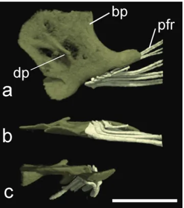
Fig. 10. HRXCT model of pelvic girdle of Hoplomyzon cardosoi, MCNG 375, holotype, 18.5 mm SL. a. Dorsal view; b. lateral view and c. frontal view. bp: basipterygium;
dp: dorsal process of basipterygium; pfr: pelvic-fin rays.
Scale bar = 1 mm.
Distribution and habitat. Known from three tributaries, which drain southwestern portions of Lake Maracaibo Basin in Zulia State, Venezuela (Fig. 12). The Caño raya at type locality is a medium size stream (~12m wide) with mostly
slow flowing white waters running over sand intercalated with riffles of fast flowing waters over pebbles; little marginal and floating vegetation.
Etymology. Hoplomyzon cardosoi is named in honor and memory of a dear colleague who prematurely passed away, Alexandre Rodrigues Cardoso, for his humbleness, positive attitude, and dedicated friendship, and furthermore for
his contributions to the taxonomy of Neotropical fishes,
including the family Aspredinidae.
Conservation status. Hoplomyzon cardosoi is only known from three localities in the southeastern tributaries of the Lake Maracaibo, with an Area of Occupancy (AOO) of 12 square kilometers, estimated considering a 2 km grid. The Maracaibo Lake has become heavily polluted in the past decades and many mangrove areas have been destroyed or
modified by coastal development and land conversion (Reis
et al., 2016). The three collecting events occurred from 1977 to 1982, and intense agriculture developed in the area since then, resulting in continuing decline in habitat quality. Based on the above evidence H. cardosoi can be categorized as Near Threatened (NT) by the IUCN criteria (IUCN, 2016).
Discussion
The new species shares most of the 27 synapomorphies
listed for Hoplomyzontini by Friel (1994). The exception
is character 24, which, in the derived condition, states that the metapterygoid contacts both the lateral ethmoid anteriorly and the quadrate posteriorly. In Hoplomyzon cardosoi the bony portion of the metapterygoid contacts the lateral ethmoid anteriorly but fails to contact quadrate or hyomandibula posteriorly (Fig. 5), as observed in other Hoplomyzon species. This condition should be taken with
caution because variation on the degree of ossification
of metapterygoid occurs within individuals of the same species, sometimes bilaterally in the same individual such as that presented by Bunocephalus minerim (Carvalho et al., 2015). Characters directly related to cartilage, such as the ascending process of Meckel’s cartilage discontinuous with the main portion of that cartilage (ch. 20) and the absence of cartilage on basipterygium anterior to the
pelvic-fin rays (ch. 100) were not observed.
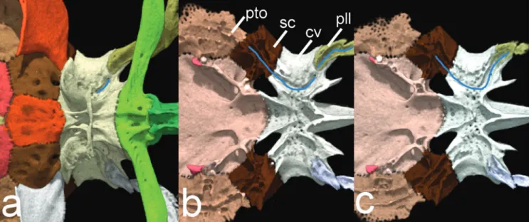
Additionally, a new synapomorphy for Hoplomyzontini
is proposed, and refers to the pathway of the posterior lateral line associated with the lateral portion of the complex
centrum of the Weberian apparatus (Fig. 11). After exiting the posterior portion of the postemporo-supracleithrum the
canal enters the parapophysis of the compound centrum,
then briefly exit ventrally (Fig. 11a), and finally re-enter
bone (Fig. 11b) before exiting posteriorly through an
imbricated, ossified lateral-line tubules (Fig. 11c). In other aspredinids the lateral line leaves the
postemporo-supracleithrum and lies lateral to the parapophyses of the
complex centrum of the Weberian apparatus, sometimes passing through laterally displaced ossified canal tubules
(Acanthocephalus: Friel, 1995: fig. 2; Pseudobunocephalus:
Friel, 2008: fig. 3; Bunocephalus: Carvalho et al., 2015:
fig. 3; Amaralia: Friel, Carvalho, 2016: fig. 4; Xyliphius: Carvalho et al., in press: fig. 9c). This unique condition was clearly observed and described with the aid of computed microtomography reconstructions, but also assessed and
evaluated within other Hoplomyzontini based on cleared
and stained specimens (see comparative material).
Hoplomyzon cardosoi shares, in part, with other putative congeners an apomorphic feature proposed by Friel (1994: ch. 13; Tab. 2.) in which the ventral surface of the premaxilla has two bony knobs presumably
associated with external fleshy papillae. The new species
has the papillae on the ventral surface of upper jaw, a condition similar to other Hoplomyzon species, but lack the bony knobs on the ventral surface of premaxilla (Fig.
4a). Therefore the external fleshy papillae and the bony
to head by membrane, and 4) pectoral-fin spine short (less than 25% SL). Hoplomyzon cardosoi shares with other species of Hoplomyzon vertebral processes not overlapping adjacent ones (Figs. 3, 4 and 9), contrasting with adjacent processes overlapping subsequent ones
in other species of Hoplomyzontini (Friel, 1994; Friel, Lundberg, 1996: fig. 4a). This feature has an ambiguous
optimization if Hoplomyzon is the sister group to remaining
Hoplomyzontini (Friel, 1994; Friel, Lundberg, 1996).
Outgroup aspredinids lack comparable vertebral processes.
Regarding the configuration of the ventral vertebral processes anterior to anal fin within Hoplomyzontini, the
new species differs from other Hoplomyzon (Tab. 2.), and
other Hoplomyzontini, by having a single paired process
associated with the vertebra 8 (n=1, MCNG 26955) or vertebrae 9 (n=2) and two single processes anterior to
anal-fin pterygiophore on vertebrae 10 and 11 (Figs. 9b-c). However, the new species shares with other Hoplomyzon
the relative posterior position of the anal-fin, in which
its pterygiophore is situated between the vertebrae
12 and 13, contrasting with an anal-fin pterygiophore
An additional putative synapomorphy for Hoplomyzon,
although not confirmed in H. atrizona, is the absence of an uncinate process on epibranchial 3 (Fig. 7). The third epibranchial bears an uncinate process in all aspredinids except the genus Pseudobunocephalus. Since both genera are not closely related (Friel, 1994), this is likely an independent loss.
An interesting apomorphic feature in Hoplomyzon cardosoi is the presence of elongated mental filaments on the ventral surface of head and cleithrum. A similar condition is found in Ernstichthys intonsus (Stewart, 1985: fig. 3), and to some degree small papillary structures are found in
the same region in other members of the Hoplomyzontini (Stewart, 1985: fig. 2b; Taphorn, Marrero, 1990: fig. 1d).
Such structures are shorter in these other hoplomyzontins representatives, but seem homologous to the additional
mental filaments that are hypertrophied in adults of H. cardosoi and presumably Ernstichthys intonsus. The
presence of these mental filaments in Aspredinidae is not
unique for hoplomyzontins, as they are also found in the genus Aspredinichthys (Mees, 1987: figs. 1-2).
Fig. 11. Sequential cut of complex vertebrae of Weberian apparatus and elements associated with posterior lateral line in ventral view of Hoplomyzon cardosoi, MCNG 375, holotype, 18.5 mm SL. a. Ventral view, b. cut at horizontal line at about dorsal limit of vertebra 5, and c. cut at horizontal line at about dorsal limit of second posterior lateral line tubule. Left side of posterior lateral line canal pathway highlighted in blue. cv: complex vertebrae; pll: posterior lateral line tubule; pto: pterotic;
Hoplomyzon cardosoi is small with a mature female measuring 18.5 mm SL, and can be categorized as miniature
(Weitzman, Vari, 1988). In Aspredinidae, miniaturization occurs not only in Hoplomyzontini (e.g., H. cardosoi, H. papillatus, Micromyzon akamai, and M. orinoco) but also in an unrelated species (e.g., Acanthobunocephalus nicoi), implying that extreme size reduction has occurred independently multiple times within the evolution of the family. Microtomography reconstruction proved to be an optimal tool for the detailed study of osteological features in
miniature fish that have well calcified skeletons, such as the
hoplomyzontins.
The high degree of endemism of fishes from the Maracaibo Basin is an aspect reflecting mostly the isolation
of this region since the rise of the Cordillera Merida in the late Miocene (Albert et al., 2006; Hardman, Lundberg, 2006). An interesting aspect of Lake Maracaibo Basin is the apparent allopatric distribution in this region of H. cardosoi and its congener H. atrizona, the former being found in southwestern and lower portions and the latter in
south-southeastern and upper portions of the basin. Lake
Maracaibo Basin, together with the Orinoco River basin, are the regions with the highest diversity of hoplomyzontins with three (H. atrizona, H. cardosoi, and Dupoyichthys sapito) and four species (H. sexpapilostoma, H. papillatus, Ernsticthys anduzei, and Micromyzon orinoco) respectively, representing the center of diversity of hoplomyzontins in South American drainages.
Tab. 2. Summary of characteristics variable within Hoplomyzon species based on examined material and literature (Stewart, 1985; Taphorn, Marrero 1990).
Character/species Hoplomyzon atrizona Hoplomyzon cardosoi Hoplomyzon papillatus Hoplomyzonsexpapilostoma
Shape of anterior margin of mesethmoid
Median furrow and undeveloped cornua
Straight and slightly developed cornua
Deep median furrow and developed cornua
Deep median furrow and developed cornua
Ventral surface of premaxilla With paired bony knobs Smooth With paired bony knobs With paired bony knobs
Premaxillary teeth Not examined Absent Present Present
Dentary teeth Present Absent Present Present
Upper lip symphyseal papilla Paired Unpaired Unpaired Paired
Rictal barbel Well developed Well developed Short Short
Dorsal-fin rays Seven Five Five Six
Anal-fin rays Six Eight Six Seven
Vertebrae and shape of ventral
pre-anal fin processes. 8-9 laterally expanded; 10-11 branched
9 laterally expanded
10-11 simple 8-9 laterally expanded; 10-11 branched 9-10 laterally expanded; 11 branched; Number of vertebrae 30-31 (n = 2) 30-32 (n = 3) 29-30 (n = 2) 29 (n = 1)
Posterior lateral line Somewhat straight on anterior
body Zig-zag on anterior body Zig-zag on anterior body
Somewhat straight on anterior body
Fig. 12. Distributions of species of Hoplomyzon based on examined museum specimens and literature records (Stewart, 1985; Taphorn, Marrero, 1990). Triangles: H. cardosoi; diamonds: H. atrizona; circles: H. papillatus and squares: H. sexpapilostoma.
47525, 5 paratypes. CUMV 78528, 1. CUMV 78542, 1. CUMV 78603, 1. CUMV 78629, 1. CUMV 78737, 1. CUMV 79746, 1.
CUMV 82559, 1. CUMV 90168, 8. FMNH 97762, 5 paratypes.
USNM 194160, 2.
Acknowledgments
We are grateful to the following people for loan
of specimens and hospitality while visiting museums: M. Sabaj, K. Luckenbill, and J. Lundberg (ANSP), C.
MacMahan, S. Mochel, and K. Swagel (FMNH), C.
Oliveira and F. Roxo (LBP), D. Taphorn and O. Castillo
(MCNG), C. Lucena and M. Lucena (MCP), S. Fisch-Müller and R. Covain (MHNG), M. de Pinna, A. Datovo
and O. Oyakawa (MZUSP); C. Dardia (CUMV), R.
Robins (UF); L. Malabarba and J. Wingert (UFRGS);
F. Carvajal (UMSS); R. Vari and S. Raredon (USNM).
We deeply acknowledge D. Taphorn for bringing
specimens of this new species to our attention and for
information on type locality. We thank J. Maisano and
M. Colbert (UT Austin) for preparation of computed
tomography x-rays and K. Luckenbill (ANSP) for help
in image editing with VGStudio Max software. This research was partially supported by Conselho Nacional
de Desenvolvimento Científico e Tecnológico (CNPq) (process number 229355/2013-7 to TPC, and processes number 305180/2010-0 and 207038/2013-9 to RER), and
Fundação de Amparo à Pesquisa do Estado do Rio Grande
do Sul - FAPERGS (process number
2361-2551/14-7 to RER). First author is currently supported with a
postdoctoral scholarship PNPD-CAPES. Thanks to A. Londoño-Burbano for translating the abstract to Spanish. We also thank John Lundberg for discussions on catfish
morphology and review of the manuscript previous to submission.
References
Albert JS, Lovejoy NR, Crampton WGR. Miocene tectonism and the separation of cis- and trans-Andean river drainages: Evidence from Neotropical fishes. J South Am Earth Sci. 2006; 21(1):14-27.
Albert JS, Petry P, Reis RE. Major biogeographic and phylogenetic
patterns. In: Albert JS, Reis RE, editors. Historical biogeography of Neotropical freshwater fishes. Berkeley: UCPress; 2011. p.21-57.
from the upper and middle rio São Francisco basins, Brazil.
Neotrop Ichthyol. 2015; 13(3):499-512.
Carvalho TP, Lundberg JG, Baskin JN, Friel JP, Reis RE. A new species of the blind and miniature genus Micromyzon Friel and Lundberg, 1996 (Silurifomes: Aspredinidae) from the Orinoco
river: describing catfish diversity using high-resolution
computed tomography. Proc. Acad. Nat. Sci. Philadelphia.
2016; 165(1):37-53.
Carvalho TP, Reis RE, Sabaj MH. Description of a new blind and
rare species of Xyliphius (Silurifomes: Aspredinidae) from the
Amazon basin using high-resolution computed tomography.
Copeia. Forthcoming 2017.
Dahdul WM, Lundberg JG, Midford PE, Balhoff JP, Lapp H, Vision TJ, Haendel MA, Westerfield M, Mabee PM. The teleost
anatomy ontology: anatomical representation for the genomics
age. Syst Biol. 2010; 59(4):369-83.
Friel JP. A phylogenetic study of the Neotropical banjo catfishes
(Teleostei: Siluriformes: Aspredinidae). [PhD Thesis]. Durham, NC: Duke University; 1994.
Friel JP. Acanthobunocephalus nicoi a new genus and species
of miniature banjo catfishes from the upper Orinoco and
Casiquiare rivers, Venezuela (Siluriformes: Aspredinidae).
Ichthyol Explor Freshw. 1995; 6(1):89-95.
Friel JP. Family Aspredinidae. In: Reis RE, Kullander SO, Ferraris
CJ, Jr., organizers. Check list of the freshwater fishes of South and Central America. Porto Alegre: Edipucrs; 2003. p.261-267.
Friel JP. Pseudobunocephalus, a new genus of banjo catfish with the description of a new species from Orinoco river system of Colombia and Venezuela (Siluriformes: Aspredinidae).
Neotrop Ichthyol. 2008; 6(3):293-300.
Friel JP, Carvalho TP. A new species of Amaralia Fowler
(Siluriformes: Aspredinidae) from the Paraná-Paraguay river basin. Zootaxa. 2016; 4088(4):531-46.
Friel JP, Lundberg JG. Micromyzon akamai, gen. et spec. nov., a small
and eyeless banjo catfish (Siluriformes: Aspredinidae) from the river channels of the lower Amazon basin. Copeia. 1996(3):641-48. Hardman M, Lundberg JG. Molecular phylogeny and a chronology of diversification for “phractocephaline” catfishes
(Siluriformes: Pimelodidae) based on mitochondrial DNA and nuclear recombination activating gene 2 sequences. Mol
Phylogenet Evol. 2006; 40(2):410-18.
Lundberg JG, Baskin JN. The caudal skeleton of the catfishes, Order Siluriformes. New York: American Museum of Natural History; 1969. (American Museum Novitates; No. 2398).
Mees GF. The members of the subfamily Aspredininae, family Aspredinidae in Surinam (Pisces, Nematognathi). Proc Kon
Ned Akad Wetensch, Ser C, Biol Med Sci. 1987; 90(2):173-92. Mungall CJ, Torniai C, Gkoutos GV, Lewis SE, Haendel MA.
Uberon, an integrative multi-species anatomy ontology.
Genome Biol. 2012; 13(1): R5[20p.].
de Pinna MCC. A phylogenetic analysis of the Asian catfish
families Sisoridae, Akysidae, and Amblypidae, with hypothesis on the relationships of the Neotropical Aspredinidae (Teleostei,
Ostariophysi). Chicago: Field Museum of Natural History;
1996. (Fieldiana, Zoology, New series; No. 84).
de Pinna MCC, Ng HH. The second ural centrum in Siluriformes
and its implication for the monophyly of superfamily Sisoroidea
(Teleostei, Ostariophysi). New York: American Museum of Natural History; 2004. (American Museum Novitates; No.
3437).
Reis RE, Albert JS, Di Dario F, Mincarone MM, Petry P, Rocha LA. Fish biodiversity and conservation in South America. J
Fish Biol. 2016; 89(1):12-47.
Sabaj MH. American Society of Ichthyologists and Herpetologists.
Standard symbolic codes for institutional resource collections in herpetology and ichthyology: an online reference. Version 6.5 [Internet]. 2016. [updated 2016 August 16] Available from:
http://www.asih.org/sites/default/files/documents/symbolic_ codes_for_collections_v6.5.pdf
Sabaj Pérez MH. Photographic atlas of fishes of Guiana Shield. Bull Biol SocWash. 2009; 17(1):53-9.
Stewart DJ. A review of the South American catfish tribe Hoplomyzontini (Pisces, Aspredinidae), with descriptions of
new species from Ecuador. Chicago: Field Museum of Natural
History; 1985. (Fieldiana, Zoology, New series; No. 25).
Taphorn DC, Marrero C. Hoplomyzon sexpapilostoma, a new
species of Venezuelan catfish (Pisces: Aspredinidae), with
comments on the Hoplomyzontini. Chicago: Field Museum of Natural History; 1990. (Fieldiana, Zoolology, New series; No. 61).
Taylor WR, Van Dyke GC. Revised procedures for staining and clearing small fishes and other vertebrates for bone and cartilage. Cybium. 1985; 9(2):107-19.
Weitzman SH, Vari RP. 1988. Miniaturization in South American freshwater fishes; overview and discussion. Proc Biol Soc Wash. 1988; 101(2):444-65.