Understanding postoperative cognitive dysfunction:
Novel insights
Dissertation presented to obtain the PhD degree in the
Faculdade de Ciências
Médicas, Universidade Nova de Lisboa.
The experimental work was conducted in the Department of Anesthesiology
and Perioperative Care, University of California San Francisco, USA under
the scientific supervision of Mervyn Maze, MB, ChB, FRCP, FRCA, FMedSci.
The internal co-supervisor at Faculdade de Ciências Médicas was Professor
Mª Emília Monteiro.
This work was financially supported by:
Table of contents
Table of contents 3
List of abbreviations 4
BACKGROUND 6
Surgery 8
Sleep 11
Cerebral Ischemia 13
Metabolic Syndrome 14
AIMS OF THE THESIS 15
METHODS AND RESULTS 16
Surgery 17
Depletion of Bone Marrow-derived Macrophages Perturbs the Innate Immune
Response to Surgery and Reduces Postoperative Memory Dysfunction 18
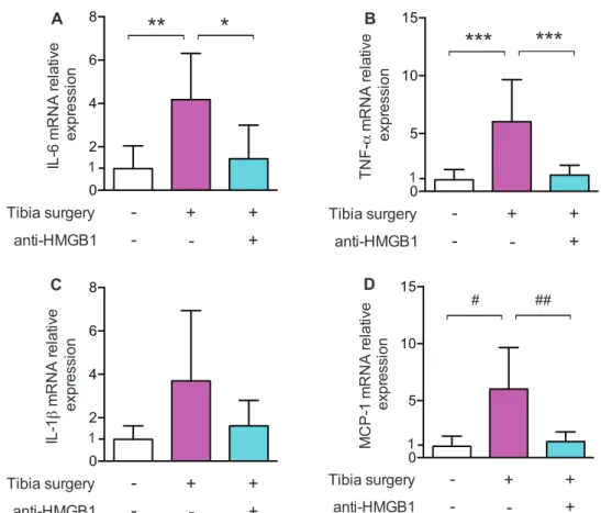
HMGB1 Initiates Postoperative Cognitive Decline by Engaging Bone
Marrow-derived Macrophages 29
The Neuroinflammatory Response of Postoperative Cognitive Decline 38
Sleep 57
Consequences of Sleep Fragmentation in the Perioperative Setting 58
Sleep and Anesthesia: Common Mechanisms of Action 70
Cerebral Ischemia 80
Bone Fracture Exacerbates Murine Ischemic Cerebral Injury 81
Metabolic Syndrome 93
Surgery Results in Exaggerated and Persistent Cognitive Decline in a Rat
Model of the Metabolic Syndrome 94
DISCUSSION AND CONCLUSION 103
TABLES 108
Table 1 – Risk Factors for Postoperative Delirium 108
Table 2 – Risk Factors for Postoperative Cognitive Dysfunction 109
REFERENCES 111
List of abbreviations
AD
Alzheimer’s disease
BBB
blood brain barrier
BZD
benzodiazepines
BM-DM
bone marrow-derived macrophage
CCR2
C-C chemokine receptor type 2
CSF
cerebral spinal fluid
DAMP
damage-associated molecular pattern
ELISA
enzyme-linked immunosorbent assay
HMGB1
high-molecular group box 1 protein
ICU
Intensive Care Unit
IKK
β
Ikappa B kinase
IL
interleukin
LPS
lipopolysaccharide
MCP-1
monocyte chemotactic protein-1
MetaS
metabolic syndrome
nAchR
nicotinic acetylcholine receptor
qPCR
quantitative polymerase chain reaction
NF-
κ
B
nuclear factor kappa B
NREM
non-rapid eye movement
PAMP
pathogen-associated molecular pattern
POCD
postoperative cognitive dysfunction
POD
postoperative delirium
PRR
pattern recognition receptors
REM
rapid eye movement
SF
sleep fragmentation
TLR
toll-like receptor
TNF
tumor necrosis factor
The scientific content of the present thesis has been included in the
publication of the following international scientific periodicals with
referees:
•
The neuroinflammatory response of postoperative cognitive decline,
Vacas S.
, Degos V., Feng X., Maze M., British Medical Bulletin,
2013;106:161-78.
http://www.ncbi.nlm.nih.gov/pubmed/23558082
•
Sleep and Anesthesia: Common Mechanisms of Action,
Vacas S
.,
Kurien P., Maze M., Sleep Medicine Clinic, Volume 8, Issue 1, March 2013
http://dx.doi.org/10.1016/j.jsmc.2012.11.009
•
Surgery Results in Exaggerated and Persistent Cognitive Decline in a
Rat Model of the Metabolic Syndrome, Feng X., Degos V., Koch L.G., Britton
S.L., Zhu Y.,
Vacas S.
, Terrando N., Nelson J., Su X., Maze M.,
Anesthesiology. 2013 May;118(5):1098-105.
http://www.ncbi.nlm.nih.gov/pubmed/23353794
•
Depletion of Bone Marrow-derived Macrophages Perturbs the Innate
Immune Response to Surgery and Reduces Postoperative Memory
Dysfunction, Degos V.,
Vacas S.
, Han Z., van Rooijen N., Gressens P., Su
H., Young W.L., Maze M., Anesthesiology. 2013 Mar;118(3):527-536.
http://www.ncbi.nlm.nih.gov/pubmed/23426204
•
Bone Fracture Exacerbates Murine Ischemic Cerebral Injury, Degos
V., Maze M.,
Vacas S.,
Hirsch J., Guo Y., Shen F., Jun K., van Rooijen N.,
Gressens P., Young W.L., Su H., Anesthesiology. 2013 Jun;118(6):1362-72.
http://www.ncbi.nlm.nih.gov/pubmed/23438676
•
HMGB1 Initiates Postoperative Coginitive Decline by Engaging Bone
Marrow-derived Machrophages,
Vacas S.
, Degos V., Tracey K.J., Maze M.,
Anesthesiology, October 2013
http://www.ncbi.nlm.nih.gov/pubmed/24162463
•
Consequences of Sleep Fragmentation in the Perioperative Setting,
Vacas S.
, Degos, V., Maze, M.,
submitted
BACKGROUND
Impairment of cognition after surgery is a disturbing reality. Postoperative
delirium (POD), listed in the
Diagnostic and Statistical Manual of Mental
Disorders
, DSM-5
2, is characterized by inattention, disorganized thinking and
altered level of consciousness with acute onset and fluctuating course. While
some patients develop POD, others develop a later onset form of
postoperative cognitive decline known as postoperative cognitive dysfunction
(POCD). POCD is diagnosed by the International Society of Postoperative
Cognitive Dysfunction as subtle deficits in one or more discrete domains of
cognition, which include attention, concentration, executive function, verbal
memory, visuospatial abstraction and psychomotor speed
3. The diagnosis
requires sensitive presurgical and postsurgical neurocognitive testing
4.
This
condition typically develops over weeks to months and is long-lasting. As a
consequence, patients can lose their livelihood or independence, which can
seriously reduce their quality of life
5-7.
It is estimated that POCD occurs in more than 10% of non-cardiac surgical
patients
8over 60 years old
9and is independently associated with poor
short-term and long-short-term outcomes including an increased risk of mortality
6,10.
Some reports describe cognitive decline persisting for up to one year after
surgery. Both because the number of major surgical interventions (requiring
anaesthesia) exceeds 230 million world-wide
11and because of the increasing
prevalence of surgical interventions in patients with comorbidities, if current
rates hold steady, we can expect that many millions of patients will run the
risk of developing POCD. This possibility raises the stakes considerably: not
only on an individual level, but also on a societal scale.
Although several risk factors have been identified
(table 1 and 2)
, the exact
pathophysiology that underlies POCD remains undefined. Data from
preclinical studies support the concept that inflammation is a possible
pathogenic mechanism for post-operative cognitive dysfunction
12-16. Increased
expression of interleukins in mice hippocampus following surgery was
associated with cognitive decline
12,17, corroborating the view that
induced neuroinflammation can result in cognitive impairment. Surgical
patients exhibit elevations of proinflammatory cytokines in both the central
nervous system and the systemic circulation, the extent of which may relate to
the degree of cognitive decline
18-20. Assuming that neuroinflammatory
changes noted postoperatively in rodents also occur in humans, reasons must
be sought why POCD is a relatively infrequent clinical event (± 10%)
5whereas neuroinflammation always occurs
12,19. Among the possibilities
include the fact that the neuroinflammatory changes are usually evanescent
and do not normally cause a long-lasting consequence in the animal
models
21. Several clinical conditions can transform the self-limiting
post-surgical neuroinflammatory response into one that is persistent
22-24. Causes
for the persistence in neuroinflammation may be due to dysfunction in the
inflammation
initiation
or
resolving
mechanisms.
Surgery
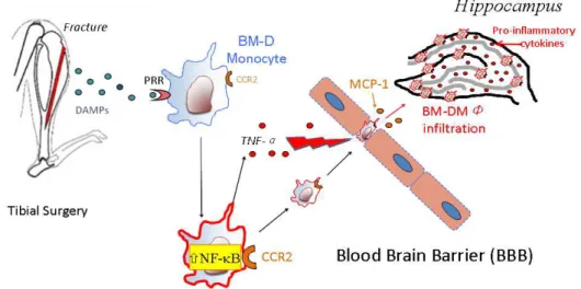
Tissue trauma, such as in surgical insult, releases damage-associated
molecular patterns (DAMPs) that are recognized by pattern recognition
receptors (PRR), which then trigger an immune response in a manner
remarkably similar to that of microbial-derived pathogen-associated molecular
patterns (PAMPs)
25-27. Among PRRs, Toll-like receptors (TLRs) are of critical
importance, recognizing various ligands (including PAMPs and DAMPs) and
activating TLR signals along different pathways, thereby increasing the
synthesis and release of pro-inflammatory mediators. Although the function of
TLR4 during lipopolysaccharide (LPS) endotoxemia
28has been deeply
explored, the pathways of infection-mediated neuroinflammation and cognitive
decline seem to be distinct from that of aseptic surgical trauma
14. One of the
most important DAMPs (released from dead or dying cells through
non-apoptotic processes
29) is high-mobility group box 1 protein (HMGB1). HMGB1
can bind and signal through a family of PRRs that are evolutionarily
conserved
30. Clinical conditions such as sepsis, arthritis, and stroke, all
release massive amounts of HMGB1
31. Both DAMPs and PAMPs converge
on NF-
κ
B to increase synthesis and release of pro-inflammatory cytokines
32,
including TNF-
α
, which disrupt blood brain barrier (BBB) integrity
12,14,15,33.
Early activation of the innate immunity through DAMPs (HMGB1 and
cytokines) will introduce the initial response to surgery resulting in
neuroinflammation and concomitant cognitive decline
(figure 1)
14.
Figure 1. Neuroinflammatory response to surgery1
Following injury, a
transient
inflammation is necessary for tissue repair
processes that promote healing. Neuroinflammation after surgery is likely to
include a pro-inflammatory phase and an anti-inflammatory phase (neural and
humoral pathways mediate the switch between these two phases)
34,35.
Regarding the
resolution
phase of the inflammatory state, this is mediated by
neural factors, termed the cholinergic anti-inflammatory pathway. Release of
acetylcholine mediates inhibition of macrophage NF-
κ
B activity by signalling
through the
α
7 subtype of nicotinic acetylcholine receptors (
α
7 nAChR).
Ultimately it inhibits synthesis and release of pro-inflammatory cytokines from
circulating immunocompetent cells
32,36,37. The neural cholinergic reflex is very
important in resolving the inflammatory pathogenesis of several diseases
including sepsis
38, rheumatoid arthritis
39, and colitis
40. Furthermore, the
cholinergic anti-inflammatory pathway also modulates the function of T
regulatory cells
41, which influences the production of anti-inflammatory
cytokines (IL-10 and IL-4)
42and alternative macrophage activation that
promotes the resolution of inflammation
43. Abnormalities of the switching
mechanism may cause a non-resolving chronic inflammatory state that could
create the circumstances for persistent cognitive decline.
Studies have shown the importance of this reflex for resolving DAMP-induced
neuroinflammation, pro-inflammatory cytokine release, neuroinflammation,
and cognitive decline; stimulating the
α
7 nAChR in macrophages, inhibited
NF-
κ
B activity while
in vivo,
α
7 nAChR agonists prevented postoperative
monocyte migration into the hippocampus as well as memory impairment
44.
Advanced age is associated with decline in cholinergic function
45, which may
be relevant in explaining the high prevalence of postoperative cognitive
dysfunction in elderly patients
44.
When inflammation does not subside, it can contribute to the pathogenesis of
diseases
46. Through a permeable BBB, CCR2-expressing bone
marrow-derived macrophages (BM-DM) are attracted, by the newly-expressed
chemokine, MCP-1, into the brain parenchyma. The macrophages synthesize
and release a variety of pro-inflammatory cytokines that interfere with
processes required for memory. Macrophage-specific Ikappa B kinase (IKK)
β
coordinates activation of NF-
κ
B; when it is deleted it prevents BBB disruption
and BM-DM infiltration into the hippocampus following surgery
44. Transgenic
mice that overexpress Hsp72 and inhibit NF-
κ
B activity have attenuation of
postoperative neuroinflammation and cognitive decline
21,47.
Learning and memory processes rely on the hippocampus, a region of the
brain that contains a large number of proinflammatory cytokine receptors
48,49.
The hippocampus has the highest density of 1 receptors, and although
IL-1
β
is required for normal learning and memory processes, higher levels can
also produce diminished cognitive function
50,51. Recent studies have
suggested a role for cytokines such as IL-1 as well as IL-6 in the genesis of
POCD
12,15,16. The relative prevalence of the TNF-
α
receptor, and other PRR,
on the endothelium of this brain region may account for its vulnerability to
systemic proinflammatory cytokines
52. Surgical trauma in animal models is
associated with the persistent activation of macrophages in the CNS
12, as well
as, high hippocampal levels of IL-1
β
, TNF-
α
and IL-6
12. These changes are
correlated with cognitive dysfunction seen in animal models (contextual fear
memory
12,14,16, spatial learning
27,53or reversal learning
26). Sub-clinical
inflammation following administration of LPS substantially increases IL1-
β
levels and cognitive deterioration after surgery
15. In addition, several studies
suggest that the marked and sustained expression of inflammation-related
enzymes, such as cyclooxygenase-2, plays an important role in secondary
events that amplify cerebral injury after ischemia
54. Patients also exhibit a
robust neuroinflammatory response to peripheral surgery with an initial rise in
pro-inflammatory cytokines in the CSF
19,20,55.
Sleep
Sleep is crucial for the repair of many types of injury and disease, especially
with regard to the central nervous and immune systems; it also has anabolic,
restorative properties that improve both neurocognitive and immune function.
During non rapid eye movement (NREM) sleep slow wave activity performs a
homeostatic function to reduce the strength of synapses that has been
acquired during wakeful activity
56. This synaptic homeostasis improves
subsequent cognitive function by allowing new changes in synaptic strength.
For example, both NREM and REM sleep are necessary for the consolidation
of learning and memory while sleep deprivation results in cognitive
dysfunction
57.
Sleep disturbance is commonly observed in the hospital setting and include
changes to sleep patterns and quality (especially sleep fragmentation), as well
as sleep architecture. Polysomnographic studies revealed extreme sleep
disruption in Intensive Care Unite (ICU) patients with decreases in total
sleep-time, altered sleep architecture (predominance of stage 1 and 2 sleep,
decreased or absent stage 3 NREM and REM sleep), and sleep
fragmentation
58,59; also, up to 50% of the total sleep-time occurred during
daytime. Studies have shown that fragmented sleep is prevalent due to
frequent arousals and awakenings, and that sleep architecture is altered with
an increase in light sleep, and a decrease in restorative slow wave sleep
60.
Environmental factors and health care practices further contribute to sleep
disruption in critically ill patients; these include disturbances like
inappropriately high noise levels, continuous ambient light, and the near
constant performance of medical tests procedure and procedures. Lack of
sleep hygiene results in cognitive dysfunction
61,62, contributes to delirium
63,
adversely affects immunity
64,65, and independently increases both morbidity
and mortality
66. Sleep disruption during hospital care has the potential to
adversely impact patients’ outcome and also provides a direct financial cost
with respect to the length of hospital stay and depletion of healthcare
resources.
Additionally, many sedative and analgesic agents potently suppress slow
wave sleep. Sedative practices have also shown to be a main causative factor
for this disruption
67,68. Anaesthetics have different action targets and
ultimately different consequences. The pivotal work of the MENDs trial
68-70indicated the benefits of a specific sedative agent, dexmedetomidine, in the
outcome of ICU population.
α
2 adrenergic agonists converge on sleep
pathways within the brainstem while those that act by modulating the
GABA
Areceptor converge at the level of the hypothalamus. Several studies
have now demonstrated the association between the use of benzodiazepine
(BZD) and increased incidence
69and duration
71of delirium in ICU patients. A
recent prospective study has also shown that patients with sleep disorders
have an increased likelihood of exhibiting postoperative delirium
24.
Despite the common occurrence of both ICU delirium and sleep disruption in
critically ill patients, a causal relationship has, as of yet, not been well
described. Still, the question remains if we are doing all in our power to avoid
the development of POCD.
Cerebral Ischemia
Stroke is the leading cause of disability in adults
72and an important risk
factor for bone fracture
73. In the United States,
≈
70,000 stroke victims suffer
from bone fracture within the first year after their stroke
74,75. A small proportion
of these stroke patients experience bone fracture within the first 24h after the
ischemic stroke, but the impact of bone fracture on acute stroke lesion is
unknown. Symptomatic pre-operative neurologic diseases, including dementia
and any disease of the central nervous system, are often considered as
exclusion criteria for POCD studies
5. Interestingly, cerebral vascular accidents
(without residual deficit) were associated with risk factors for POCD,
suggesting a pre-operative ischemic brain insult could influence the possibility
of POCD
5.
Metabolic Syndrome
Roughly 25% of the 45 million surgical patients in the US have MetaS
76, a
constellation of conditions whose precise definition and diagnostic criteria
continue to evolve
77. MetaS, comprising of insulin resistance, visceral obesity,
hypertension, and dyslipidemia increases the risk of postoperative
complications contributing to a significant higher mortality rate
78-81. While each
of the subphenotypes that define the MetaS have a strong genetic
component, lifestyle factors that contribute to this cluster of conditions include
sedentary behaviour and a diet with a high caloric content from saturated fats
and/or simple carbohydrates
82. Many complications of MetaS (including
atherosclerosis) are inflammatory in nature and the pathologic metabolism in
adipose stores may be the source of pro-inflammatory adipokines
83.
Conversely,
with little adiponectin to attenuate activation of the transcription
factor NF-
κ
B in macrophages, expression of genes for pro-inflammatory
cytokines are increased
84; up-regulated NF-
κ
B activity in morbid obesity can
be rectified with adiponectin
85. Recent evidence indicates patients suffering
from MetaS may be particularly susceptible to POCD
23,80.
AIMS OF THE THESIS
Studies have sought to identify factors that may contribute to POCD, which
include surgery, in-patient care factors, and patient-related factors. This work
considers the possible role of inflammation in the development of
postoperative cognitive dysfunction in the setting of underlying systemic
diseases/conditions.
The specific aims are:
1) To identify the role of hippocampal recruitment of BM-DM in the
pathogenesis of POCD.
2) To determine the mechanism by which HMGB1 regulates the activation
and trafficking of circulating BM-DM to the brain.
3) To investigate the contribution of perioperative SF to the neuroinflammatory
and cognitive responses of surgery.
4) To determine whether bone fracture, shortly after ischemic stroke,
enhances stroke-related injuries by augmenting the neuroinflammatory
response.
5) To investigate whether surgery induces a more severe and persistent form
of cognitive decline in a rat model of MetaS.
METHODS AND RESULTS
Surgery
Depletion of Bone Marrow-derived Macrophages Perturbs the Innate
Immune Response to Surgery and Reduces Postoperative Memory
Dysfunction
Participated in study design, conducted qPCR and ELISA experiments and participated in
data analysis and interpretation and final preparation of the manuscript.
"OFTUIFTJPMPHZ7t/P 527 .BSDI ABSTRACT
Background: According to rodent models of postoperative cognitive decline, activation of the innate immune response following aseptic surgical trauma results in the elaboration of hippocampal proinflammatory cytokines, which are capable of disrupting long-term potentiation, the neurobiologic cor-relate of memory. The authors hypothesize that hippocampal
recruitment of bone marrow–derived macrophages plays a causal role in these processes, resulting in memory dysfunction. Methods: Clodrolip injection (liposomal formulation of clodronate) before stabilized tibial fracture under general anesthesia was used to deplete bone marrow–derived mac-rophages. Systemic inflammation and neuroinflammation were studied on postoperative day 1, and memory in a fear-trace conditioning paradigm was assessed on postoperative day 3. CX3CR1GFP/+ CCR2RFP/+ mice were used to identify
bone marrow–derived macrophages.
Results: Clodrolip effectively depleted splenic CCR2+ bone marrow–derived macrophages. It also attenuated the surgery-induced increase of interleukin-6 in the serum and the hippocampus, and prevented hippocampal infiltration of CCR2+ cells without affecting the number of CX3CR1+ microglia. It did not alter the surgery-induced increase in hippocampal monocyte chemoattractant protein-1, the recruitment signal for CCR2+ cells. Clodrolip prevented surgery-induced memory dysfunction, as evidenced by a sig-nificant increase in freezing time (29% [95% CI, 21–38%] vs. 48% [95% CI, 38–58%], n = 20, P = 0.004), but did not affect memory in nonsurgical mice.
Conclusion: Depletion of bone marrow–derived mac-rophages prevents hippocampal neuroinflammation and
Depletion of Bone Marrow–derived Macrophages
Perturbs the Innate Immune Response to Surgery and
Reduces Postoperative Memory Dysfunction
Vincent Degos, M.D., Ph.D.,* Susana Vacas, M.D.,† Zhenying Han, M.D.,‡ Nico van Rooijen, Ph.D.,§ Pierre Gressens, M.D., Ph.D.,‖ Hua Su, M.D.,# William L. Young, M.D.,** Mervyn Maze, M.B., Ch.B.‡
* Associate Researcher, Center for Cerebrovascular Research, Department of Anesthesia and Perioperative Care, University of California, San Francisco, San Francisco, California, INSERM, U676, Hôpital Robert Debré, and Department of Anesthesiology and Criti-cal Care, Groupe Hospitalier Pitié-Salpêtrière, Assistance Publique-Hôpitaux de Paris, Université Pierre et Marie Curie, Paris, France. † Postdoctoral Fellow, Department of Anesthesia and Perioperative Care, University of California, San Francisco, San Francisco, Cali-fornia, and Program for Advanced Medical Education, Lisbon, Por-tugal. ‡ Assistant Researcher, Center for Cerebrovascular Research, Departments of Anesthesia and Perioperative Care, University of California, San Francisco, San Francisco, California. § Professor, Vrije Universiteit, VUMC, Department of Molecular Cell Biology, Faculty of Medicine, Amsterdam, The Netherlands. ‖ Professor, INSERM, U676, Hôpital Robert Debré, Paris, France. # Associate Professor, Center for Cerebrovascular Research, Departments of Anesthesia and Perioperative Care, University of California, San Francisco, San Francisco, California. ** Professor and Vice Chair, Center for Cere-brovascular Research, Departments of Anesthesia and Perioperative Care, Neurological Surgery, and Neurology, University of California, San Francisco, San Francisco, California. †† Professor and Chair, Department of Anesthesia and Perioperative Care, University of California, San Francisco, San Francisco, California.
Received from the Department of Anesthesia and Perioperative Care, University of California, San Francisco, San Francisco, Cali-fornia. Submitted for publication July 26, 2012. Accepted for pub-lication December 7, 2012. Supported by grant Nos. R01NS027713, P01NS044155, and R21NS070153 from the National Institutes of Health, Bethesda, Maryland; and AHA10GRNT3130004 from the American Heart Association, Dallas, Texas. Additional support was received from the University of California, San Francisco, San Francisco, California; INSERM, Paris, France; Société Française d’Anesthésie Réanimation, Paris, France; Fondation des Gueules Cassées, Paris, France; and Institut Servier, Paris, France. Dr. van Rooijen provided complimentary clodronate and control liposomes.
Address correspondence to Dr. Degos: Department of Anes-thesia and Perioperative Care, University of California, San Fran-cisco, 1001 Potrero Avenue, Box 1363, San FranFran-cisco, California 94110. [email protected]. Information on purchasing reprints may be found at www.anesthesiology.org or on the masthead page at the beginning of this issue. ANESTHESIOLOGY’S articles are made freely accessible to all readers, for personal use only, 6 months from the cover date of the issue.
Copyright © 2013, the American Society of Anesthesiologists, Inc. Lippincott Williams & Wilkins. Anesthesiology;118:527–
What We Already Know about This Topic
tAnimal models of postoperative cognitive dysfunction implicate an innate immune response, with increased circulating inflammatory mediators and migration of bone marrow–derived cells into the brain
tInhibitors of inflammatory mediators reduce postoperative memory deficits in mice but also reduce wound healing
What This Article Tells Us That Is New
tIn mice, treatment with a drug that depletes bone marrow– derived macrophages reduced circulating inflammatory mediators after surgical orthopedic injury, reduced migration of immune cells into the brain, and reduced postoperative memory deficits
◆ This article is accompanied by an Editorial View. Please see: Sanders RD, Avidan MS: Postoperative cognitive trajectories in adults: The role of inflammatory processes. ANESTHESIOLOGY
2013; 118:484–6.
"OFTUIFTJPMPHZ 528 Degos et al.
Macrophages and Postoperative Memory Dysfunction
memory dysfunction after experimental tibial fracture. These
data suggest that the hippocampal recruitment of bone marrow–derived macrophages is a necessary mechanism in murine postoperative cognitive dysfunction. Interventions designed to prevent its activation and/or migration into the brain may represent a feasible preemptive strategy.
A
CUTE postsurgical memory deterioration leads to per-sistent cognitive decline1 that can result in considerable morbidity and increased mortality.2 The specter of memory dysfunction, including acute delirium, postoperative decline, and dementia, is a source of anxiety for patients and their fam-ilies.3 Knowledge about the molecular and cellular pathways involved in postoperative memory dysfunction may provide a launching pad for the development of biomarkers to identify the most vulnerable patients as well as preventive strategies.Using a murine model of aseptic surgical trauma with a long-bone fracture, we previously demonstrated that postop-erative cognitive decline requires the engagement of the innate immune response. This engagement includes increased
sys-temic expression of alarmins and proinflammatory cytokines such as interleukin (IL)-6 in the blood4,5; increased ratio of CD11b+ cells corresponding to macrophages/microglia cells,4 and specifically the ratio of CCR2+ bone marrow-derived macrophages6; and elaboration of proinflammatory cytokines that are capable of disrupting hippocampal long-term poten-tiation, a neurobiologic correlate of learning and memory.3,7–10
Strategies designed to block the effect of
proinflamma-tory cytokines with IL-1 receptor antagonist (anakinra) or tumor necrosis factor (TNF)-α antibody (etanercept) pre-vented murine postoperative memory dysfunction.4,5These
interventions also prevented inflammation-dependent wound healing.11,12
In this study, we tested the hypothesis that mediation of postoperative memory decline requires recruitment of sys-temic bone marrow–derived macrophages into the brain, using a specific pharmacologic strategy to acutely deplete systemic phagocytes before an aseptic surgical trauma with an experimental tibial fracture.
Materials and Methods
Animals
All experimental procedures involving animals were approved by the University of California, San Francisco Institutional Animal Care and Use Committee, and conformed to National Institutes of Health guidelines. Twelve 8- to 12-week-old CCR 2RFP/+CX3CR1GFP/+ male mice6,13 (fig. 1A) were used to identify bone marrow–derived macrophages. CCR2 and CX3CR1 are acronyms for chemokine (C-C motif) receptor 2 (whose cognate ligand is monocyte chemoattractant protein [MCP]-1) that is highly expressed in bone marrow–derived macrophages, and CX3C chemokine receptor 1 (CX3CR1, fractalkine receptor) that is highly expressed in resident microglia. Eighty-nine
Fig. 1. Study design and splenic macrophage depletion with clodrolip. (A) First experiment with 12 CCR2RFP/+CX3CR1GFP/+ mice divided into two groups of six mice each treated with intraperitoneal (IP) injection of clodrolip versus CT-lip 1 h before the tibia fracture model and 25 h before tissue collection. (B) Second experiment with 24 C57BL/6J mice divided into four groups of six mice treated with IP injection of clodrolip versus CT-lip 1 h before the tibia fracture and sacrificed 12 or 24 h after the tibia fracture. (C) Third experiment with 70 C57BL/6J mice divided into four groups treated with IP injection of CT-lip versus clodrolip 1 h before the tibia fracture (20 mice per group) versus sham procedure (15 mice per group). The training session of the memory test was performed 30 min after the IP injection and 30 min before surgery, and the context session was performed 72 h after surgery. CT-lip = control liposome.
"OFTUIFTJPMPHZ 529 Degos et al.
PERIOPERATIVE MEDICINE
wild-type male mice (C57BL/6J, 10–12 weeks old) were purchased from The Jackson Laboratory (Bar Harbor, ME):
29 for the cytokine expression (fig. 1B) and 70 for the behavior tests (fig. 1C). Mice did not experience unexpected lethality in the study and were euthanized according to our institutional animal care and use committee guidelines.
In Vivo Systemic Phagocyte Depletion with Clodrolip
Clodrolip is a liposomal formulation of clodronate (dichlo-romethylene bisphosphonic acid), a nontoxic bisphospho-nate. Liposomes are lipid vesicles consisting of concentric phospholipid bilayers surrounding aqueous compartments. In this case, liposomes are used as “Trojan horses” encapsu-lating clodronate, which are then ingested and digested by phagocytes, followed by an intracellular release and accumu-lation of clodronate. At a certain intracellular concentration, clodronate induces apoptosis of the phagocytes. Clodro-nate liposomes were obtained from clodroClodro-nateliposomes. org** (Vrije Universiteit, Amsterdam, The Netherlands) at
a concentration of 7 mg/ml and prepared as described previ-ously.14,15 Clodrolip (200 μl, approximately 100 mg/kg) was injected intraperitoneally 60 min before the bone fracture. Control animals received 200 μl of control liposomal solu-tion (CT-lip). No intraperitoneal or extraperitoneal damage was observed after clodrolip intraperitoneal administration.
Long-bone Fracture with Tibia Fracture Surgery
Anesthesia was induced and maintained with isoflurane by inhalation. We used a dedicated chamber for induction with 5% isoflurane for 3 min, and the operation was performed under 2% isoflurane for 10–12 min. Under aseptic surgical conditions, an open tibial fracture of the right hind limb with intramedullary fixation was performed as described pre-viously.4 Body temperature was maintained at 37° ± 0.5°C using a thermal blanket throughout the surgical procedure, and analgesia was provided by injection of buprenorphine (0.3 mg in 100 μl of saline). Sham mice for bone fracture (sham group) received the same anesthesia and analgesia as the bone fracture mice.
Measurement of IL-6 in Serum
Mouse blood was collected using cardiac puncture under general anesthesia (isoflurane, 3%) in separate cohorts 12 and 24 h after the bone fracture procedure (fig. 1B). Blood samples were centrifuged at 1300 rpm for 10 min at room temperature, and the serum was collected and frozen at −80°C. IL-6 is secreted by bone marrow–derived macro-phages in response to alarmins,16 and the IL-6 level in the serum is increased within the first 24 h after the tibia frac-ture.4,5The IL-6 level in the serum is also associated with the postoperative memory dysfunction phenotype5 and is affected by clodrolip in response to lipopolysaccharide infu-sion.17 For these reasons, we decided to quantify IL-6 levels
in the serum of mice exposed to clodrolip or CT-lip using the IL-6 enzyme-linked immunosorbent assay kit (KMC0062; Invitrogen, Grand Island, NY). Results are expressed as fold increase compared with that measured in five control mice that did not receive any treatment or surgery.
Measurement of Cytokines in the Hippocampus
The hippocampi of the mice were collected rapidly under a dissecting microscope, 12 and 24 h after the tibia fracture (fig. 1B), and placed in RNAlater solution (Qiagen, Valencia, CA). To avoid blood contamination, mice were perfused with saline for 5 minutes before sample collection. Total RNA was extracted using the RNeasy Lipid tissue Kit (Qiagen) treated with recombinant DNase I using a RNase-Free Dnase set (Qiagen), and reverse-transcribed to complementary DNA with a High Capacity RNA to Complementary DNA Kit (Applied Biosystems, Bedford, MA). TaqMan Fast Advanced Master Mix (Applied Biosystems) and gene-specific primers and probes used for quantitative polymerase chain reaction are as follows: β-actin (NM_007393.1), IL-6 (Mm00446190_m1), TNF-α (Mm00443258_m1), IL-1β
(Mm01336189_m1), and MCP-1 (Mm00441242_m1). Quantitative polymerase chain reaction was performed using StepOnePlus (Applied Biosystems). Each RNA sample was run in triplicate, and relative gene expression was calculated using the comparative threshold cycle (∆CT) method and normalized to β-actin. Results are expressed as fold increase compared with that observed in five control mice that did not receive any treatment or surgery.
Quantification of Bone Marrow–derived Macrophages and Microglia
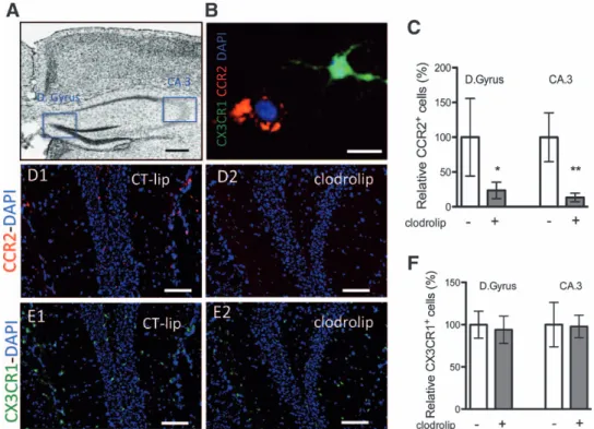
Twenty-four hours after the tibia fracture surgery, the brain and spleen of the CCR2RFP/+CX3CR1GFP/+ mice were collected
after intracardiac perfusion with paraformaldehyde 4% (fig. 1A). Spleen and brain (bregma, −1.0 to −1.4 mm, corre-sponding to interaural 2.7 to 2.3 mm in coronal orientation) were sectioned into 20-μm-thick slices and mounted with Vectashield DAPI (Vector Laboratories, Burlingame, CA). The expression of CCR2-RFP and CX3CR1-GFP cells was assessed using confocal images, performed with a Spectral Confocal microscope (Nikon Instruments, Melville, NY) using three laser lines (405, 488, and 561 nm). Z-stacks were rendered into a three-dimensional image using the NIS-Elements AR 3.0 software (Nikon), and the expression of CCR2-RFP and CX3CR1-GFP cells was quantified using ImageJ (National Institutes of Health, Bethesda, MD), with three different photographs per mouse taken with a 20× objective. Data are expressed as relative cell percentages nor-malized to the average value of the CT-lip group.
Behavioral Test for Hippocampus-dependent Memory with Trace Fear Conditioning
Fear conditioning is used to assess memory in rodents, which are trained to associate a conditional stimulus, such as a con-ditioning chamber, with an aversive, unconditional stimulus, ** http://www.clodronateliposomes.org/ashwindigital.
asp?docid=26. Accessed January 7, 2013.
"OFTUIFTJPMPHZ Degos et al.
Macrophages and Postoperative Memory Dysfunction
such as a foot shock. Freezing behavior is an indicator of aver-sive memory that is measured when subjects are reexposed to the conditional stimulus. With this model, lesions of the hip-pocampus disrupt recall of fear responses to the presentation of the context, resulting in a diminution in freezing.18,19
For this study, we used a previously published para-digm.4–6,20 Briefly, the behavioral study was conducted using a conditioning chamber (Med Associates, Inc., St. Albans, VT) and an unconditional stimulus (two periods of foot shock of 0.75 mA during 2 s). An infrared video camera, mounted in front of the chamber, captured motion speed (Video Freeze; Med Associates).
All of the animals underwent the same training session, regardless of the specific intervention, and received their training 30–40 min after the liposomal intraperitoneal injec-tions (whether clodrolipid or CT-lip) that occurred 30 min before surgery (fig. 1C). Three days after conditioning, mice were returned to the same chamber where training had occurred for a context test. During the context test, mice were exposed just to the context and no tones or foot shocks were delivered. Freezing was recognized by the software as a total lack of movement, excluding breathing and movement of vibrissae (linear detection with a minimal freeze duration of 20 frames corresponding to 0.7 s and a motion threshold of 20 arbitrary units).4–6,20 Decrease in the percentage of time spent freezing indicated impairment of memory.
Body Weight and Maximal Motion Speed
The body weight of the animals was measured 3 days after surgery, following assessment of freezing behavior. An infra-red video camera (Video Freeze) captuinfra-red and quantified motion speed during the context test, and the maximal motion speed was recorded for each mouse.
Statistical Analysis
Data are presented as mean ± 95% CI. Normality was tested with the d’Agostino–Pearson omnibus normality test. Equality of variances was tested with the F test. For two-sample comparisons, Student t tests were used (using the Welch correction if necessary); Mann–Whitney U tests were used if data were not normally distributed. For comparisons of more than two groups, means were compared using one-way ANOVA followed by Student t tests with a Bonferroni-corrected alpha level.
We used the two-way ANOVA procedure to determine whether or not time and treatment were significant factors in predicting IL-6 concentration in the serum, and IL-6, IL-1β, TNF-α, and MCP-1 messenger ribonucleic acid (mRNA) expression in the hippocampi. Given the highly skewed nature of the mRNA expression, we checked the distribution of the residuals. We applied a log transforma-tion (ln[X]) to the response of the mRNA expression before performing analysis to better adhere to the ANOVA model’s assumptions of normally distributed residuals and homosce-dasticity of residuals.
For the behavior tests, animals were tagged and allocated randomly to each group before any treatment, and research-ers were blinded to the group assignment that was revealed only after the analysis phase. A repeated measures ANOVA was performed to determine whether treatment (CT-lip and clodrolip) and the three time periods (baseline, first shock, and second shock) were significant predictors of percentage freezing time during the training session.
For this study, our primary outcome was percentage of freezing time during the context session. Based on previous freezing time data,4 we estimated that a sample of 18 C57BL/6J surgical mice per group was necessary to demonstrate a 20% increase in percentage freezing time, with 80% power at the 0.017 alpha level (after adjusting for three comparisons) to find a significant difference between clodrolip and CT-lip.
A two-tailed value of P < 0.05 was considered statistically significant for two-group comparisons, and the significance threshold was adjusted for multiple comparisons with a Bonferroni correction. Prism 5 (GraphPad Software, Inc., La Jolla, CA) was used to conduct the statistical analyses.
Results
Clodrolip Depletes Splenic Bone Marrow–derived Macrophages and Prevents Hippocampal Bone Marrow– derived Macrophage Infiltration
Using CCR2RFP/+CX3CR1GFP/+ mice (fig. 1A), in which
RFP+ bone marrow–derived macrophages and GFP+ resi-dent microglia can be tracked,6,13 we found that clodrolip depleted splenic macrophages and surgery-induced bone marrow–derived macrophage infiltration into the hippo-campus. The CCR2+ cells, which are mainly present in the splenic red pulp (fig. 2A), decreased by 96% in the clodro-lip-exposed mice (fig. 2B) (95% CI, 95–97%, P < 0.001). As shown in figure 3, the number of CCR2+ cells was also significantly reduced in the hippocampi of clodrolip-treated mice compared with CT-lip–treated mice 24 h after surgery (decrease of 76% for the dentate gyrus and 87% in the cornu ammonis 3). However, clodrolip treatment did not change the number of CX3CR1+ cells in the dentate gyrus and cornu ammonis 3 hippocampal regions (fig. 3).
Clodrolip Reduces Systemic and Hippocampal Proinflammatory Cytokines
We previously showed that proinflammatory cytokines in the blood and hippocampus increased within the first day after surgery.4 To test whether clodrolip treatment would reduce the proinflammatory cytokines, we studied serum and hip-pocampal expression 12 and 24 h after surgery (fig. 1B). Twelve hours after surgery, the rise in IL-6 in the serum was significantly attenuated in mice exposed to clodrolip (two-way ANOVA, P = 0.004 for the treatment, P = 0.003 for the time effect, and P = 0.19 for interaction) (fig. 4).
Between 12 and 24 h after surgery, the increase in mRNA hippocampal expression of IL-6, TNF-α, and IL-1 induced by
"OFTUIFTJPMPHZ Degos et al.
PERIOPERATIVE MEDICINE
Fig. 2. Effects of systemic macrophage depletion with clodrolip on the CCR2+ splenic cells. (A) Representative photographs of spleen section showing CCR2+ cell repartition mainly in the red pulp (RP) and less in the white pulp (WP), 24 h after tibia fracture. Top photographs are of low magnification (scale bar = 100 μm) and bottom photographs are highly magnified images (scale bar = 50 μm) in the CT-lip and the clodrolip mice. (B) Quantification of the relative percentage of CCR2+ cells in the spleen after clodrolip (n = 6, ***P < 0.001 with unpaired Student t test). CCR2 = chemokine (C-C motif) receptor 2; CT-lip = control liposome; DAPI = 4’,6’-diamidino-2-phenylindole; IP = intraperitoneal (bars = mean ± 95% CI).
Fig. 3. Effects of systemic macrophage depletion with clodrolip on the hippocampal CCR2+ and CX3CR1+ cells. (A) Representa-tive photographs of the section of interest corresponding to bregma, −1.2 mm (scale bar = 500 μm), showing the dentate gyrus (D.Gyrus) and the cornu ammonis subdivision 3 (CA.3). (B) Representative highly magnified photograph (scale bar = 20 μm) of a ramified CX3CR1+ green cell and an amoeboid CCR2+ red cell in the hippocampus. (C) Bar graph shows quanti
fication of the relative percentage of CCR2+ cells in the dentate gyrus and the CA.3 regions after clodrolip treatment (n = 6, signi
ficant F test for both comparisons, *P = 0.02; **P = 0.002 with unpaired Student t tests with Welch’s correction). (D) Representative photo-graphs of the dentate gyrus hippocampal sections in the CT-lip (D1) and the clodrolip (D2) mice (scale bar = 100 μm) showing the decrease of CCR2+ cells after clodrolip treatment. (E) Representative photographs of the dentate gyrus hippocampal section in the CT-lip (E1) and the clodrolip (E2) mice (scale bar = 100 μm) showing the absence of CX3CR1+ cell depletion after clodrolip treatment. (F) Bar graph shows quantification of the relative percentage of CCR2+ cells in the dentate gyrus and the CA.3 regions after clodrolip (n = 6, P = 0.51 for dentate gyrus and P = 0.51 for CA.3). CT-lip = control-liposome; CCR2 = chemokine (C-C motif) receptor 2; DAPI = 4’,6’-diamidino-2-phenylindole (bars = mean ± 95% CI).
"OFTUIFTJPMPHZ Degos et al.
Macrophages and Postoperative Memory Dysfunction
surgery returned to almost baseline values at 24 h (fig. 5). Clo-drolip exposure significantly inhibited the surgery-induced increased expression of mRNA IL-6 (two-way ANOVA, P < 0.001 for the treatment, P = 0.002 for the time effect, and P =
0.51 for interaction), and interacted with the time-dependent decrease for TNF-α (two-way ANOVA, P = 0.03 for inter-action). Clodrolip treatment did not change IL-1β mRNA expression (two-way ANOVA, P = 0.42 for the treatment, P < 0.001 for the time effect, and P = 0.66 for interaction) (fig. 5).
Systemic macrophages are recruited into tissues by the chemoattractant MCP-1 that binds to CCR2, which is expressed on the surface of bone marrow–derived macro-phages.21,22 Following surgery, MCP-1 mRNA expression increases and is unaffected by prior exposure to clodrolip
(two-way ANOVA, P = 0.64 for the treatment, P < 0.001 for the time effect, and P = 0.67 for interaction) (fig. 5D).
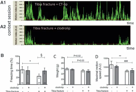
Clodrolip Prevents Surgery-induced Memory Impairment
During the preoperative training period, learning was simi-lar in the clodrolip-exposed and the control (nonexposed) groups, with the percentage of freezing being highly associ-ated with time (fig. 6). During the context session, surgery significantly decreased percentage of freezing time in compar-ison with the sham group (52% [95% CI, 41–63%] vs. 29% [95% CI, 21 to 37%], P = 0.0012); preoperative exposure to clodrolip resulted in significantly greater freezing time than in the nonexposed surgical cohort (29% [95% CI, 21–38%] vs. 48% [95% CI: 38–58%], P = 0.004), reaching a level simi-lar to that observed in the sham-operated clodrolip-exposed mice (49% [95% CI, 36–63%], P = 0.86) (fig. 7, A and B).
Clodrolip did not affect the body weight of the mice 3
days after the injection (fig. 7C). As for maximal motion speed, the clodrolip-treated groups were no different from
the CT-lip groups, even though the maximal motion speed of the surgical groups was significantly slower than the sham groups (fig. 7D).
Discussion
In this study, we report for the first time that bone marrow– derived macrophages are required in the pathogenesis of the neuroinflammatory and memory dysfunction induced by surgery. Also, we report that a possible hippocampal signal through MCP-1 is involved in the recruitment of bone marrow–derived macrophages to this brain region. Data from rodent surgical models have provided insight into the neuroinflammatory basis for postoperative cognitive decline. This usually transient process appears to be part of a
motivational system that reorganizes the organism’s priorities to facilitate recovery. To date, we have established a pivotal early role for the proinflammatory cytokine TNF-α,6 and our study demonstrated that hippocampal infiltration of bone marrow–derived macrophages also plays a role in the initiation of neuroinflammation.
Hippocampal Infiltration of Bone Marrow–derived Macrophages after Surgery
Monocyte infiltration into the brain is mainly described in acute brain injuries such as stroke23 and traumatic brain inju-ries,24 as well as chronic inflammatory brain injuries such as multiple sclerosis.13 Using long-bone fracture as a surrogate for a peripheral orthopedic surgical insult, we previously reported that CCR2+ cells were present in the hippocam-pus.6 Because microglia can also express CCR2 under certain conditions,25,26 we could not ascertain whether these CCR2-expressing cells arose from the resident macrophage popula-tion (microglia) or through an infiltrapopula-tion from outside of the central nervous system. Using clodrolip to specifically deplete the systemic pool of phagocytes, including bone marrow– derived macrophages, we were able to demonstrate that the CCR2+ cells in the hippocampus are a result of the recruit-ment of bone marrow–derived macrophages into the brain.
For passage into the brain, monocytes are required to overcome the blood–brain and/or blood–cerebral spinal fluid barrier27,28; these barriers can be disrupted by direct acute brain injury.29,30 Interestingly, after peripheral surgery, the blood–brain barrier is disrupted, although there is no discernible brain lesion.6 Now we show that after surgery, the hippocampus expresses MCP-1, which is capable of attracting CCR2+-expressing cells migrating through the disrupted blood–brain barrier. This increased expression of MCP-1
is unaffected by clodrolip treatment, indicating that bone
marrow–derived macrophages are not a self-perpetuating source of this chemoattractant for its own recruitment. Future understanding of the source and the triggers for hippocampal MCP-1 following peripheral surgery may result Fig. 4. Effects of systemic macrophage depletion with
clodro-lip on the IL-6 serum concentration after the tibia fracture 12 and 24 h after tibia fracture in the clodrolip and CT-lip groups. Data are normalized to the average value of the control mice group that did not receive any treatment or any surgery and expressed as fold time increase (n = 6 per group and per tim-ing, two-way ANOVA, P = 0.004 for the treatment effect, P = 0.003 for the time effect, and P = 0.19 for interaction). CCR2 = chemokine (C-C motif) receptor 2; CT-lip = control liposome; DAPI = 4’,6’-diamidino-2-phenylindole; IL = interleukin-6; IP = intraperitoneal (boxes and circles = mean ± 95% CI).
"OFTUIFTJPMPHZ Degos et al.
PERIOPERATIVE MEDICINE
in interventional strategies designed to prevent recruitment of bone marrow–derived macrophages into the brain.
Hippocampal Bone Marrow–derived Macrophage Infiltration and Memory Dysfunction
Our recent data suggest that transient hippocampal inflam-mation is the key element in postoperative memory dys-function because (1) hippocampal areas are known to be involved in memory tasks7; (2) hippocampal neuroin-flammation profile correlates with the level of memory dysfunction4,5; and (3) hippocampal neuroinflammation leads to long-term potentiation disruption.31,32 Now we report here that the absence of bone marrow–derived mac-rophage infiltration, produced by systemic depletion by clodrolip, decreases surgery-induced hippocampal inflam-mation and memory dysfunction. Therefore, postoperative bone marrow–derived macrophage recruitment into the
hippocampus plays a key role in the initiation of postopera-tive memory dysfunction.
In the context of postoperative cognitive decline, determining which cells are involved in the initiation of the inflammation response is important because, when exaggerated, this could overwhelm resolving responses and produce persistent postoperative cognitive decline. Earlier, we described that surgical trauma induces systemic release of alarmins (i.e., high-mobility group protein 1) and proinflammatory cytokines (i.e., TNF-α and IL-6).4,5 Improving our knowledge of cellular and molecular initiation mechanisms will allow insight into an ex vivo bioassay to prospectively determine whether patients are at risk.
Limitations of the Study
We used experimental tibial fracture to generate animal postoperative memory acute dysfunction. With this model, we traumatized the bone marrow directly, which could play Fig. 5. Clodrolip effect and early kinetic of the mRNA hippocampal expression of IL-1β, TNF-α, IL-6, and MCP-1 after tibia fracture. (A) Hippocampal mRNA expression of IL-6 relatively expressed as fold increase compared with control brain expression 12 and 24 h after tibia fracture in the CT-lip and the clodrolip-treated mice (n = 6 per group and per timing, two-way ANOVA, P = 0.0003 for the treatment effect, P = 0.0023 for the time effect, and P = 0.51 for interaction). (B) Hippocampal mRNA expression of TNF-α relatively expressed as fold increase compared with control brain expression 12 and 24 h after tibia fracture in the CT-lip and the clodrolip-treated mice (n = 6 per group and per timing, two-way ANOVA, P = 0.06 for the treatment, P < 0.0001 for the time effect, and P = 0.035 for interaction). (C) Hippocampal mRNA expression of IL-1β relatively expressed as fold increase compared with control brain expression 12 and 24 h after tibia fracture in the CT-lip and the clodrolip-treated mice (n = 6 per group and per timing, two-way ANOVA, P = 0.42 for the treatment effect, P < 0.0001 for the time effect, and P = 0.66 for interaction). (D) Hippocampal mRNA expression of MCP-1 relatively expressed as fold increase compared with control brain expression 12 and 24 h after tibia fracture in the CT-lip and the clodrolip-treated mice (two-way ANOVA, P = 0.64 for the treatment, P = 0.0005 for the time effect, and P = 0.67 for interaction). CT-lip = control liposome; IL = interleukin; MCP-1 = monocyte chemoattractant protein-1; mRNA = messenger ribonucleic acid; TNF = tumor necrosis factor (boxes = mean ± 95% CI and data are presented with a logarithmic scale).
"OFTUIFTJPMPHZ Degos et al.
Macrophages and Postoperative Memory Dysfunction
a key role. However, other models that did not damage bone marrow with a splenectomy33 also showed that surgery gen-erated postoperative cognitive dysfunction.
Fibrin is deposited in the hippocampus after tibia frac-ture,6 suggesting that the blood–brain barrier becomes dis-rupted and may allow the passage of clodrolip to act directly on the microglia population.34 However, we found that the systemic administration of clodrolip acts only on the num-ber of CCR2+ cells without significantly affecting the num-ber of CX3CR1+ cells (fig. 3); if clodrolip has an effect on
microglia, it may be to functionally modify them. For this reason, we cannot exclude the possibility that clodrolip does not affect the function of microglia, and that microglia do
not play a key role in postoperative cognitive dysfunction. For this study, we used a pharmacologic strategy to quickly deplete the pool of systemic macrophages. However, because clodrolip is highly toxic for monocytes and macrophages,35 it can increase the risk of postsurgical infections, generating a phenotype of its own. With a single dose, we did not observe loss of weight or other signs of sickness within the Fig. 6. Clodrolip effect on the training session. (A) Representative record of a training session showing the motion (motion index expressed in arbitrary units) of the mouse according to the time. The two green bars represent the two shocks and the three
red rectangles represent the 40-s periods used to quantify the baseline, first, and second shock freezing responses. Bar graph (B) quantifies the percentage of freezing time (n = 35), and two-way ANOVA shows a significant effect of time (P < 0.0001), no significant effect of treatment (P = 0.68), and no interaction (P = 0.61).
Fig. 7. Depletion of systemic macrophages reduces surgery-induced memory dysfunction. (A) Representative records of con-text sessions of tibia fracture mice treated with CT-lip (A1) and with clodrolip (A2) showing the motion (motion index expressed in arbitrary units) of the mice according to the time. (B) Quantification of the freezing time percentage according to the four groups (n = 15–20, *P = 0.0012 and §P = 0.004, respectively, with one-way ANOVA and Bonferroni post hoc analysis). (C) Quantifi ca-tion of the body weight in the four groups 3 days after surgery (n = 15–20, P = 0.02 and P = 0.03, respectively, not significant after adjustment for multiple comparisons with one-way ANOVA and Bonferroni post hoc analysis and no significant effect of the clodrolip treatment. (D) Quantification of the maximal motion speed in the four groups 3 days after surgery (n = 15–20, **P = 0.012 and ###P = 0.0003, respectively, with one-way ANOVA and Bonferroni post hoc analysis and no significant effect of the clodrolip treatment; bars = mean ± 95% CI). AU = arbitrary units; CT-lip = control-liposome.
"OFTUIFTJPMPHZ Degos et al.
PERIOPERATIVE MEDICINE
3 days. We performed a very short-term study, focusing on the acute exaggeration phase of neuroinflammation, and did not perform any long-term study with clodrolip. Clodrolip should be considered as a tool for mechanistic studies but cannot be proposed for clinical therapy.
To distinguish whether these recruited cells were resident or recruited systemic macrophages, we used CCR2RFP/+CX3 CR1GFP/+ mice. CCR2 is receptor for MCP-1 and is mainly
expressed in bone marrow–derived monocytes–macrophages. We previously showed that CD11b+ macrophages–microglia cells were recruited in the hippocampus after tibial fracture.4 In this study, however, we did not determine that CCR2+ cells were only bone marrow–derived macrophages. Further study on the role of other systemic phagocytes, including neutrophils, in our phenotype will be of most interest.
Hippocampal cytokine expression was performed with mRNA and not with protein. This is a limitation, but we considered that the potential extravasation from blood may affect protein levels. Indeed, in this model we found
blood–brain barrier leakage after the tibia fracture,6 and the increase of circulating IL-6 protein in the serum could con-taminate the hippocampal samples with passive movement in the parenchyma (this phenomenon could be amplified by the perfusion itself ). By analyzing mRNA expression in the brain collected after purging blood from the vessels, we ensured that hippocampal cells were the source of proinflam-matory cytokines.
In conclusion, we showed in this study that bone mar-row–derived macrophage activation after experimental tibial fracture is directly involved in the tibia fracture–induced hippocampal bone marrow–derived macrophage infiltration and animal memory dysfunction. Understanding the cellular and biologic pathways involved in postoperative cognitive decline is a key element in designing interventions to pre-vent this disease. Reducing activation and/or migration of innate immune cells, such as systemic macrophages, into the brain represents a viable preemptive strategy.
The authors thank members of the University of California, San Francisco Center for Cerebrovascular Research and the Maze Labo-ratory for their support; Niccolo Terrando Ph.D., Assistant Profes-sor, Karolinska Institute, Stockholm, Sweden, for assistance with setting up memory tests; and Israel F. Charo, M.D., Associate Direc-tor, University of California, San Francisco, Gladstone Institute, San Francisco, California, for providing the CCR2RFP/+CX3CR1GFP/+ mice.
References
1. Saczynski JS, Marcantonio ER, Quach L, Fong TG, Gross A,
*OPVZF4,+POFT3/$PHOJUJWFUSBKFDUPSJFTBGUFSQPTUPQFSB UJWFEFMJSJVN/&OHM+.FEo
.POL5(8FMEPO#$(BSWBO$8%FEF%&WBOEFS"B.5
Heilman KM, Gravenstein JS: Predictors of cognitive dysfunc UJPO BGUFS NBKPS OPODBSEJBD TVSHFSZ "/&45)&4*0-0(: 2008;
o 5FSSBOEP/#S[F[JOTLJ.%FHPT7&SJLTTPO-*,SBNFS+) -FVOH+..JMMFS#-4FFMFZ887BDBT48FJOFS.8:BGGF ,:PVOH8-9JF;.B[F.1FSJPQFSBUJWFDPHOJUJWFEFDMJOF JOUIFBHJOHQPQVMBUJPO.BZP$MJO1SPDo $JCFMMJ.'JEBMHP"35FSSBOEP/.B%.POBDP$'FMENBOO .5BLBUB.-FWFS*+/BODIBIBM+'BOTFMPX.4.B[F. 3PMFPGJOUFSMFVLJOCFUBJOQPTUPQFSBUJWFDPHOJUJWFEZTGVOD UJPO"OO/FVSPMo
5FSSBOEP / .POBDP $ .B % 'PYXFMM #. 'FMENBOO . .B[F.5VNPSOFDSPTJTGBDUPSBMQIBUSJHHFSTBDZUPLJOFDBT DBEFZJFMEJOHQPTUPQFSBUJWFDPHOJUJWFEFDMJOF1SPD/BUM"DBE 4DJ64"o
5FSSBOEP/&SJLTTPO-*3ZV+,:BOH5.POBDP$'FMENBOO .+POTTPO'BHFSMVOE.$IBSP*'"LBTTPHMPV,.B[F.
Resolving postoperative neuroinflammation and cognitive
EFDMJOF"OO/FVSPMo -ZODI."-POHUFSNQPUFOUJBUJPOBOENFNPSZ1IZTJPM3FW o ,BUTVLJ)/BLBJ4)JSBJ:"LBKJ,,JTP:4BUPI.*OUFSMFVLJO CFUBJOIJCJUTMPOHUFSNQPUFOUJBUJPOJOUIF$"SFHJPOPGNPVTF IJQQPDBNQBMTMJDFT&VS+1IBSNBDPMo
#FMMJOHFS '1 .BEBNCB 4 4JHHJOT (3 *OUFSMFVLJO CFUB JOIJCJUTTZOBQUJDTUSFOHUIBOEMPOHUFSNQPUFOUJBUJPOJOUIF SBU$"IJQQPDBNQVT#SBJO3FTo
$VOOJOHIBN"+.VSSBZ$"0/FJMM-"-ZODI."0$POOPS ++*OUFSMFVLJOCFUB *-CFUBBOEUVNPVSOFDSPTJTGBDUPS 5/'JOIJCJUMPOHUFSNQPUFOUJBUJPOJOUIFSBUEFOUBUFHZSVT
in vitro./FVSPTDJ-FUUo
*TIJEB : ,POEP 5 ,JNVSB " .BUTVTIJNB , .VLBJEB / "CTFODFPG*-SFDFQUPSBOUBHPOJTUJNQBJSFEXPVOEIFBMJOH BMPOHXJUIBCFSSBOU/'LBQQB#BDUJWBUJPOBOEBSFDJQSPDBM TVQQSFTTJPO PG 5('CFUB TJHOBM QBUIXBZ + *NNVOPM o
3VZTTFO8JUSBOE " (PTTFD - 4BMMJPU $ -VD . %VDMPT . (VJHOBSE4%PVHBEPT.$PNQMJDBUJPOSBUFTPGTVSHJDBM
procedures performed in rheumatic patients receiving tumor
OFDSPTJT GBDUPS BMQIB CMPDLFST $MJO &YQ 3IFVNBUPM o
4BFEFSVQ / $BSEPOB "& $SPGU , .J[VUBOJ . $PUMFVS "$ 5TPV$-3BOTPIPGG3.$IBSP*'4FMFDUJWFDIFNPLJOFSFDFQ UPSVTBHFCZDFOUSBMOFSWPVTTZTUFNNZFMPJEDFMMTJO$$3SFE nVPSFTDFOUQSPUFJOLOPDLJONJDF1-P40OFF 7BO 3PPJKFO / 4BOEFST " -JQPTPNF NFEJBUFE EFQMFUJPO
of macrophages: Mechanism of action, preparation of lipo TPNFTBOEBQQMJDBUJPOT+*NNVOPM.FUIPETo WBO 3PPJKFO / #BLLFS + 4BOEFST"5SBOTJFOU TVQQSFTTJPO
PG NBDSPQIBHF GVODUJPOT CZ MJQPTPNFFODBQTVMBUFE ESVHT 5SFOET#JPUFDIOPMo
-PU[F .5 5SBDFZ ,+ )JHINPCJMJUZ HSPVQ CPY QSPUFJO ).(#/VDMFBSXFBQPOJOUIFJNNVOFBSTFOBM/BU3FW *NNVOPMo
(BDB+(1BMFTUSBOU%-VLFT%+0MBVTTPO.1BSLFS8%BWJT 3%+S1SFWFOUJPOPGBDVUFMVOHJOKVSZJOTXJOF%FQMFUJPOPG
pulmonary intravascular macrophages using liposomal clo ESPOBUF+4VSH3FTo
,JN ++ 'BOTFMPX .4 .PEBMJUZTQFDJmD SFUSPHSBEF BNOFTJB PGGFBS4DJFODFo
1IJMMJQT 3( -F%PVY +& %JGGFSFOUJBM DPOUSJCVUJPO PG BNZH EBMB BOE IJQQPDBNQVT UP DVFE BOE DPOUFYUVBM GFBS DPOEJ UJPOJOH#FIBW/FVSPTDJo
5FSSBOEP/3FJ'JEBMHP"7J[DBZDIJQJ.$JCFMMJ..B% .POBDP$'FMENBOO..B[F.5IFJNQBDUPG*-NPEVMB UJPOPOUIFEFWFMPQNFOUPGMJQPQPMZTBDDIBSJEFJOEVDFEDPH OJUJWFEZTGVODUJPO$SJU$BSF3
:PTIJNVSB 5 3PCJOTPO &" 5BOBLB 4 "QQFMMB & ,VSBUTV + -FPOBSE &+ 1VSJmDBUJPO BOE BNJOP BDJE BOBMZTJT PG UXP IVNBO HMJPNBEFSJWFE NPOPDZUF DIFNPBUUSBDUBOUT + &YQ .FEo
$IBSP *' .ZFST 4+ )FSNBO " 'SBODJ $ $POOPMMZ "+ $PVHIMJO 43 .PMFDVMBS DMPOJOH BOE GVODUJPOBM FYQSFTTJPO PGUXPNPOPDZUFDIFNPBUUSBDUBOUQSPUFJOSFDFQUPSTSFWFBMT BMUFSOBUJWFTQMJDJOHPGUIFDBSCPYZMUFSNJOBMUBJMT1SPD/BUM "DBE4DJ64"o
4IJDIJUB5 )BTFHBXB & ,JNVSB" .PSJUB 3 4BLBHVDIJ 3 5BLBEB * 4FLJZB 5 0PCPTIJ ) ,JUB[POP 5 :BOBHBXB 5