103 103103 103 103 Mem Inst Oswaldo Cruz, Rio de Janeiro, Vol. 100(2): 103-110, April 2005
The Genus
Cyclospora
(Apicomplexa: Eimeriidae), with a
description of
Cyclospora schneideri
n.sp. in the snake
Anilius
scytale scytale
(Aniliidae) from Amazonian Brazil – A Review
Ralph Lainson
Departamento de Parasitologia, Instituto Evandro Chagas, Av. Almirante Barroso 492, 66090-000 Belém, PA, Brasil
A review is made of the recorded species of the coccidian genus Cyclospora and major events leading up to the discovery of C. cayetanensis, which is responsible for serious outbreaks of diarrhoea in man and is one of the aetiological agents of “traveller’s diarrhoea”. Humans appear to be the specific hosts, with the entire life-cycle in the intestine: to date there is no convincing evidence that the disease is a zoonosis. A description is given of oocysts and endogenous stages of C. schneideri n.sp., in the snake Anilius scytale scytale. Sporulation is exogenous and completed after about one week at 24-26°. Mature oocysts 19.8 × 16.6 (15.1 × 13.8-25.7 × 20.1), shape-index 1.2 (1.0-1.3): no oocyst residuum or polar bodies. Oocyst wall a single colourless, smooth layer with no micropyle: it is rapidly deformed or broken. Sporocysts 13.6 × 9.4 (11.3 × 8.3-15.1 × 9.9), shape-index 1.4 (1.2-1.5) with an inconspicuous Stieda body. Sporozoites 11-13 × 2.5-3. Endogenous stages are intracytoplasmic in the epithelial cells of the small intestine and with the characters of the Eimeriorina.
Key words:Cyclospora - cyclosporiasis- Cyclospora cayetanensis - Homo sapiens - Cyclospora schneideri n.sp. oocysts -endogenous stages - Anilius scytale - snakes -Brazil
Financial support: The Wellcome Trust, London, grant 066445 E-mail: [email protected]
Received 21 May 2004 Accepted 25 November 2004 Background history
Description of the coccidian Cyclospora cayetanensis of humans in 1992naturally resulted in an explosion of papers on this parasite, largely clinical and epi-demiological. It is not the object of the present paper to attempt a revision of the 200 or more publications that ensued, admirably compiled by Steve Upton (2001), but rather to discuss the history of the genus, the species recorded in non-human hosts and the major events that led up to the discovery of Cyclospora in man. A descrip-tion is given of a new species encountered in the Brazilian snake Anilius scytale scytale.
The type species of the genus Cyclospora, C. glomericola, was described in the millipede Glomeris (Diplopoda) by Aimé Schneider in 1881 and, to date, ap-pears to be the only species encountered in an inverte-brate host. Previously, Eimer (1870) had noted the pres-ence of a parasite with cyclosporan morphology in the intestine of the mole Talpa europaea, but gave it no name, and it remained for Schaudinn (1902) to give a full de-scription of the life cycle of this parasite, which he named C. caryolytica. Tanabe (1938) later described the devel-opment of what he considered to be the same species in another mole referred to as Mogera wogura coreana from Japan.
Schaudinn described the asexual and sexual stages of C. caryolytica in both the small and large intestine of the mole, where they develop within the nucleus of the epi-thelial cells. With the growth of these stages the nucleus
disintegrates and the host cell becomes a mere sac con-taining the parasite: heavy infection may result in a fatal enteritis. Schaudinn noted two types of meronts and sug-gested that the one producing larger merozoites gave rise to the macrogamonts, while the other gave smaller mero-zoites which were destined to become microgamonts. Tanabe (1938) was unable to demonstrate more than a single type of meront for what he considered to be C. caryolytica in M. wogura coreana and suggested that Schaudinn was dealing with a mixed infection of that para-site and the merogonic stages of a concomitant Eimeria infection. The concensus of opinion now is that such morphologically different asexual stages merely represent different generations of meronts rather than a sexual di-morphism: Lainson (1965), for example, noted three differ-ent types of merozoites produced during the asexual divi-sion of C. niniae in the snake Ninia sebae sebae. In typi-cal eimeriid fashion, the microgamont of C. caryolitica produces a large number of flagellated microgametes and, following fertilization of the macrogamonts and develop-ment of a resistant membrane around the zygote, the re-sulting oocysts are expelled, unsporulated, in the faeces. Sporulation is completed in 4-5 days, with the formation of two sporocysts, each containing two sporozoites and a sporocystic residuum.
1 0 4 1 0 41 0 4
1 0 41 0 4 The Genus Cyclospora • Ralph Lainson
the liver. Oocysts entering the intestine with the bile are voided in the faeces and exogenous sporulation is com-pleted in about two weeks.
Duszynski and Wattam (1988b) re-described the oo-cysts of C. talpae in T. europaea from England and, in addition, noted that some oocysts were present which differed from those of C. talpae in minor details (princi-pally in size). Whether or not they belonged to yet an-other species of Cyclospora was not decided.
Ford and Duszynski (1988, 1989) turned their atten-tion to faecal samples from other members of the Insectivora and encountered three further species of Cyclospora. C. megacephali was described in the “east-ern mole” Scalopus aquaticus, and both C. ashtabulensis and C. parascalopi in the “hairy-tailed mole” Parascalops breweri. The site of development in these animals was not ascertained.
Finally, Ford et al. (1990) gave the name of C. angimu-rinensis to oocysts they found in the faeces of the heteromyid rodent Chaetodipus hispidus from the US, and Northern Mexico. Once again, the site of endog-enous development was not determined.
Human cyclosporiasis
In 1979 Ashford published a paper recording the pres-ence of what were most probably oocysts of Cyclospora in the faeces of three patients in Papua New Guinea, two of whom were suffering from diarrhoea. The oocysts con-tained two sporocysts, but Ashford was unable to iden-tify the parasite to generic level due to difficulties in de-termining the number of sporozoites in the sporocysts. This important finding, strangely overlooked in much of the literature, was followed by a series of publications on similar findings in patients, most of whom were suffering from acute “traveller’s diarrhoea” acquired in areas with poor standards of hygiene, or in immunocompromised (Aids) patients (Soave et al. 1986, Long et al. 1990, Hart et al. 1990, Shlim et al. 1991). Long et al. (1991) proposed the term “cyanobacterium-like bodies” (CLB) for the cysts, because of a superficial ultrastructural resemblance to unicellular members of the blue-green algae. Bendall et al. indicated the coccidial nature of the cyanobacterium-like bodies in the 1993 edition of the Lancet, illustrated by photomicrographs of the sporulated oocysts containing two sporocysts. It was at the 41st annual meeting of the American Society of Tropical Medicine and Hygiene in 1992, however, that Ortega and colleagues finally pre-sented unpublished evidence showing that the sporo-cysts each contained two sporozoites, and that the para-site was, therefore, a member of the genus Cyclospora. The name C. cayetanensis was later proposed by these authors in an abstract of this presentation (Ortega et al. 1992), although Ashford et al. (1993) questioned the va-lidity of the name in view of what they considered to be an inadequate written description of the parasite, and the absence of illustrations. Subsequently, Ortega et al. (1994) described the light and electron microscope morphology of the oocysts in detail and the specific name C. cayetanensis nowremains in firm usage.
Growing epidemiological evidence pointed to the transmission of C. cayetanensis by way of contaminated
food or water and to the fact that the parasite enjoyed a world-wide distribution in various countries of Central and South America, Asia, Africa and Europe, and in Aus-tralia. The remarkable efficiency of transmission and the medical importance of human cyclosporiasis first became apparent, however, following a series of explosive out-breaks of acute diarrhoea among large numbers of guests at a number of social events in the US and Canada, during the years 1996-1998 (Chambers et al. 1996, Herwaldt & Ackers 1997, Herwaldt & Beach 1999). Careful in-vestigations finally traced the source of infection to un-washed raspberries imported from Guatemala and, during studies in that country (Bern et al. 1999), an infection-rate of 2.3% was found in the stools from 5552 persons exam-ined in governmental health centres and hospitals. Posi-tive faeces were most commonly found in children and persons suffering from gastroenteritis, and the authors noted a seasonality of cyclosporiasis. Thus, an infec-tion-rate of 3.8% in May rose to a peak of 6.7% in June, and subsided to zero during the period August to No-vember of the same year. A study of raspberry farm work-ers and family membwork-ers showed 6 of 182 pwork-ersons (3.29%) to be passing oocysts. Finally, in a case-control analysis of 68 infected individuals, 62 (91%) admitted drinking un-treated water about two weeks before the onset of illness (Bern et al. 1999).
Further outbreaks of human cyclosporiasis among participants at social events in the US were attributed to the consumption of fresh basil or lettuce in salads and side-dishes (Anon. 1997a, b, Lopez et al. 2001). In Ger-many, 34 persons developed acute diarrhoea diagnosed as cyclosporiasis, and the source of infection was again considered to be salad side-dishes of lettuce, imported from southern Europe, spiced with fresh leafy herbs (Döller et al. 2002). Epidemiological investigations in endemic areas of other countries revealed oocysts of C. ca-yetanensis on green leafy vegetables, in sewage water, and even in tap-water (Ortega et al. 1997a, Sturbaum et al. 1998, Sherchand et al. 1999, El-Naga 1999, Cam et al. 2001).
The source of C. cayetanensis in water and food
1 0 5 1 0 5 1 0 5 1 0 5 1 0 5 Mem Inst Oswaldo Cruz, Rio de Janeiro, Vol. 100(2), April 2005
chickens, ducks, turkeys, pigeons, pigs, cattle, horses, goats, dogs, cats, and guinea-pigs in Haiti, despite their living in or near houses with human infection: they con-cluded that domestic animals were not a reservoir of C. cayetanensis and that man appears to be the only host. Yai et al. (1997) reported oocysts that appeared to be those of C. cayetanensis in two dogs from São Paulo, Brazil, but Carollo et al. (2001) found no signs of infection in 140 stray dogs they examined in the same area.
To date, then, available evidence suggests that hu-mans are the specific hosts of C. cayetanensis and the sole source of oocysts, following their faecal contamina-tion of food and water. Supporting this is histological evidence showing that the entire life-cycle of the parasite takes place in the human intestine (Sun et al. 1996, Ortega et al. 1997b), and the striking host specificity shown by other primate species of Cyclospora, namely C. cer-copitheci and C. colobi of monkeysand C. papionis of baboons, even when there is a geographical overlap of the hosts and parasites (Eberhard et al. 1999a, 2001). To what extent such specificity extends to Cyclospora spe-cies recorded in other hosts is uncertain. The genus would appear to be particularly common in snakes (Table), and this review extends the list of ophidian hosts with the description of a new species in the snake Anilius scytale scytale (Aniliidae).
A. scytale scytale, is a relatively small, burrowing “pipe-snake” found in the Guianas, North Brazil, Venezuela and Amazonian Colombia, Ecuador and Peru. It is harmless, but because of its vivid red colour with black rings it is often mistaken for the venomous coral snake Micrurus. Seldom seen above ground, it is most commonly found when flushed outduring heavy rain.
Only two of these snakes were available for study, in December, 1990 and January 2004, both from the state of Pará, North Brazil. They were passing coccidial oocysts in their faeces and sporulation was in both cases com-pleted in approximately one week in 2% aqueous potas-sium dichromate solution (K2Cr2O7) at 24-26º. Oocysts were measured using normal light microscopy, an eye-piece micrometer and the oil immersion lens. Dimensions are given in µmas means, followed by the range in paren-theses, the shape index (ratio of length/width) and the number measured (n). Tissues for histology were fixed in 10% buffered neutral formalin and embedded in paraffin wax: sections were cut at 4 µm. Photo-micrographs were made with a Zeiss “Photomicroscope III” and Kodak TMX100 film.
Cyclospora schneideri n.sp. (Figs 1-18)
Description of the oocyst - Mature forms (Figs 14-18) ovoid to subspherical or, more rarely, spherical: 19.8 × 16.6 (15.1 ×13.8-25.7 × 20.1), shape index 1.2 (1-1.3), n = 100. No oocyst residuum or polar bodies. Oocyst wall approxi-mately 0.5-1 thick, colourless, smooth, and apparently of a single layer: no micropyle or striations. It is fragile and soon becomes highly deformed. The two dizoic sporo-cysts average 13.6 × 9.4 (11.3 × 8.3-15.1 × 9.9), shape index 1.4 (1.2-1.5), n = 77: there is an inconspicuous nipple-like Stieda body. The sporozoites measure 11-13.0 × 2.5-3, n =
13, and curve slightly around a sporocystic residuum of fine granules and larger globules. Refractile bodies were not detected.
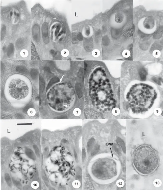
Endogenous stages - These develop, in conspicuous parasitophorous vacuoles, in the cytoplasm of the epi-thelial cells of the small intestine. In histological sections of the infected gut, most of the developing parasites were clearly located above the host cell nucleus, but it is pos-sible that others commenced development below and, with growth, eventually positioned themselves between or above the nuclei. Mature meronts (Figs 1, 2) were scanty but appeared to be of a single, small type of approximately 16 ×14 (n = 4): they produce 6-12 merozoites, measuring an average of 10 × 2.5 (n = 4) and segmentation leaves no residuum. Growing microgamonts (Figs 8, 9) reach up to 33 × 26 and contain many bulky, heavily stained nuclei distributed predominantly at the periphery of the para-site. Mature forms (Figs 10, 11) have a residuum contain-ing a few big vacuoles, and shed a large number of micro-gametes measuring approximately 5.0 × 1. Young macro-gamonts (Figs 3-5) have a poorly staining nucleus con-taining a densely scon-taining karyosome (Fig. 4). Mature forms (Fig. 7) may reach up to 24 × 20 (15 × 10 - 24 × 20), n = 10, and contain very prominent small and large wall-forming bodies (Figs 6, 7). The wide size range of the mature macrogamonts results in an equally wide range in the size of the zygotes and the oocysts. This at first gave the impression that both snakes were infected with two different species of Cyclospora. Demonstration of a smooth gradation between the dimensions of the small-est and the largsmall-est oocysts, however, militates against this possibility.
Sporulation - Exogenous, in approximately one week. Host - The snake Anilius scytale scytale (Linnaeus), (Aniliidae).
Type locality - Capanema, state of Pará, North Brazil. Type material - Oocysts in 10% buffered formalin, histo-logical sections of the endogenous stages and phototypes in the Department of Parasitology, Instituto Evandro Chagas and the Muséum National d’Histoire Naturelle, Paris: Accession No. 2257.
Prevalence - Uncertain. Only two A. s. scytale were exam-ined, and both were infected.
Pathology -No apparent pathology.
Etymology - The specific name is in honour of Aimé Schneider, who founded the genus Cyclospora.
ophid-1 0 6 1 0 61 0 6
1 0 61 0 6 The Genus Cyclospora • Ralph Lainson
TABLE
Recorded species of Cyclospora
Species Hosts Oocysts (µm) Endogenous stages
C. glomericola Glomeris sp. (Diplopoda) 25-36 × 9-10 Not described: oocysts in lumen of gut Schneider 1881
C. caryolytica Talpa europaea, Mogera wogura 18 × 12.5 Intranuclear in epithelial cells of small
Schaudinn 1902 coriana (Insectivora: Talpidae) a and large intestine
C. viperae Coluber scalaris, Coronella austriaca, 16.8 × 10.5 Intracytoplasmic in epithelial cells of
Phisalix 1923 Natrix viperinus, Vipera aspis the intestine
(Reptilia: Ophidia)
C. babaulti Vipera berus (Reptilia: Ophidia) 17 × 10 Intracytoplasmic in epithelial cells of
Phisalix 1924b the intestine
C. scinci Scincus officinalis (Reptilia: 10 × 7.0 Intracytoplasmic in epithelial cells of
Phisalix 1924d Squamata: Scincidae) the intestine
C. tropidonoti Natrix natrix, Natrix stolata 17 × 10 Intracytoplasmic in epithelial cells of
Phisalix 1924c (Reptilia: Ophidia) the intestine
C. zamenis Coluber viridiflavus viridiflavus 17 × 10 Intracytoplasmic in epithelial cells of
Phisalix 1924e (Reptilia: Ophidia) intestine
Cyclospora sp. Hemidactylus frenatus 16.9-30.3 × Uncertain
Yamamota 1933 (Reptilia: Squamata: Gekkonidae) 16.7-26
C. niniae Ninia sebae sebae (Reptilia: Ophidia) 14.6 × 13.3 Intracytoplasmic in epithelial cells of
Lainson 1965 intestine
C. cercopitheci Cercopithecus aethiops (Primates) 8-10 × 8-10 Not ascertained Eberhard et al. 1999a
C. talpae Talpa europaea (Insectivora: Talpidae) 15-18 × 10-12 Intranuclear in epithelial cells of bile
Pellerdy & Tanyi 1968 ducts and capillary sinusoids of the liver
C. megacephali Scalopus aquaticus 18.5 × 15.7 Not ascertained Ford & Duszynski 1988 (Mammalia: Insectivora)
C. ashtabulensis Parascalops breweri 18.0 × 14.3 Not ascertained Ford & Duszynski 1989 (Mammalia: Insectivora)
C. parascalopi Parascalops breweri 16.5 × 13.6 Not ascertained Ford & Duszynski 1989
C. angimurinensis Chaetodipus hispidus 19-24 × 16-22 Not ascertained Ford et al. 1990 (Mammalia: Rodentia)
C. cayetanensis Homo sapiens (Primates) 8.6 × 8.6 Intracytoplasmic in epithelial cells of
Ortega et al. 1994 the intestine
C. colobi Colobus guereza (Primates) 8-9 × 8-9 Not ascertained Eberhard et al. 1999a
C. papionis Papio anubis (Primates) 8-10 × 8-10 Not ascertained Eberhard et al. 1999a
C. schneideri n.sp. Anilius scytale scytale 19.8 × 16.6 Intracytoplasmic in epithelial cells of
(present paper) (Reptilia: Ophidia) the intestine
a: opinions are divided regarding the taxonomy of Japanese moles, with some authors considering that Mogera wogura coreana is a synonym of Talpa micrura coreana and vice versa(Nowak & Paradiso 1983, Duszynski & Wattam 1988a).
ian Cyclospora species remains uncertain, but the wide zoological difference and wide geographical separation of A. s. scytale and the Europeancolubrids and vipers makes it most unlikely that C. schneideri n.sp., is conspe-cific with any one of the four species described by Phisalix. I have commented on the similarity of the oocysts of the four species of Cyclospora named by Phisalix (Lainson 1965) and suggested that C. babaulti, C. tropidonoti and C. zamensis might be synonyms of C viperae. Duszynski
1 0 7 1 0 7 1 0 7 1 0 7 1 0 7 Mem Inst Oswaldo Cruz, Rio de Janeiro, Vol. 100(2), April 2005
Table until such time as conclusive evidence is provided with which to sink them. Their re-examination by DNA analysis might confirm or refutetheir present taxonomic status, and in this respect it is of interest that Eberhard et
al. (1999a) considered that the three species C. cerco-pitheci, C. colobi, and C. papionis of non-human pri-mates could not be separated by morphology of their oo-cysts but only at molecular level.
1 0 8 1 0 81 0 8
1 0 81 0 8 The Genus Cyclospora • Ralph Lainson
Figs 14 -16: Cyclospora schneideri n.sp., of the snake Anilius scytale scytale. Three mature, living oocysts, showing collapse of the fragile oocyst wall, the two sporozoites (sp) in each of the two sporocysts, sporocystic residuum (sr) and Stieda body (st). Bar = 10 µm. Normal light miscroscopy.
Figs 17, 18: Cyclospora schneideri n.sp., of the snake Anilius scytale scytale. Line drawings of living, mature oocysts, showing undeformed appearance (17) and later collapse of the oocyst wall (18); the latter is the same oocyst as that shown in the photograph in Fig. 15.
It is now becoming apparent that species of Cy-clospora infect a wider range of hosts than previously supposed and it should come as no surprise that reptiles are hosts of Cyclospora when one considers the wide host range of other members of the Eimeriidae. Thus, spe-cies of Eimeria are recorded in millipedes, centipedes, coleopterans, fish, amphibians, reptiles, birds and mam-mals (Levine 1988); the type species of Isospora, I. rara, was described in the gastropod Limax sp., and other spe-cies are common parasites of amphibia, reptiles, birds and mammals (including man); Caryospora species are found in reptiles, birds and mammals. By light microscopy, it is clearly difficult to determine the number of sporozoites in a sporocyst of an oocyst as small as that of C. cayetanensis (diameter 8.6 µm), and other Cyclospora species of simi-lar size may well have been erroneously assigned to the genus Isospora or Sarcocystis. However, due to the much larger size of the oocysts of both C. niniae and C.
schneideri n.sp. (14.6 ×13.3 and 19.8 ×16.6, respectively), it has not been difficult to determine the dizoic nature of their sporocysts, particularly when they are seen in an end-on position (Figs 15, 18).
ACKNOWLEDGEMENTS
To Constância M Franco and Manoel C de Souza for tech-nical assistance. Histological sections were prepared by Walter M Campos.
REFERENCES
Anon 1997a. Outbreak of cyclosporiasis northern Virginia -Washington, D.C. - Baltimore, Maryland Metropolitan Area. Can Comm Dis Rept 23: 166-168.
Anon 1997b. Update: outbreaks of cyclosporiasis – United States and Canada, 1997. Can Comm Dis Rept 23: 143-144.
1 0 9 1 0 9 1 0 9 1 0 9 1 0 9 Mem Inst Oswaldo Cruz, Rio de Janeiro, Vol. 100(2), April 2005
Ashford RW, Warhurst DC, Reid GDF 1993. Human infection with cyanobacterium-like bodies. Lancet 341: 1034.
Bendall RP, Lucas S, Moody A, Tovey G, Chiodini PL 1993. Diarrhoea associated with cyanobacterium-like bodies: a new coccidian enteritis of man. Lancet 341: 590-592.
Bern C, Hernandez B, Lopes MB, Arrowood MJ, Alvarez De Mejia M, Merida AM De, Hightower AW, Venczel L, Herwaldt BL, Klein RE 1999. Epidemiologic studies of
Cyclospora cayetanensis in Guatemala. Emerg Inf Dis 5: 766-774.
Bern C, Ortega Y, Checkley W, Roberts JM, Lescano AG, Cabrera L, Verastegui M, Black RE, Sterling C, Gilman RH 2002. Epidemiologic differences between cyclosporiasis and cryptosporidiosis in Peruvian children. Emerg Inf Dis 8: 581-585.
Cam PD, Sorel N, Dan LC, Larker E, Tassin S, Barber JP, Miegeville M 2001. A new contribution to the epidemio-logical survey of Cyclospora cayetanensis in Hanoï water supplies (Vietnam): a 12-month longitudinal study. Med Mal Infect 31: 591-600.
Carollo MCC, Amato Neto V, Braz LMA, Kim Dowoong 2001. Pesquisa de oocistos de Cyclospora sp., em fezes de cães da Grande São Paulo, Estado de São Paulo, Brasil. Rev Soc Bras Med Trop 34: 597-598.
Chambers J, Somerfeldt S, Mackey L, Nichols S, Ball R, Rob-erts D, Dufford N, Reddick A, Gibson J 1996. Outbreaks of
Cyclospora cayetanensis infection – United States, 1996.
MMWR 45: 549-551.
Döller PC, Dietrich K, Filipp N, Brockmann S, Dreweck C, Vonthein R, Wagner-Wiening C, Wiedermann A, 2002. Cyclosporiasis outbreak in Germany associated with the consumption of salad. Emerg Inf Dis 8: 992-994.
Duszynski DW, Wattam AR 1988a. Coccidian parasites (Apicomplexa: Eimeriidae) from Insectivores V. Ten forms from the moles of Japan (Euroscoptor, Mogera spp.). J Protozool 35: 55-57
Duszynski DW, Wattam AR 1988b. Coccidian parasites (Apicomplexa: Eimeriidae) from insectivores IV. Four new species in Talpa europaea from England. J Protozool 35: 58-62.
Duszynski DW, Upton SJ, Couch L 1999. The Coccidian Ge-nus Cyclospora. Available at: <htpp://biology.unm.edu/bi-ology/coccidia/cyclo.html> Accessed on 2 April, 2004.
Eberhard ML, Nace EK, Freeman AR 1999a. Survey for
Cyclospora cayetanensis in domestic animals in an endemic area in Haiti. J Parasitol 85: 562-563.
Eberhard ML, Njenga MN, Silva AJ Da, Owino D, Nace EK, Won KY, Mwenda JM 2001. A survey for Cyclospora spp., in Kenyan primates, with some notes on its biology. J Parasitol 87: 1394-1397.
Eberhard ML, Silva AJ Da, Lilley BG, Pieniazek NJ 1999b. Morphologic and molecular characterization of new
Cyclospora species from Ethiopian monkeys: C. cercopitheci
sp.n., C. colobi sp.n., and C. papionis sp.n. Emerg Inf Dis 5: 651-658.
Eimer T 1870. Ueber die ei-und Kugelförmigen sogenannten Psorospermien der Wirbelthiere, A Stuberr’s Verlang-shandlung,Würzburg, Germany.
El-Naga IFA 1999. Studies on a newly emerging protozoal patho-gen: Cyclospora cayetanensis. J Egypt Soc Parasitol 29: 575-586.
Ford PL, Duszynski DW 1988. Coccidian parasites from in-sectivores VI. Six new species from the Eastern mole
Scalopus aquaticus. J Protozool 35: 223-226.
Ford PL, Duszynski DW 1989. Coccidian parasites (Api-complexa: Eimeriidae) from insectivores VII. Six new spe-cies from the hairy-tailed mole Parascolops breweri. Para-sitology 75: 508-513.
Ford PL, Duszynski DW, McAllister CT 1990. Coccidia (Apicomplexa) from heteromyid rodents in the Southwest-ern United States, Baja California, and NorthSouthwest-ern Mexico, with three new species from Chaetodipus hispidus. J Parasitol 76: 325-331.
García López HL, Rodríguez Tovar LE, Garza CEM La, 1996. Identification of Cyclospora in poultry [Correspondence].
Emerg Inf Dis 2: 356-357.
Hart AS, Ridinger MT, Soundarajan R, Peters CS, Swiatlo AL, Kocka FE 1990. Novel organism associated with chronic diarrhoea in AIDS.Lancet 335(8682):169-170
Herwaldt BL, Ackers ML 1997. An outbreak in 1996 of cyclosporiasis associated with imported raspberries. New Engl J Med 336: 1548-1556.
Herwaldt BL, Beach MJ 1999. The return of Cyclospora in 1997: another outbreak of cyclosporiasis in North America associated with imported raspberries. Ann Int Med 130: 210-220.
Lainson R 1965. Parasitological studies in British Honduras II.
Cyclospora niniae sp.n. (Eimeriidae: Cyclosporinae) from the snake Ninia sebae sebae (Colubridae). Ann Trop Med Parasitol 59: 159-163.
Levine ND 1988. The Protozoan Phylum Apicomplexa, Vol. I, CRC Press, Boca Raton, FL, 203 pp.
Long EG, Ebrahimzadeh A, White EH, Swisher B, Callaway CS 1990. Alga associated with diarrhea in patients with ac-quired immunodeficiency syndrome and in travellers. Clin Microbiol 28: 1101-1104.
Long EG, White EH, Carmichael WW, Quinlisk PM, Raja R, Swisher BL, Daugharty, Cohen MT 1991. Morphological and staining characteristics of a cyanobacterium-like organ-ism associated with diarrhea. J Inf Dis 164: 199-202.
Lopez AS, Dodson DR, Arrowood MJ, Orlandi PA Jr, Silva AJ Da, Bier JW, Hanauer SD, Kuster Rl. Oltman S, Baldwin MS, Won KY, Nace EM, Eberhard ML, Herwaldt BL 2001.Outbreak of cyclosporiasis associated with basil in Missouri in 1999. Clin InfDis 32: 1010-1017.
Mohamed HA, Molyneux DH 1990. Developmental stages of
Cyclospora talpae in the liver and bile duct of the mole
Talpa europaea. Parasitology 101: 345-350.
Nowak RM, Paradiso JC 1983. Walker’s Mammals of the World, 4th ed., Vol. I, Johns Hopkins Univ. Press, Baltimore, MD.
Ortega YR, Gilman RH, Sterling CR 1994. A new coccidian parasite (Apicomplexa: Eimeriidae) from humans. J Parasitol 80: 625-629.
1 1 0 1 1 01 1 0
1 1 01 1 0 The Genus Cyclospora • Ralph Lainson
and clincal findings in patients with cyclosporiasis and a description of intracellular parasite life-cycle stages. J Inf Dis 176: 1584-1589.
Ortega YR, Roxas CR, Gilman RH, Miller NJ, Cabrera L, Taquiri C, Sterling CR 1997a. Isolation of Cryptosporidium parvum
and Cyclospora cayetanensis from vegetables collected in markets of an endemic region of Peru. Am J Trop Med Hyg 57: 683-686.
Ortega YR, Sterling CR, Gilman RH, Carna VA, Diaz F 1992.
Cyclospora cayetanensis: a new protozoan pathogen of humans. Abstract 289 in Proceedings of the 41st Annual Meeting of the American Society of Tropical Medicine and Hygiene. J Am Soc Trop Med Hyg (Suppl.):210.
Pellérdy L, Tanyi Z 1968. Cyclospora talpae sp.n. (Protozoa: Sporozoa) from the liver of Talpa europaea. Folia Parasitol (Praha) 15: 275-277.
Phisalix M 1923. Coccidiose intestinale de la vipère aspic à
Cyclospora viperae n.sp. Bull Soc Path Exot 16: 637-642.
Phisalix M 1924a. Note complémentaire sur Cyclospora viperae, coccidie parasite de l’intestinde la vipère aspic. Bull Soc Path Exot 17: 559-562.
Phisalix M 1924b. Coccidiose intestinale de Vipera berus à
Cyclospora babaulti nov.sp. Bull Soc Path Exot 17: 868-871.
Phisalix M 1924c. Cyclospora tropidonoti nov.spec., coccidie intestinale de la couleuvre à collier. Bull Soc Path Exot 17: 871-873.
Phisalix M 1924d. Coccidiose intestinale du Scincus officinalis
Laur., à Cyclospora scinci nov.sp. Bull Mus Natn Hist Nat Paris 30: 100-101.
Phisalix M 1924e. Cyclospora zamenis nov.sp., coccidie à localisation intestinale de Zamenis viridiflavus Lacép. Bull Mus Natn Hist Nat Paris 30: 501-502.
Phisalix M 1933. Coccidiose intestinale à Cyclospora viperae
chez la couleuvre vipérine, la couleuvre lisse et la couleuvre à échelons. Bull Soc Path Exot 26: 415-420.
Schaudinn F 1902. Studien über krankheitserregende Protozoen I. Cyclospora caryolitica Shaud., der Erreger der perniciösen
Enteritis des Maulwurfs. Arb K Gesundheitsamte 18: 378-416.
Schneider A 1881. Sur les psorospermies oviformes ou coccidies. Espécies nouvelles ou peu connues. Arch Zool Exp Gen 9: 387-404.
Sherchand TB, Cross JH, Jimba M, Sherchand S, Shrestha MP 1999. Study of Cyclospora cayetanensis in health care fa-cilities, sewage water and green leafy vegetables in Nepal.
South As J Trop Med Pub Hlth 30: 58-63.
Shlim DR, Cohen MT, Eaton M, Rajah R, Long EG, Unger BL 1991. An algal-like organism associated with an outbreak of prolonged diarrhea among foreigners in Nepal. Am J Trop Med Hyg 45: 383-389.
Soave R, Dubey JP, Ramos LJ, Tummings M 1986. A new intestinal pathogen? Clin Res 34: 533 (Abstract).
Sturbaum GD, Ortega YR, Gilman RH, Sterling CR, Cabrera L, Klein DA 1998. Detection of Cyclospora cayetanensis in wastewater. Appl Environ Microbiol 64: 2284-2286.
Sun T, Ilardi CF, Asnis D, Bresciani AR, Goldenberg S, Roberts B, Terchberg S 1996. Light and electronmicroscopic identi-fication of Cyclospora species in the small intestine: evi-dence of the presence of asexual life cycle in human host.
Am J Clin Path 105: 216-220.
Tanabe M 1938. On three species of coccidia of the mole, Mogera wogura coreana Thomas, with special reference to the life history of Cyclospora caryolytica. Keijo J Med 9: 21-52.
Upton SJ 2001. Cyclospora cayetanensis. Available at: < http: //www.ksu.edu/parasitology/cyclospora/cyclospora.html >. Accessed on 2 April, 2004.
Yai IEO, Bauab AR, Hirschfeld MPM, Oliveira ML De. Damaceno JT 1997. Brief communication: the first two cases of Cyclospora in dogs, São Paulo, Brazil. Rev Inst Med Trop São Paulo 39: 177-179.
Yamamoto K 1933. Studien über die Kokzidien [In Japanese, with German summary]. Fukuoka Acta Med 26: 40-43.
Zerpa R, Uchima N, Huicho L 1995. Cyclospora cayetanensis