online | memorias.ioc.fiocruz.br
Individual serological follow-up of patients with suspected or
confirmed abdominal angiostrongyliasis
Penélope E Palominos, Rose Gasnier, Rubens Rodriguez1, Aventino A Agostini1, Carlos Graeff-Teixeira/+
Laboratório de Parasitologia Molecular, Instituto de Pesquisas Biomédicas, Pontifícia Universidade Católica do Rio Grande do Sul (PUCRS) and Laboratório de Biologia Parasitária, Faculdade de Biociências da PUCRS, Avenida Ipiranga 6690, 2o andar, Sala 20, 90690
900 Porto Alegre, Brasil 1Instituto de Patologia de Passo Fundo, Universidade de Passo Fundo, Passo Fundo, RS, Brasil
Abdominal angiostrongyliasis (AA) is a zoonotic nematode infection caused by Angiostrongylus costaricensis, with widespread occurrence in the Americas. Although the human infection may be highly prevalent, morbidity is low in Southern Brazil. Confirmed diagnosis is based on finding parasitic structures in pathological examination of biop-sies or surgical resections. Serology stands as an important diagnostic tool in the less severe courses of the infection. Our objective is to describe the follow up of humoral reactivity every 2-4 weeks up to one year, in six individuals with confirmed (C) and ten suspected (S) AA. Antibody (IgG) detection was performed by ELISA and resulted in gradually declining curves of reactivity in nine subjects (56%) (4C + 5S), that were consistently negative in only three of them (2C + 1S) after 221, 121 and 298 days. Three individuals (2C + 1S) presented with low persistent reacitivity, other two (1C + 1S) were serologically negative from the beginning, but also presenting a declining tendency. The study shows indications that abdominal angiostrongyliasis is usually not a persistent infection: although serological negativation may take many months, IgG reactivity is usually declining along time and serum samples pairing may add valuable information to the diagnostic workout.
Key words: abdominal angiostrongyliasis - Angiostrongylus costaricensis - eosinophilic gastroenteritis - zoonosis
Abdominal angiostrongyliasis (AA) is a nematode infection caused by Angiostrongylus costaricensis. Wild
rodents are the definitive hosts and larval stages develop
in terrestrial mollusks (Morera 1971). The parasitosis occurs mainly in the Americas, from Southern USA to Northern Argentina and Southern Brazil (Ubelaker & Hall 1979, Agostini et al. 1984, Demo & Pessat 1986).
Imported infections have been diagnosed in USA, Eu
-rope and probably in Africa (Baird et al. 1987, Silvera et
al. 1989, Vázquez et al. 1993). Accidental human infec-tion may present as acute abdominal disease that usually
affects the ileo-cecum transition with an inflammatory
reaction mainly produced by secreted antigens and the presence of eggs in small capillaries. The disease may
complicate with intestinal obstruction due to extensive eosinophilic infiltration and intestinal perforation, peri -tonitis and sepsis, secondary to arterial thrombosis and necrosis (Céspedes et al. 1967). Detection of parasite structures in examination of biopsies or surgical
speci-mens allows the definitive diagnosis. In the absence of intra-arterial worms or eggs, histological findings such as severe eosinophilic infiltration, granulomatous reaction and eosinophilic vasculitis lead to a suspected diagnosis
(Graeff-Teixeira et al.1991a). Serology is essential for the diagnostic workout in uncomplicated clinical cases, since
Finantial support: PUCRS (FABIO, IPB and Hospital São Lucas), CNPq (477782/2004-3, 307872/2004-1), FAPERGS
+ Corresponding author: graeteix@pucrs.br Received 19 October 2007
Accepted 11 February 2008
fecal elimination of parasitic structures have never been reported. Previous evaluations of IgE, IgM, IgA or iso -types of IgG anti-crude adult worm antigens did not
re-sult a better diagnostic tool than an IgG-ELISA currently in use (Geiger et al. 2001). Our objective is to report the
follow up of IgG anti-A. costaricensis production in pa-tients from the endemic area in Southern Brazil.
PATIENTS, MATERIAL AND METHODS
Patients - A number of pathology laboratories and
clinical services in the Brazilian southernmost state, Rio Grande do Sul, are included in a vigilance network looking for suspected or confirmed new cases of AA, ac
-cording to previously described histopathological criteria (Graeff-Teixeira et al. 1991a). Briefly, the detection of
parasite structures (intra-arterial worms or eggs) is the
mainstay for a confirmed diagnosis, while the intense eo
-sinophillic inflammatory response in the intestinal wall, the eosinophilic vasculitis (especially in arteries) and
granulomatous reaction (including granulomatous arteri-tis) support the suspicion of A. costaricensis infection (Agostini et al. 1984). From 1999 to 2003, six patients
with confirmed abdominal angiostrongyliasis (group C)
and ten with suspected diagnosis (group S) had a serolog-ical follow-up for 298 ± 156 (media ± standard deviation) and 409 ± 212 days, respectively (Table). The collection of blood samples were scheduled as follows: one initial
sample immediately after the diagnosis and samples ev
-ery two weeks for three months and ev-ery month up to
one year. Time zero was the date of the surgical
proce-dure. Large variation in the intervals for sample collec
-uled by the research protocol. Serum was immediately
separated, eventually stored locally at -20°C and kept in
ice for transportation, never longer than two days, before final storage at -80°C at PUCRS laboratories.
The parasite and ELISA - A. costaricensis, Santa
Rosa and Nova Itaberaba strains, are maintained in vivo
at the laboratory through passages in Swiss mice or Oli-goryzomis nigripes and Biomphalaria glabrata (Esteio
strain). Details of antigen preparation and ELISA meth -odology are described elsewhere (Graeff-Teixeira et al. 1997, Geiger et al. 2001). In brief, the immunoenzymatic assay was performed using crude female worm antigens
with sensitivity and specificity of 76% and 91%, respec
-tively. Results of the ELISA are expressed as a ratio: average optical density from duplicates/cut-off value as
described by Geiger et al. (2001).
Bioethics - Informed consent was obtained from all adult participants and from parents or legal guardians of minors, with the name of the appropriate institutional
re-view board having approved the project. The investigation protocol was approved by the ethical committee
(CEP-PUCRS, 15 december 1999) and performed according to Brazilian regulation (Resolução MS-CNS 196/96).
RESULTS
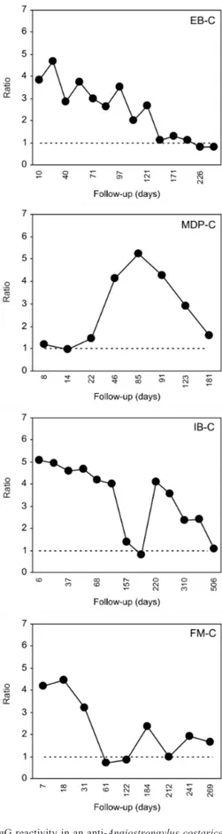
The individual IgG reactivity is shown graphically in
Figs 1, 2, 3 and 4. Nine of them (4 from group C and
5 from group S) presented gradually declining curves,
although only three (2 from group C and 1 from group
S) had a consistently negative ELISA after 221, 121 and
298 days (214 ± 89) (Table). Two patients (NGP-C and
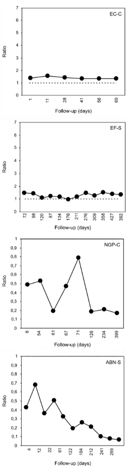
ABN-S) were serologically negative from the beginning,
but both showed a tendency to reduce the reactivity, es
-pecially ABN-S (Fig. 4). Other two patients (EC-C and EF-S) were persistently positive at very low antibody ti
-ters (Fig. 4). Some individuals (MDP-C in Fig. 1; MF-S and IW-S in Fig. 3) presented a low initial reactivity level
before a sharp increase at 53 ± 10 days. IB-C (Fig. 1) showed a striking reduction at 157 days and another peak
of reactivity at 220 days with gradual decline up to 506
days. In spite of clinical follow-up data not being
pre-sented because they were not available all the time from most of the patients, convalesce was usually symptom-free and all of them survived.
DISCUSSION
Sero-epidemiological studies have demonstrated ac -cidental human infection with A. costaricensis to be very
frequent, while morbidity is usually very low
(Graeff-Teixeira et al. 2005). Severe cases requiring surgical treat -ment for perforation and/or intestinal obstruction are rare in Southern Brazil as can be illustrated by the low number
of cases with definitive diagnosis in the present study. As indicated by a preliminary evaluation, antibody
response gradually decreases along time, indicating that
the worms do not survive long time in humans (Geiger
et al. 2005). The inappropriateness of the classic wisdom that the infection courses with high morbidity in a less adapted host, is illustrated by the situation in human AA, where cycle is not completed and adult worms do not
survive a long time, but infection only rarely manifests its more extreme “virulence” or “pathogenicity”. This situation fits in the proposition of alternative models to describe host-parasite coevolutionary status, like the
TABLE
Length of serological follow-up in 16 patients with suspected or confirmed abdominal angiostrongyliasis in Southern Brazil Days before consistent Total days
Initialsa Age (years) Sex negative serology of follow-up
Suspected diagnosis
MTD - S 49 f - 188
IW - S 26 f - 236
CP- S 79 f 221 287
ABN - S 13 f 1b 300
OB - S 27 m 121 378
EF - S 44 f - 392
LG - S 32 f - 458
MF - S 35 f - 459
DS - S 55 m - 538
LW - S 30 m - 842
mean ± standard deviation 39 ± 19 409 ± 212
Confirmed diagnosis
EC - C 18 f - 69
MDP - C 48 f - 181
FM - C 10 f - 300
EB - C 49 m 298 330
NGP - C 32 f 1 399
IB - C 48 m - 506
mean ± standard deviation 35 ± 18 298 ± 156
Fig. 1: IgG reactivity in an anti-Angiostrongylus costaricensis an-tibody detection ELISA. Four patients (MTD-S, OB-S, LW-S and CP-S) with suspected diagnosis (S). OB-S shows a very sharp decline and persistent negative serology from 121 days onwards.
Fig. 4: IgG reactivity in an anti-Angiostrongylus costaricensis antibody detection ELISA. Very low reactivity: patients EC-C and EF-S persis -tently above the cut-off; NGP-C and ABN-S below the cut-off (notice that ratio 1 is the top value in “y” axis). Although a formal negative result in ELISA, especially patient ABN-S shows a declining tendency in IgG reactivity.
“titer-time curve” of parasite transmission forms made available for the infection of new hosts, thereby favoring parasite species survival in nature (Garnik 1992).
Because of the difficulties for detection of L1 in
stools, serology is a key diagnostic methodology to
re-veal the presence sub-clinical or asymptomatic courses
of AA. Serology may also help the follow-up of patients leading to early detection of complications requiring surgical treatment. One patient (MDP-C, Fig. 1) had a
dramatic increase in reactivity coincident with a recur
-rence of severe intestinal inflammatory lesions leading to a second surgical intervention. IB-C (Fig. 1) also showed
a second peak which was not related to new lesions, sug-gesting reinfection. On the other hand, a decrease in anti-body titer is apparently a good prognostic sign.
Several patterns of reactivity may be delineated from
these data: (1) a predominant gradually decreasing
reac-tivity (EB-C, Fig. 1 and OB-S, Fig. 2); persistently posi
-tive in (2) high (LG-S and MF-S, Fig. 3) and (3) low titers (DS-S, Fig. 3; EC-C and EF-S, Fig. 4); and (4) delayed peak reactivity (MDP-C, Fig. 1; MF-S and IW-S, Fig. 3).
The similarity of reactivity curves in several pat
-terns both from confirmed and suspected abdominal an
-giostrongyliasis add validation to clinical and histopatho
-logical criteria as previously established (Agostini et al.
1984, Graeff-Teixeira et al. 1991a).
The persistence of antibodies for many months,
ei-ther in high or low titers, was not previously detected, since all patients in the evalution by Geiger et al. (2001) were serologically negative from three months onwards.
Although rare, the most intriguing situations are the
per-sistent high titers, like the curve from LG-S (Fig. 3) or
LW-S (Fig. 2), suggesting persistence of infection for a longer than expected time.
Part of the problems of sensitivity of immunological
methods may be explained by the low responders.
Vari-ability and diversity of antibody response to a parasite in -fection should be taken in consideration when diagnostic
methods are under analysis. More extensive observations
with clinical, parasitological and serological follow-up
are required for better evaluation of prognostic value of
serological and other molecular methods in abdominal angiostrongyliasis.
ACKNOWLEDGEMENTS
To Ingrid Porto and Juliano Romanzini for technical help. Serum samples collections were made possible through coop-eration from the following Institutions and individuals: Ana CA Silva, Antonio C Laitano, Caroline P Andrade, Fabiane Rigotti, Fabiane W Cidade, Maria F Pellicioli, Márcia CF Silva, Pablo B Bubols, Paula B Terraciano, Rafael L Maurer, Renata Ben, Roberta K Barbosa (FABIO/IPB PUCRS); Aven -tino A Agostini and Rubens Rodriguez (Instituto de Patologia de Passo Fundo); Ana M Gaiger (Lab Pat Reunidos, P.Alegre); Alex Guerini (Hospital Nonoai); Altair Collares (Hospital de Sarandi); Altair Copatti and Glaé Lameira (Hospital Santo Antônio, Três de Maio); Anencir F Silva, Irmã Romana, Ro -sane Perios and Nídia Perios (Hospital Cândido Godoi); Au -ristela Barros (SM Saúde, Planalto), Carlos Reichel (FAMED, PUCRS), Carlos Muneroli and Tânia Otto (Hospital Comu-nitário de Carazinho); Clóvis Bringuenti (Laboratório Vital, Nonoai); Eglê de Almeida (Hospital Cristo Redentor, Marau);
Gilberto Mayer (Hospital São Vicente, Passo Fundo); Janice P Zanella (Laboratório Sta Maria, Planalto); João Mesquita (Hospital Cesar Santos, Passo Fundo); José HM Silva (Lab das Clínicas, Passo Fundo), Paulo Sommer and Ursula Bockwin-kel (Hosp Porto Xavier); Paulo Valiatti (Lab Fontoura Xavier) and Laboratório Biolabor (Marau).
REFERENCES
Agostini AA, Marcolan AM, Lisot JMC, Lisot JUF 1984. An-giostrongilíase abdominal. Estudo anátomo-patológico de qua -tro casos observados no Rio Grande do Sul, Brasil. Mem Inst
Oswaldo Cruz79: 443-445.
Baird JK, Neafie RC, Lanoie L, Connor DH 1987. Abdominal an-giostrongylosis in an african man: case study. Am J Trop Med Hyg 37: 353-356.
Cespedes R, Salas J, Mekbel S, Troper L, Mullner F, Morera P 1967. Granulomas entericos y linfaticos con intensa eosinofilia tisular producidos por un estrongilideo (Strongylata): I. Patologia. Acta Med Costarric 10: 235-255.
Demo OJ, Pessat OAN 1986. Angiostrongilosis abdominal. Primer caso humano encontrado em Argentina. Prensa Med Argentina 73: 732-738.
Garnik E 1992. Parasite virulence and parasite-host coevolution: a reappraisal. J Parasitol 78: 381-386.
Geiger SM, Laitano AC, Sievers-Tostes C, Agostini AA, Schulz-Key H, Graeff-Teixeira C 2001. Detection of the acute phase of abdominal angiostrongyliasis with a parasite-specific IgG enzyme linked im-munosorbent assay. Mem Inst Oswaldo Cruz 96: 515-518.
Graeff-Teixeira C, Camillo-Coura L, Lenzi HL 1991a. Histopatho-logical criteria for diagnosis of abdominal angiostrongyliasis. Parasitol Res 77: 606-611.
Graeff-Teixeira C, Camillo-Coura L, Lenzi HL 1991b. Clinical and epidemiological studies on abdominal angiostrongyliasis in southern Brazil. Rev Inst Med Trop Sao Paulo 33: 375-380.
Graeff-Teixeira C, Ferreira-da-Cruz MF, Agostini AA, Camillo-Coura L 1997. Standartization of na immunoenzymatic assay (ELISA) for seroepidemiology of abdominal angiostrongyliasis.
Trop Med Internat Health 2: 254-260.
Graeff-Teixeira C, Goulart AH, Brum CO, Laitano AC, Sievers-Tostes C, Zanini GM, Bered PL, Morassutti A, Geiger S, Abrahms-Sandi E, Oliveira FTS, Maurer RL, Aguiar LF, Garrido CT, Silva ACA, Rodriguez R, Schultzk-Key H, Agostini AA 2005. Longitudinal clinical and serological survey of abdominal angiostrongyliasis in Guaporé, southern Brazil, from 1995 to 1999. Rev Soc Bras Med Trop 38: 310-315.
Morera P 1973. Life history and redescription of Angiostrongylus costaricensis Morera and Céspedes, 1971. Am J Trop Med Hyg 22: 613-621.
Silva ACA, Graeff-Teixeira C, Zaha A 2003. Diagnosis of abdominal angiostrongyliasis by PCR from sera of patients. Rev Inst Med Trop Sao Paulo 45: 295-297.
Silvera CT, Ghali VS, Roven S, Heiman J, Gelb A 1989. An- An-giostrongyliasis: a rare cause of gastrointestinal hemorrhage. Am
J Gastroenterol 84: 329-332.
Ubelaker JE, Hall NM 1979. First Report of Angiostrongylus costari-censis Morera & Céspedes, 1971 in the United States. J Parasitol 65: 307.