Rektus Kası Endometriozisi / Rectus Muscle Endometriosis
Endometriosis in the Rectus Abdominis Muscle: A Case Report
Rektus Abdominis Kası Yerleşimli Endometriozis: Olgu Sunumu
DOI: 10.4328/JCAM.2053 Received: 16.09.2013 Accepted: 01.10.2013 Printed: 01.10.2013 J Clin Anal Med 2013;4(suppl 2): 126-8 Corresponding Author: Soner Düzgüner, Ministry of Health, Dr. Sami Ulus Women’s Health Teaching and Research Hospital, Babur Street No:44 (06080) Altındağ, Ankara, Turkey. T.: +90 3123056000 F.: +90 3123170353 E-Mail: [email protected]
Özet
Rektus abdominis kası yerleşimli endometriozis nadir görülen bir hastalıktır. Kli-niğimizde rektus abdominis kası yerleşimli endometriozis tanısı ile cerrahi tedavi uyguladığımız olgunun klinik, radyolojik, histopatolojik özelliklerini değerlendirdik.
Anahtar Kelimeler
Ekstrapelvik Endometriozis; Rektus Abdominis Kası Yerleşimli Endometriozis; Skar Dokusunda Yerleşmiş Endometriozis
Abstract
Endometriosis involving the rectus abdominis muscle is a very rare disease. We present a case in whom endometriosis was found in the rectus abdominis muscle and discuss the imaging indings and histopathological correlation.
Keywords
Extrapelvic Endometriosis; Rectus Muscle Endometriosis; Scar Endometriosis Taşcı Yasemin1, Gelişen Orhan2, Düzgüner Soner3, Gökçin Hakan4, Balin Ipek Nur2
¹Department of Obstetrics and Gynecology, Zekai Tahir Burak Women’s Health Teaching and Research Hospital, ²Department of Obstetrics and Gynecology, Etlik Zübeyde Hanım Women’s Health Teaching and Research Hospital, ³Department of Obstetrics and Gynecology, Dr. Sami Ulus Women’s Health Teaching and Research Hospital,
4Department of General Surgery, Etlik Zübeyde Hanım Women’s Health Teaching and Research Hospital, Ankara, Turkey
| Journal of Clinical and Analytical Medicine
| Journal of Clinical and Analytical Medicine
Rektus Kası Endometriozisi / Rectus Muscle Endometriosis
Introduction
Endometriosis is a benign gynecological condition character-ized by endometrial tissue outside the uterine cavity and can occur at intra- or extrapelvic localizations. Endometriosis in-volving out of the pelvis is account for 12% of all endometriosis [1]. Endometrial implants have been reported in many unusual sites outside the pelvis including the abdominal wall. The ab-dominal wall endometriosis usually results from implantation of endometrial cells during laparotomy. Surgical scar endome-triosis has an incidence of 0,03% to 0,4% [2]. We present a rare case that endometriosis involving the rectus abdominis muscle.
Case Report
A 23-year-old woman was presented with persistent right lower abdominal pain and abdominal mass ongoing for 2 years, but worsening in the last 3 months. She reported that the size of the mass seemed to enlarge with her menses, and it become more painful. Her history included two cesarean sections 5 and 2 years before. On physical examination, a palpated solid and tender mass measuring 3x2 cm was found at her well-healed Pfannenstiel scar close to the right side. Routine hematologic and biochemical results were normal. Preoperative CA125 level was 45.0 U/ml. There was no previous history of endometrio-sis. Pelvic examination and pelvic ultrasonography were nor-mal. High resolution ultrasound of the abdominal wall using 12 MHz linear transducer has revealed 21x21 mm heterogeneous hypoechogenic mass within the distal part of the right rectus abdominis muscle [Figure 1]. Based on the indings of lower
abdomen physical examination and supericial ultrasonography, we decided to surgical intervention. At operation, the mass with a dimension of 3 x 3 cm located inside the rectus muscle that contained dark red coloured luid was removed including the surrounding ibers of the rectus abdominis and covering fascia of the rectus sheath. Fascial defect of the abdominal wall was repaired by using polypropylene mesh. There were no indings of pelvic endometriosis on abdominal exploration. Histological sections revealed endometrial glands surrounded by stroma and embedded in ibrous connective tissue between the muscle ibers with clear surgical resection margins (Figure 2). CA125 level were normal range ater the operation. The patient was
discharged from hospital on the 2nd postoperative day un-eventfully.
Discussion
The extrapelvic endometrial implants have been detected in various organs. Abdominal wall endometriosis, also known as scar endometrioma, is a rare site of localization, usually oc-curring ater previous cesarean section or pelvic surgery. It is thought to result from mechanical transplantation of endome-trial tissue into scars during abdominal surgery [3]. The other possible theories in the development of extrapelvic endometrio-sis are retrograde menstruation, venous or lymphatic metasta-sis and metaplasia [4]. In present case report, endometriometasta-sis was found within the body of the rectus abdominis muscle at the right corner of the surgical scar. It is an unusual and rare lo-calization for endometriosis since it was irst described in 1984 by Amato and Levitt [5].
The cesarean section is the operation most frequently associ-ated with abdominal wall endometriosis with an incidence that is reported as 0.03% to up to 1% [6]. Similar cases faced ater hysterotomy, hysterectomy, early amniocentesis, appendecto-my or episiotoappendecto-my have also been reported [7,8]. The reason of this situation may be explained as transportation of endome-trial tissue to an ectopic site irstly and then ectopic cells may induce metaplasia of the surrounding cells into the endometrial tissue [1]. It is also known that during pregnancy high level of serum progesterone impact negatively on endometriotic tissue growth and the implanted endometrium should be less prone to implant and proliferate at the ectopic site. A 20% of abdominal wall endometriosis is not associated with a previous surgery [9]. In this case possible mechanism can be explained as venous or lymphatic metastasis of endometrial cells [9].
The classical symptoms of abdominal wall endometriosis are cyclic pain associated with a palpable mass adjacent to a surgi-cal scar. However, approximately 30% of the patients complain about constant pain in the scar area or some cases may present as an asymptomatic mass in the region of a surgical scar [10]. The mean period between the surgery and symptoms is about 18 months, but up to ten years ater surgery [10] which was 2
Figure 1. Ultrasound of sot tissues of the abdominal wall shows a heterogenous hypoechogenic mass.
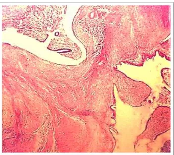
Figure 2. Histopathology showing endometrial glands and stroma (black arrow) in muscle tissue
Journal of Clinical and Analytical Medicine | 127
| Journal of Clinical and Analytical Medicine
Rektus Kası Endometriozisi / Rectus Muscle Endometriosis
years in our case.
Ultrasonography may be helpful in deining the cystic or solid nature of the mass and even exclude other intra-abdominal pa-thologies such as hernia. Suture granuloma, abscess, neuroma, desmoids tumor, hematoma, lymphadenopathy, subcutaneous cyst and metastatic cancer should be considered for diferen-tial diagnosis. Excision and histopathological evaluation of the mass is required for deinitive diagnosis. Computed tomography guided needle aspiration may be used for selected cases, but it has been associated to an increased risk of recurrence [11]. The treatment of choice for rectus abdominis endometriosis is a wide local excision of the lesion with surgically negative mar-gins. The use of synthetic mesh may be necessary for adequate closure of the abdominal wall. Recurrence of endometriosis in this area has rarely been reported but inadequate excision can lead to recurrence. GnRH agonists may also provide a tempo-rary reduction in size of the endometriotic lesion, but it has been recommended only in cases with coexisting pelvic endometrio-sis [12].
In conclusion, endometriosis of the rectus abdominis muscle should be considered in the diferential diagnosis in cases with painful abdominal wall mass with and without a surgical history.
Competing interests
The authors declare that they have no competing interests.
References
1. Dordevic M, Jovanovic B, Mitrovic S, Dordevic G, Radovanovic D, Sazdanovic P. Rectus abdominis muscle endometriosis ater cesarean section- Case report. Acta Clin Croat 2009; 48: 439-443.
2. Blanco RG, Parithivel VS, Shah AK, Gumbs MA, Schein M, Gerst PH. Abdominal wall endometriomas. Am J Surg 2003; 185(6): 596-598.
3. Giannella L, La Marca A, Ternelli G, Menozzi G. Rectus abdominis muscle endo-metriosis: Case report and review of the literature. J Obstet Gynaecol Res 2010; 36(4): 902-906.
4. Gunes M, Kayikcioglu F, Ozturkoglu E, Haberal A. Incisional endometriosis ater cesarean section, episiotomy and other gynecologic procedures. J Obstet Gynaecol Res 2005; 13(5): 471-475.
5. Amato M, Levitt R. Abdominal wall endometrioma: CT indings. J Comput Assist Tomogr 1984; 8: 1213-1214.
6. Hensen JH, Van Breda AC, Puylaert JB. Abdominal wall endometriosis: Clinical presentation and imaging features with emphasis on sonography. Am J Roent-genol 2006; 186: 616-620.
7. Thapa A, Kumar A, Gupta S. Abdominal wall endometriosis: report of a case and how much we know about it? Internet J Surg 2007; 9: 123-128.
8. Loverro G, Mei L, Vicinio M, Cormio G, Selvaggi L. Umblical endometriosis. J Gynecol Surg 2001; 17: 65-68.
9. Horton JD, Dezee KJ, Ahnfeldt EP, Wagner M. Abdominal wall endometriosis: A surgeon’s perspective and review of 445 cases. Am J Surg 2008; 196: 207-212. 10. Picod G, Boulanger L, Bounoua F, Leduc F, Duval G. Abdominal wall endome-triosis ater cesarean section: report of iteen cases. Gynecol Obstet Fertil 2006; 34(1): 8-13.
11. Coeman V, Sciot R, Van Breuseghem I. Rectus abdominis endometriosis: a re-port of two cases. Br J Radiol 2005; 78: 68-71.
12. Papaziogas T, Papaziogas B, Kabaroudis A, Galanis I, Lazaridis Ch, Souparis A, Alexandrakis A. Endometriosis of the rectus abdominis muscle ater cesarean section. Eur Surg 2002; 34(3)203-205.
How to cite this article:
Yasemin T, Orhan G, Soner D, Hakan G, Nur Bİ. Endometriosis in the Rectus Ab-dominis Muscle: A Case Report. J Clin Anal Med 2013;4(suppl 2): 126-8.
| Journal of Clinical and Analytical Medicine
128