455
Rev Bras Cir Cardiovasc | Braz J Cardiovasc Surg
Rev Bras Cir Cardiovasc 2014;29(3):455-8 Molinari GJDP, et al. - Avoiding pitfalls of intraoperative peripheral
endovascular surgery with the aid of OsiriX: expanding the use of virtual
luoroscopy
RBCCV 44205-1575 DOI: 10.5935/1678-9741.20140094
Avoiding pitfalls of intraoperative peripheral
endovascular surgery with the aid of OsiriX:
expanding the use of virtual luoroscopy
Como evitar armadilhas do intraoperatório em cirurgia endovascular periférica com o auxílio do OsiriX:
ampliando o uso da luoroscopia virtual
Giovani José Dal Poggetto Molinari
1, MD, Andréia Marques de Oliveira Dalbem
1, MD, Ana Terezinha
Guillaumon
1, MD, MsC, PhD
1Universidade Estadual de Campinas, Campinas, SP, Brazil.
This work was carried out in Hospital de Clínicas da Universidade Estadual de Campinas (Unicamp) Centro de Referência em Alta Complexidade em Cirurgia Endovascular, Campinas, SP, Brazil.
No inancial support.
Correspondence adress:
Giovani José Dal Poggetto Molinari
Universidade Estadual de Campinas - Unicamp/Cidade Universitária Zeferi-no Vaz - Rua Vital Brasil, 251 - Barão Geraldo - Campinas, SP, Brazil Zip Code: 13083-888 Mail box: 6142
E-mail: [email protected]
Article received on June 14th, 2014 Article accepted on July 21st, 2014
HOW TO DO IT
Abstract
We have shown how the analysis of the angiotomography reconstruction through OsiriX program has assisted in endovascular perioperative programming. We presented its application in situations when an unexpected existence of metallic overlapping artifact (orthopedic osteosynthesis) compromised the adequate visualization of the arterial lesion during the procedure. Through manipulation upon OsiriX software, with assistance of preview under virtual luoroscopy, it was possible to obtain the angles that would avoid this juxtaposition. These angles were reproduced in the C-arm, allowing visualization of the occluded segment, reducing the need for repeated image acquisitions and contrast overload, allowing the continuation of the procedure.
Descriptors: Fluoroscopy. User-Computer Interface. Mul-tidetector Computed Tomography. Endovascular Procedures.
Resumo
Temos demonstrado como a análise da reconstrução da
an-giotomograia utilizando o programa OsiriX tem auxiliado na
programação perioperatória endovascular. Apresentamos aqui sua aplicação em situação em que a existência de artefato metálico
(osteossíntese ortopédica) comprometia a adequada visibilização
de lesão arterial durante o procedimento. Pela manipulação da
angiotomograia no software OsiriX e com o auxílio das imagens sob luoroscopia virtual foi possível obter-se automaticamente uma angulação que evitasse esta justaposição. Os ângulos foram
reproduzidos no arcoscópio, o que permitiu expor o segmento
ocluído, reduzindo a sobrecarga de contraste e de repetidas
tomadas, permitindo a continuação do procedimento.
Descritores: Fluoroscopia. Interface Usuário-Computador.
Tomograia Computadorizada Multidetectores. Procedimentos
Endovasculares.
INTRODUCTION
Prior to computed tomography (CT) scans with multi-channel detector technology (implemented only from 1998 -
when the irst 4-channel equipment became available), the as -sessment of peripheral arterial disease by CT scan could only be performed only in a small segment of vascular system[1,2].
456
Rev Bras Cir Cardiovasc | Braz J Cardiovasc Surg
Rev Bras Cir Cardiovasc 2014;29(3):455-8 Molinari GJDP, et al. - Avoiding pitfalls of intraoperative peripheral
endovascular surgery with the aid of OsiriX: expanding the use of virtual
luoroscopy
(with placement of implantable system for osteosynthesis -
ixation with plate and pins) with no major complications.
For 3 months, she developed pain, paresthesia, coldness of the limb and progressive strength loss, with recent recrudes-cence. Physical examination presented a preserved sensibili-ty, decreased motor function and slowed perfusion. The val-ues of segmentar pressures registered in the left arm through a continuous wave doppler assessment were reduced, with a brachy-brachial index of 0,55 (N.V.: 0,9-1,1), reinforcing the hypothesis of an arterial involvement of this territory.
She underwent CT angiography exam of the supra-aortic
trunks and left upper limb. It was identiied an image of inter -rupted contrast, suggestive of occlusion of left subclavian artery
(near the metallic prosthesis) with reilling in its distal part. The
axial sections had impairment of appropriate visualization of the artery due to streak artifacts produced by the osteosynthesis, reason why it was opted for the intraoperative arteriography, for purposes of establishing an exclusive endovascular treatment (recanalization and covered stent implantation).
During surgical procedure, there was dificulty in the correct visualization of the lesion (ie., unilled portion of the
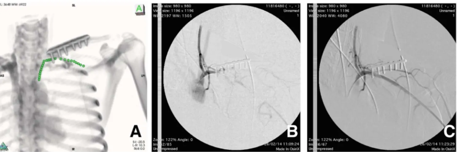
artery by intravascular contrast) due to the overlapping of radiopaque metallic rod included in the angiographic image (Figure 1A). After some attempts to capture new images
pos-terior to the angular repositioning of the luoroscopy unit,
it was decided to resort to auxiliary study of CT in OsiriX
software. At irst, the manipulation of the axial images was
performed, marking the proximal and distal portions of the subclavian artery with the point tool (Figure 1B). A
three-di-mensional volume reconstruction under virtual luoroscopy
was obtained, where it was able to identify an overlap of this demarcation by the orthopedic prosthesis (Figure 1C). Abbreviations, acronyms & symbols
CT Computed Tomography
Fig. 1 - A) Intraoperatory image of digital subtraction angiography, which displays an overlapping caused by the orthopedic metallic rod over the arterial thrombosed segment. B) Study of the tomographic axial view, where marks with the OsiriX’s point tool were performed, over the estimated trajectory of the left subclavian artery. C) Three-dimensional-by-volume reconstruction under virtual luoroscopy, after marking with points. It was possible to identify, in fact, the overlapping of the structures and the need of the C-arm angular repositioning intraoperatively.
the irst, CT angiography is less invasive, less expensive and
exposes patients to lower doses of radiation[4].
However, the study of CT angiography generates a mas-sive number of digital data and unwieldy, which interpreta-tion and discriminainterpreta-tion of changes are best evaluated when post processed in software, such as multi-planar
reconstruc-tion, volume rendering and maximum intensity projection[4]. We have shown how the analysis of tomographic
recon-struction of ine sections through the OsiriX program has aid -ed in perioperative programming of endovascular abdominal aneurysms, in prediction of the intra-luminal placement of angiographic catheters and in correction of angular
position-ing of the luoroscopy unit[5,6].
We also reported how concepts of angulation and Virtual Fluoroscopy used on OsiriX (from multichannel CT angiogra-phy) are used to obtain the best intraoperative angle of visual exposure of peripheral artery, in situations where the existence
of artifacts (like metallic orthopedic ixation) compromises its
proper visualization during endovascular procedures.
CASE REPORT
Female patient, 24 years old, assisted at the Outpatient Clinic for High Complexity Endovascular Surgery at Univer-sity of Campinas, with history of motorcycle trauma (fall) for
6 months, causing injury with fracture of clavicle, 3rd and 4th
457
Rev Bras Cir Cardiovasc | Braz J Cardiovasc Surg
Rev Bras Cir Cardiovasc 2014;29(3):455-8 Molinari GJDP, et al. - Avoiding pitfalls of intraoperative peripheral
endovascular surgery with the aid of OsiriX: expanding the use of virtual
luoroscopy
By rotational manipulation of three-dimensional image, it
was possible to obtain an angle that would avoid this juxtapo -sition (Figure 2A). The angles were reproduced in the C-arm, allowing visualization of the exposed occluded segment, al-lowing the rest of the procedure to follow (Figure 2B). There-fore, we proceeded to the artery recanalization with coated stent (Gore Viabahn 7x50) which was performed without any complications (Figure 2C).
RESULTS AND DISCUSSION
Until recently, digital subtraction arteriography was the modality of choice for evaluating patients with suspected
vascular injury. However, due to the wide access of multi -channel exams, it has rapidly replaced the conventional angi-ography in the diagnosis of vascular trauma[7].
In a meta-analysis by Jens et al.[8] sensitivity and
speci-icity for vascular injury (when compared to subtraction ar -teriography or surgery) was 99.2% and 96.2% respectively.
This justiies why all service centers should have a trauma
tomography as an accurate method for diagnosis of vascular
injury in extremity trauma, by reserving the conventional an
-giography for interventional or when the irst is inconclusive.
Vascular abnormalities in the upper limbs are found less frequently than those affecting the lower extremities. Arterial imaging studies of the upper limbs is usually performed in trauma victims with ischemic symptoms of upper extremi-ties, prep planning of complex vascular reconstructions and dialysis access as well as follow up of endovascular or surgi-cal procedures[9].
Despite the many advantages, CT angiography also has its limitations. It can be ineffective in the diagnosis if the contrast reaches the studied segment too soon or too late at the period of image acquisition by the device. Another scenario in the diagnosis of arterial lesions after trauma is the presence of artifacts caused by metallic fragments
on the periphery of the vessel, such as projectiles from irearms. These streaks make it impossible to analyze the
correct image[7].
Anyway, it is important to remember that in any tomo-graphic analysis is necessary to review the images of axial
views to conirm the indings presented in reconstructions and deine the possible presence of these or other artifacts
that simulate disease[9].
In our case the analysis of the image in axial views
showed these streaks artifacts, compromising the injury ex
-tension evaluation, even after adjusted values of windowing
(brightness and contrast). Therefore it was decided to supple-ment with subtraction arteriography intraoperatively. Thus, it would be possible to estimate, based on the gap of
intravas-cular contrast ill of the left subclavian artery, the extent of
the lesion, the size and required length of the stent(covered stent graft).
However when facing the radiopaque image produced by
the orthopedic ixation of the clavicle, it prevented the per -fect view of the angiographic image due to its overlapping and radiopacity. To continue with the procedure without vi-sualization of the arterial segment with intravascular contrast
reill would increase risk of false paths, extensive dissections
and the failure of the intervention.
458
Rev Bras Cir Cardiovasc | Braz J Cardiovasc Surg
Rev Bras Cir Cardiovasc 2014;29(3):455-8 Molinari GJDP, et al. - Avoiding pitfalls of intraoperative peripheral
endovascular surgery with the aid of OsiriX: expanding the use of virtual
luoroscopy
Authors’ roles & responsibilities
GJDPM Analysis and/or interpretation of data; statistical analysis;
inal manuscript approval; conception and design of
the study; conduct of operations and/or experiments; manuscript drafting or critical review of its content AMO Analysis and/or interpretation of data; conduct of
procedures and/or experiments
ATG Analysis and/or interpretation of data; manuscript drafting and review of its content
We used the angiotomography previously performed in attempt to simulate the optimum viewing angle of the oc-cluded segment to be recanalyzed. The OsiriX point tool marks permanently a voxel from a three-dimensional image -
which is deined as the representation of a pixel (the smallest
point of a digital image) in a volumetric grid originated from a series of pictures - and is reproduced in a three-dimension-al-by-volume reconstruction. Doing so, it’s possible to ob-tain any information of spatial positioning of this mark, such as the path of the occluded subclavian artery in this case.
Thus, when marking with points the trajectory of the artery,
it was possible to visualize it in any sort of reconstruction, axial, three-dimensional-by-volume or multiplanar. Through rotational manipulation of the three-dimensional-by-vol-ume image, we could illustrate the studied artery avoiding overlapping the osteosynthesis plaque. The angles obtained
automatically from the software were reproduced in the lu -oroscopy unit, allowing the correct visualization of the an-giographic image. Thereafter, it was possible to reduce the need for repeated angiographic takes in attempt to obtain the best exposure of the artery, avoiding iodinated contrast overload. Therefore we proceeded to the recanalization of the thrombosed segment with placement of a covered stent with therapeutic success (which can be observed by control an-giography). The patient’s clinical improvement was evident,
veriied with the complete resolution of the symptoms.
The use of OsiriX as a complementary tool allows doc-tors to assist in the preparation of surgeries (as endovascular)
extending it beyond the ield of diagnostic radiology. These
tasks can be easily incorporated into the armamentarium of the surgeon to avoid pitfalls and unforeseen situations that
are identiied intraoperatively, increasing the operatory risk
and oftentimes leading to intervention failure.
REFERENCES
1. Brockmann C, Jochum S, Sadick M, Huck K, Ziegler P, Fink C, et al. Dual-energy CT angiography in peripheral arterial occlusive disease. Cardiovasc Intervent Radiol. 2009;32(4):630-7.
2. Hiatt MD, Fleischmann D, Hellinger JC, Rubin GD. Angiographic imaging of the lower extremities with multidetector CT. Radiol Clin North Am. 2005;43(6):1119-27.
3. Kock MC, Dijkshoorn ML, Pattynama PM, Myriam Hunink
MG. Multi-detector row computed tomography angiography of peripheral arterial disease. Eur Radiol. 2007;17(12):3208-22.
4. Walls MC, Thavendiranathan P, Rajagopalan S. Advances in
CT angiography for peripheral arterial disease. Cardiol Clin. 2011;29(3):331-40.
5. Molinari GJDP, Dalbem AMO, Menezes FH, Guillaumon AT.
Proposal of renal artery’s ostial projection under virtual geometric
correction in infrarenal aneurysms: initial results of a pilot study. Rev Bras Cir Cardiovasc. 2014;29(1):78-82.
6. Molinari GJDP, Dalbem AMO, Guillaumon AT. The use of virtual resources for pre operatory study in correction of infrarenal aneurysms: exploring the OsiriX’s potential. Rev Bras Cir Cardiovasc. Rev Bras Cir Cardiovasc. 2014;29(2):279-84.
7. Jens S, Kerstens MK, Legemate DA, Reekers JA, Bipat S, Koelemay MJ. Diagnostic performance of computed tomography
angiography in peripheral arterial injury due to trauma: a
systematic review and meta-analysis. Eur J Vasc Endovasc Surg. 2013;46(3):329-37.
8. Kock MC, Dijkshoorn ML, Pattynama PM, Myriam Hunink
MG. Multi-detector row computed tomography angiography of peripheral arterial disease. Eur Radiol. 2007;17(12):3208-22.
9. Waliszewska M, Jakubiak A, Guzinski M, Sasiadek M. Application of the 64-slice computed tomography as a diagnostic method in acute posttraumatic ischaemia of the upper limbs - 3 case reports. Pol J Radiol. 2010;75(2):94-7.
10. Walls MC, Thavendiranathan P, Rajagopalan S. Advances in