Mariana Marquezan(a) Margareth Maria Gomes de Souza(a)
Mônica Tirre de Souza Araújo(a) Lincoln Issamu Nojima(a) Matilde da Cunha Gonçalves Nojima(a)
(a) Pedodontics and Orthodontics Department, Federal University of Rio de Janeiro (UFRJ), Rio de Janeiro, RJ, Brazil.
Corresponding author:
Matilde da Cunha Gonçalves Nojima E-mail: [email protected]
Received for publication on May 24, 2011 Accepted for publication on Jul 20, 2011
Is miniscrew primary stability
influenced by bone density?
Abstract: Primary stability is absence of mobility in the bone bed af-ter mini-implant placement and depends on bone quality among other factors. Bone quality is a subjective term frequently considered as bone density. The aim of this preliminary study was to evaluate bone density in two bovine pelvic regions and verify the primary stability of minis-crews inserted into them. Forty bone blocks were extracted from bovine pelvic bones, 20 from iliac and 20 from pubic bone, all of them contain-ing cortical bone about 1 mm thick. Half of the sections extracted from each bone were designated for histological evaluation of bone density (trabecular bone area - TBA) and the other half for bone mineral density (BMD) evaluation by means of central dual-energy X-ray absorptiometry (DEXA). Then, twenty self-drilling miniscrews (INP, São Paulo, Brazil)
1.4 mm in diameter and 6 mm long were inserted into the bone blocks used for BMD evaluation. Peak implant insertion torque (IT) and pull-out strength (PS) were used for primary stability evaluation. It was found that iliac and pubic bones present different bone densities, iliac bone be-ing less dense considerbe-ing BMD and TBA values (P > 0.05). However, the miniscrew primary stability was not different when varying the bone type (P < 0.05). IT and PS were not inluenced by these differences in bone density when cortical thickness was about 1 mm thick.
Descriptors: Bone and Bones; Bone Density; Orthodontic Anchorage Procedures.
Introduction
Primary stability is absence of mobility in the bone bed after implant or mini-implant placement.1,2 It is achieved by mechanical contact
be-tween the miniscrew surface and bone3 and depends on the
characteris-tics of devices4,5, insertion technique4 and bone quality and quantity of
the receptor site.4,6-8
The primary stability plays an important role in the successful second-ary stability of miniscrews, since lack of immediate stability can lead to progressive mobility of the device and its subsequent loss.9 In clinical use,
the initial stability of miniscrews is also considered essential, because of immediate or early load applied on them in many patients.10 It has been
suggested that if initial mechanical retention of the mini-implant is not observed, it should be replaced by a thicker device, or its insertion site should be changed.11 Primary stability has traditionally been assessed by
the practitioner through manual veriication.12 Several other less
tive methods are described in the literature: histo-logical (BIC - bone to implant contact), which as-sesses the percentage of bone to implant contact; mechanical, which assesses insertion and removal torque or pullout strength of mini-implants, and the percussion method (Periotest value). However, there is still no gold standard to assess the primary stabil-ity of miniscrews.13
The term “bone quality” is not clearly deined in the literature. This includes physiological and structural aspects and degree of bone tissue miner-alization.14 Aspects such as bone metabolism, cell
turnover, maturation, intracellular matrix and vas-cularity have also been emphasized.1 Nevertheless,
the role of each of these aspects is not completely understood.14 In Implant dentistry, the most
accept-ed classiication of bone quality has been the one proposed by Lekholm and Zarb.15 This was based
on the amount of cortical and trabecular bone shown in preoperative radiographs. This classiica-tion, however, depends on the operator’s subjectiv-ity during radiographic evaluation.
A less subjective method for evaluating cortical and trabecular bone quality is to verify bone mineral density (BMD).14 Bone densitometry is taken as the
gold standard for quantifying BMD in
Endocrinolo-gy and TraumatoloEndocrinolo-gy.16 The bone mineral content of
tissue is measured and divided by the area of tissue to obtain bone mineral density. Another parameter of bone quality evaluated in implant dentistry is tra-becular bone density. For this measurement, how-ever, the percentage of trabecular bone area in the total biopsy area is calculated. The trabecular bone area (TBA) instead of mineral content is evaluated. For TBA analysis, histological and morphometrical methods are considered the gold standard.1
Considering the above, the aim of this study was to evaluate the primary stability of miniscrews in-serted in two bovine pelvic regions with different densities, to verify the inluence of bone density on stability.
Methodology
The sample comprised 40 bone sections extract-ed from bovine pelvis (Bos taurus), Angus lineage. Ten pelvic bones were obtained from a Slaugh-terhouse (registered with ANVISA – the Brazil-ian Health Surveillance Agency) immediately after slaughter. From each bone, two small bone sections were taken from the gluteal wing of the iliac and from the pubic bone (Figure 1). Tissue sections were removed by means of a trephine bur (8 mm in
diam-Figure 1 - Macroscopic view of the right hemi pelvis. (a) caudal view: the arrow indicates the gluteal wing of the iliac bone. (b) medial view: the arrow indicates the caudal portion of the pubic bone.
eter × 20 mm long, Sin Implantes, São Paulo, Brazil) adapted to a low speed motor (Beltec LB100, Ara-raquara, Brazil) under irrigation. The bone sections were taken from a region in which cortical bone was about 1 mm thick (measured with an orthodontic caliper, Odin, Ortho-pli, Philadelphia, USA). One of the two bone sections taken from each region was used to measure bone mineral density and evaluate primary stability. These samples were immersed in sterile physiological solution and stored by freezing (−20 ºC) until the tests were performed. The other section removed from each bone was used for his-tomorphometric analysis. These samples were im-mersed in 10% buffered formalin solution for 2 days for ixation.
Bone mineral density evaluation (BMD)
The bone mineral content of specimens was measured and divided into areas to obtain bone mineral density by means of central dual-energy X-ray absorptiometry (DEXA) (GE/Lunar Prodigy, Madison, USA), calibrated for small animals. To perform the exam, the bone blocks were thawed at room temperature and were put into plastic boxes (6 × 11 × 4 cm) containing raw rice to simulate soft tissue during irradiation. After this the samples were irradiated by DEXA for 30 seconds.17
Histomorphometric evaluation
After being immersed in 10% buffered formalin solution for 2 days, the samples were decalciied in Morse solution18 (equal parts of 50% formic acid
and 20% sodium citrate - Vetec Química Fina Ltda., Rio de Janeiro, Brazil) by immersion for 7 days and then embedded in parafin. Longitudinal sections were cut into 5-µm slices and stained with picro-sirius for histologic evaluation. Histomorphometric analysis of bone samples was performed using Im-age J software (National Institute of Mental Health, Bethesda, USA). Digitized photomicrographs (mi-croscope Nikon Eclipse E600, magniication ×40, camera DS-U2, Nikon Corporation, Tokyo, Japan) were taken and analyzed by the same examiner (ICC = 0.971). The histomorphometric evaluation result was given as a percentage of trabecular bone area (TBA).
Primary stability evaluation
Primary stability was evaluated by measuring in-sertion torque (IT) and pull-out strength (PS). Twen-ty miniscrews (INP, São Paulo, Brazil) 1.4 mm in diameter and 6 mm long were inserted into the bone blocks used for BMD evaluation. This was done with the use of a manual placement key connected to a digital torque meter (Lutron TQ-8800, Taipei, Taiwan), to allow the measurement of peak implant placement torque. The values were recorded in New-ton centimeter (Ncm). After this, the pull-out test, which consists of extracting the miniscrew from bone at a constant velocity, was performed to evalu-ate the maximum force required to remove it.19 The
mechanical test was performed in a universal test machine (Emic DL 2000, São José dos Pinhais, Bra-zil), using a 500 kgf load cell at a crosshead speed of 0.05 mm per second19 to remove the miniscrew. The
maximum pull-out strength was recorded.
Statistical analysis
The data were evaluated using the Statistical Package for Social Sciences (version 17, SPSS Inc., Chicago, USA). The values obtained were tabulated and submitted to descriptive analysis. The normality and homogeneity of variables were veriied by Shap-iro-Wilk and Levene’s tests. Intergroup comparisons of mean values were performed by the paired T-test at a level of signiicance of 5%.
Results
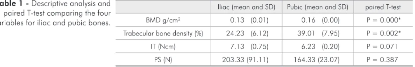
Under light microscopy, the histological sections revealed the presence of trabecular bone with os-teocytes and marrow spaces illed with fat marrow. The marrow spaces were larger in Iliac bone. De-scriptive statistics and the paired T-test results are shown in Table 1.
Discussion
Pelvic bone has previously been used in studies with miniscrews.4,20,21,22 During exploration of this
bone, it was observed that some characteristics such as color, texture and drill resistance differed in its various regions. Pubic bone was darker and more re-sistant when compared with iliac bone. Therefore, these two regions of the pelvic bone were chosen for this study. The BMD results showed that they are less dense than human jaw bones, as previously relat-ed in the literature by Devlin et al.,23 indicating the
following values: maxilla anterior region = 0.55 g/ cm²; maxilla posterior region = 0.31 g/cm²; man-dible = 1.11 g/cm²; and by Choel et al.,24
indicat-ing values for dentate mandible = 0.604 g/cm²; and edentulous mandible = 0.521 g/cm². However, the two cited studies presented a large variation in val-ues for mandibular BMD. Trabecular bone density evaluation, however, showed that the TBA value for pubic bone was similar to the results previously de-scribed by Aksoy et al.25 for maxilla (= 38.20 ± 9.65)
and for mandible (= 44.08 ± 14.97). The results of this study showed that the iliac and the pubic bones (of which the pelvis is composed) present different bone qualities: bone mineral density and trabecular bone density. These characteristics, however, had no inluence on the primary stability of mini-implants inserted in bone when the cortical was 1 mm thick.
Mean values for IT ranged from 6.23 to 7.13 Ncm, representing adequate primary stabil-ity according to Motoyoshi et al.,26 who stated that
these values should range from 5 to 10 Ncm.
Pull-out strength values ranged from 164.33 to 203.33 N, being within the range found by Huja et al.19 in a study with dog jaws: 134.5 N, for
ante-rior mandible, and 388.3 N for posteante-rior mandible. Nevertheless, no landmark for adequate pull-out strength value was found in the literature.
A previous study evaluated the inluence of BMD on the primary stability of miniscrews and, despite methodological differences, found a similar result.27
No correlation was found between BMD, veriied
Figure 2 - Micrograph of iliac (a) and pubic (b) bones (picrosirius, 40×, bars= 100 µm). Note that the marrow spaces are larger in iliac bone (a).
Iliac (mean and SD) Pubic (mean and SD) paired T-test
BMD g/cm² 0.13 (0.01) 0.16 (0.00) P = 0.000*
Trabecular bone density (%) 24.23 (6.12) 39.01 (7.95) P = 0.002*
IT (Ncm) 7.13 (0.75) 6.23 (0.20) P = 0.071
PS (N) 203.33 (91.11) 164.33 (23.07) P = 0.387
* Indicates statistical significant difference at α = 0.05%
Table 1 - Descriptive analysis and paired T-test comparing the four variables for iliac and pubic bones.
by cone beam computed tomography, and minis-crew stability, assessed by placement torque. The authors also investigated the inluence of cortical bone and found that cortical thickness and cortical BMD were positively correlated with miniscrew sta-bility.27 No studies evaluating the inluence of TBA
on miniscrew stability were found.
Two hypotheses were formulated to explain the results of the present study. The irst is that the pres-ence of a cortical thickness of 1 mm in all of the specimens had an important inluence on miniscrew stability, masking the inluence of bone mineral den-sity and trabecular denden-sity. Cortical thickness has been related to primary stability of miniscrews and implants.5,12,28,29,30 However, there is a lack of
stud-ies isolating these two factors: bone density and cortical thickness. The second hypothesis is that the difference in bone quality veriied statistically may not be clinically relevant. A bigger difference be-tween BMD and TBA values in bones could perhaps relect differences in mini-implant stability.
Despite the limitations of this in vitro study, it can be inferred that, in clinical practice, a corti-cal thickness of 1 mm is suficient to guarantee the primary stability of miniscrews, as previously
sup-posed by Motoyoshi et al.,30 even when there are
variations in BMD and TBA values.
Further research is suggested isolating the corti-cal effect and increasing the difference in density be-tween different types of bone.
Conclusions
• Iliac and pubic bones present different BMD and TBA values, the iliac being less dense when con-sidering the two parameters;
• Miniscrew primary stability was not inluenced by these differences in bone density.
Acknowledgements
The authors would like to express their grateful appreciation to the following entities and persons: CAPES (Cordenação de Aperfeiçoamento de Pessoal de Nível Superior) and FAPERJ (Fundação de Am-paro à Pesquisa do Estado do Rio de Janeiro) for the inancial support provided; INP (Sistema de Im-plantes Nacionais e de Próteses) for the donation of the miniscrews; Frigoríico Silva for the donation of the bones; doctors Maria Lucia Fleiuss Farias and Laura Maria Carvalho Mendonça for their support during bone mineral density analysis.
References
1. Molly L. Bone density and primary stability in implant ther-apy. Clin Oral Implants Res. 2006 Oct;17 Suppl 2:124-35. 2. Javed F, Romanos GE. The role of primary stability for
suc-cessful immediate loading of dental implants. A literature review. J Dent. 2010 Aug;38(8):612-20.
3. Gedrange T, Hietschold V, Mai R, Wolf P, Nicklisch M, Har-zer W. An evaluation of resonance frequency analysis for the determination of the primary stability of orthodontic palatal implants. A study in human cadavers. Clin Oral Implants Res. 2005 Aug;16(4):425-31.
4. Wilmes B, Rademacher C, Olthoff G, Drescher D. Parameters affecting primary stability of orthodontic mini-implants. J Orofac Orthop. 2006 May;67(3):162-74.
5. Song YY, Cha JY, Hwang CJ. Mechanical characteristics of various orthodontic mini-screws in relation to artificial corti-cal bone thickness. Angle Orthod. 2007 Nov;77(6):979-85. 6. Trisi P, De Benedittis S, Perfetti G, Berardi D. Primary
stabil-ity, insertion torque and bone density of cylindric implant ad modum Branemark: Is there a relationship? An in vitro study. Clin Oral Implants Res. 2011Nov;22(5):567-70.
7. Freudenthaler JW, Haas R, Bantleon HP. Bicortical titanium screws for critical orthodontic anchorage in the mandible: a preliminary report on clinical applications. Clin Oral Implants Res. 2001 Aug;12(4):358-63.
8. Cheng SJ, Tseng IY, Lee JJ, Kok SH. A prospective study of the risk factors associated with failure of mini-implants used for orthodontic anchorage. Int J Oral Maxillofac Implants. 2004 Jan-Feb;19(1):100-6.
9. Mischkowski RA, Kneuertz P, Florvaag B, Lazar F, Koebke J, Zoller JE. Biomechanical comparison of four different minis-crew types for skeletal anchorage in the mandibulo-maxillary area. Int J Oral Maxillofac Surg. 2008 Oct;37(10):948-54. 10. Melsen B, Costa A. Immediate loading of implants used for
orthodontic anchorage. Clin Orthod Res. 2000 Feb;3(1):23-8. 11. Garfinkle JS, Cunningham LL Jr., Beeman CS, Kluemper GT,
Hicks EP, Kim MO. Evaluation of orthodontic mini-implant anchorage in premolar extraction therapy in adolescents. Am J Orthod Dentofacial Orthop. 2008 May;133(5):642-53. 12. Merheb J, Van Assche N, Coucke W, Jacobs R, Naert I,
implant stability. Clin Oral Implants Res. 2010 Jun;21(6):612-7.
13. Cehreli MC, Kokat AM, Comert A, Akkocaoglu M, Tek-demir I, Akca K. Implant stability and bone density: assess-ment of correlation in fresh cadavers using conventional and osteotome implant sockets. Clin Oral Implants Res. 2009 Oct;20(10):1163-9.
14. Bergkvist G, Koh KJ, Sahlholm S, Klintstrom E, Lindh C. Bone density at implant sites and its relationship to assessment of bone quality and treatment outcome. Int J Oral Maxillofac Implants. 2010 Mar-Apr;25(2):321-8.
15. Lekholm U, Zarb G. Patient selection and preparation. In: Branemark PI, Zarb G, Albrektsson T, editors. Tissue-in-tegrated prostheses: osseointegration in clinical dentistry. Chicago: Quintessence; 1985. p. 199-209.
16. Carey JJ, Delaney MF, Love TE, Richmond BJ, Cromer BA, Miller PD, et al. DXA-generated Z-scores and T-scores may differ substantially and significantly in young adults. J Clin Densitom. 2007 Oct-Dec;10(4):351-8.
17. Pithon MM, Andrade ACDV, Rodrigues VB, Santos RL. Influ-ence of the immunossuppressant tracolimus (FK-506) on the flexural strength of femur: a study in rats. Rev Bras Ortop. 2010;45(3):286-89.
18. Morse A. Formic acid-sodium citrate decalcification and bu-tyl alcohol dehydration of teeth and bones for sectioning in paraffin J Dent Res. 1945;24(3):11.
19. Huja SS, Litsky AS, Beck FM, Johnson KA, Larsen PE. Pull-out strength of monocortical screws placed in the maxillae and mandibles of dogs. Am J Orthod Dentofacial Orthop. 2005 Mar;127(3):307-13.
20. Wawrzinek C, Sommer T, Fischer-Brandies H. Microdam-age in cortical bone due to the overtightening of orthodontic microscrews. J Orofac Orthop. 2008 Mar;69(2):121-34. 21. Wilmes B, Su YY, Drescher D. Insertion angle impact on
pri-mary stability of orthodontic mini-implants. Angle Orthod. 2008 Nov;78(6):1065-70.
22. Su YY, Wilmes B, Honscheid R, Drescher D. Application of a wireless resonance frequency transducer to assess pri-mary stability of orthodontic mini-implants: an in vitro
study in pig ilia. Int J Oral Maxillofac Implants. 2009 Jul-Aug;24(4):647-54.
23. Devlin H, Horner K, Ledgerton D. A comparison of maxillary and mandibular bone mineral densities. J Prosthet Dent. 1998 Mar;79(3):323-7.
24. Choel L, Duboeuf F, Bourgeois D, Briguet A, Lissac M. Tra-becular alveolar bone in the human mandible: a dual-energy x-ray absorptiometry study. Oral Surg Oral Med Oral Pathol Oral Radiol Endod. 2003 Mar;95(3):364-70.
25. Aksoy U, Eratalay K, Tozum TF. The possible association among bone density values, resonance frequency measure-ments, tactile sense, and histomorphometric evaluations of dental implant osteotomy sites: a preliminary study. Implant Dent. 2009 Aug;18(4):316-25.
26. Motoyoshi M, Hirabayashi M, Uemura M, Shimizu N. Rec-ommended placement torque when tightening an orthodontic mini-implant. Clin Oral Implants Res. 2006 Feb;17(1):109-14. 27. Cha JY, Kil JK, Yoon TM, Hwang CJ. Miniscrew stability
evaluated with computerized tomography scanning. Am J Orthod Dentofacial Orthop. 2010 Jan;137(1):73-9.
28. Pithon MM, Nojima LI. Evaluation of the primary stability of orthodontic miniscrew in different regions of maxilla and mandible of pigs. Innovations Implant Journal - Biomaterials and Esthetics. 2007;2(4):58-63.
29. Motoyoshi M, Inaba M, Ono A, Ueno S, Shimizu N. The ef-fect of cortical bone thickness on the stability of orthodontic mini-implants and on the stress distribution in surrounding bone. Int J Oral Maxillofac Surg. 2009 Jan;38(1):13-8. 30. Motoyoshi M, Yoshida T, Ono A, Shimizu N. Effect of cortical