Zigomar Hideo Fecchio Nasser HORIUCHI(a)
Yara Teresinha Correa SILVA-SOUSA(a)
Walter RAUCCI-NETO(a)
Fuad Jacob Abi RACHED-JUNIOR(a)
Aline Evangelista SOUZA-GABRIEL(b)
Silvio Rocha Corrêa da SILVA(a)
Edson ALFREDO (a)
(a)Universidade de Ribeirão Preto – UNAERP, School of Dentistry, Dentistry Department, Ribeirão Preto, SP, Brazil.
(b)Universidade de São Paulo – USP, School of Dentistry of Ribeirão Preto, Department of Restorative Dentistry, Ribeirão Preto, SP, Brazil.
Effect of thermoplastic filling techniques
on the push-out strength of root sealing
materials
Abstract: This study evaluates the effect of two thermoplastic obturation systems (MicroSeal and Obtura II) on bond strength of different sealers to intraradicular dentin. Sixty root canals of human
canines were prepared using ProTaper rotary iles (crown-down
technique) and irrigated with 2.5% sodium hypochlorite and 17%
EDTA. The root canals were illed by MicroSeal, Obtura II, or lateral
compaction techniques using AH Plus and Epiphany SE. 1.5 mm
thick root slices were subjected to the push-out test. ANOVA and
Tukey’s test showed that the bond strength values (MPa) observed in the groups obturated with MicroSeal (2.96 ± 2.72) and Obtura II
(2.68 ± 2.18) did not signiicantly differ from each other (p > 0.05) but were signiicantly higher than that observed in the group obturated with lateral condensation (2.01 ± 1.48; p < 0.05). There were no statistically signiicant differences in strength (p > 0.05) among the root canal thirds (cervical: 2.44 ± 2.03; middle: 2.50 ± 2.27; and apical: 2.70 ± 2.34). Adhesive failures were predominant (60%) in all groups. In conclusion, MicroSeal and Obtura II techniques, using AH plus sealer, increased the resistance to displacement of the illing material, when compared with lateral compaction. Moreover, when used with Epiphany SE, these obturation systems did not affect the bond strength
of the material to root dentin.
Keywords: Endodontics; Dental Bonding; Root Canal Obturation.
Introduction
Several reports have shown that incomplete apical and coronal
three-dimensional luid-tight seals are the main cause of endodontic
treatment failure.1,2,3,4 According to De-Deus et al.5 and Gulsahi et al.,6 the material should completely ill the root canal, and the ideal outcome is a high volume of core material, generally gutta-percha, and a small quantity of sealer, to prevent growth of residual bacteria and inhibit
introduction of new bacteria.
Different illing techniques have been proposed in an attempt to
obtain compact sealing properties.7,8,9,10,11 Among these techniques,
lateral-compaction with gutta-percha points provides adequate apical sealing. However, the eficiency of the technique mainly depends on
the type of sealer used.1 Therefore, thermoplastic canal illing methods are indicated to allow condensation of warm gutta-percha in all the root
Declaration of Interests: The authors certify that they have no commercial or associative interest that represents a conflict of interest in connection with the manuscript.
Corresponding Author: Walter Raucci-Neto
E-mail: [email protected]
DOI: 10.1590/1807-3107BOR-2016.vol30.0001
Submitted: Apr 23, 2015
canal irregularities.9 Moreover, these techniques allow correction of failures during the illing process
without requiring complete removal of material.1,3,12
The MicroSeal (AnalyticEndodontics/Kerr, Orange,
USA) is a thermomechanical filling technique that comprises placement of a laterally compacted master
gutta-percha cone and additional gutta-percha to backill the canal. Modiications of this technique have been proposed to enable illing of root canals with complex anatomy. These include use of accessory gutta-percha
points and vertical compaction with a plugger to increase
adaptation of the gutta-percha to the root canal.3,13
The efficiency of injectable, thermoplasticized gutta-percha in illing canal irregularities has already
been recognized.14 Placement of a master gutta-percha
cone in the apical part of the root canal prior to injection
has been suggested to prevent overilling and apical
extrusion.15,16 The core-carrier technique, which uses
heat-softened alpha phase gutta-percha encased in a plastic carrier, provides excellent adaptation of the
material to the prepared canal walls and irregularities.17 It is recommended that lateral-compaction be performed using ultrasonically energized spreaders producing frictional heat and plasticized gutta-percha to produce
a homogeneous root illing.14
Various obturation techniques present with various distinctive features; therefore, the null hypothesis of
this study was to compare the push out bond strengths
of MicroSeal and Obtura II (Obtura Corporation, Fenton, USA) obturation techniques using epoxy-based (AH Plus) and methacrylate-based (Epiphany) sealers.
Methodology
This study was approved by the Ethical Committee
of Universidade de Ribeirão Preto – UNAERP, SP, Brazil
(protocol 071/09).
Maxillary human canines stored in 0.1% thymol solution were washed in running water for 24 h to eliminate residues, and sixty teeth were selected on the basis of their roots, which should be completely formed, present a single canal without calciications
or accentuated curvature. The crowns were removed
at the cementoenamel junction with a water-cooled diamond disk (KG-Soresen, Barueri, Brazil), and the
roots were trimmed coronally to a uniform length of
18 mm. A #10K-ile (Dentsply-Maillefer, Ballaigues,
Switzerland) was passively introduced into each root canal to conirm the working length (17 mm). Teeth
with laterally displaced foramina and/or canal length less than 17 mm were replaced.
T he ca n a ls were i n st r u mented w it h t he
ProTaper-Universal (Dentsply-Maillefer, Ballaigues, Switzerland). The cervical-third was prepared with the instrument SX and the middle and apical-thirds were prepared with the instruments S1, S2, F1, F2, F3, F4 and F5 that were attached to the handpiece
of a rotary system activated by an electric engine
(Endo-Plus-VK-Driller, São Paulo, Brazil). The canals were irrigated with 2 mL of 2.5% NaOCl at each ile change using NaviTip needles coupled to plastic syringes (Ultradent-Products Inc., South Jordan, USA). As inal lush 17% EDTA (Farmácia de manipulação da Terra, Ribeirão Preto, Brazil) was used for 3 min, followed by lushing with 10 mL of distilled water and dried with absorbent paper points (Tanari-Tanariman Industrial Ltda., Manacapuru, Brazil).
The root canals were separated according to
the following groups (n = 20): lateral-compaction;
Obtura II and MicroSeal. Each group was divide into
2 subgroups, according to the sealer used: Epiphany SE (Pentron-Clinical-Technologies, LLC, Wallingford, USA) a methacrylate resin-based sealer and AH Plus (De Trey-Dentsply, Konstanz, Alemanha) an epoxy resin-based sealer.
Both sealers were manipulated according to the
manufactures instructions and inserted into root
canal with a lentulo spiral attached to a low-speed handpiece (Dabi-Atlante, Ribeirão Preto, Brazil) to
avoid bubble formation.
Lateral-compaction: after a #50 gutta-percha cone (Dentsply-Herpo, Petrópolis, Brazil) was itted to the working length, cold lateral-compaction was performed using a nickel–titanium inger spreader, (Hylex, size medium, Hygenic Corporation, Akron, USA) and matching accessory gutta-percha cones (Dentsply-Herpo, Petrópolis, Brazil), beginning from
the lingual part of the canal and then continued around the canal periphery until the spreader could
only penetrate 2–3 mm into the canal. Approximately 16–20 accessory gutta-percha cones were used in each
Obtura II: a plugger that penetrates 3 mm short of
the working length was used for binding point. After
sealer insertion, the #50 master cone was placed into
the canal and the heated plugger maintained only
the apical gutta-percha. Backill of the canal was accomplished using warm gutta-percha injection. Obtura II was set at 200°C and the needle placed into the root canal against the apical gutta-percha for 5 s before extruding the gutta-percha. The mass of gutta-percha forced the needle coronally to the canal oriice. A plugger was used to compact the gutta-percha at the oriice level.
MicroSeal: after sealer insertion, the master cone (MicroFlow #50) was coated with sealer and inserted into the root canal. A. 04 taper MicroSeal-Spreader (Analytic-Endodontics, Glendora, USA) was seated alongside the master cone and the MicroFlow-Cartridge containing ultra-low fusing gutta-percha was attached to MicroFlow-Syringe and inserted into a MicroSeal-Heater at 90°C for 45 s. The MicroFlow-Syringe was removed from the heater and a. 04 taper condenser was coated with plasticized gutta-percha by inserting into the
syringe and applying steady pressure as the condenser was withdraw from the syringe. The condenser was
rotated at 5000 rpm for 10 s to insure the complete illing of plasticized gutta-percha into the canal. A plugger was used to compact the gutta-percha at the oriice level.
For Epiphany SE subgroups, the sealer photoactivation was performed for 40 s with the light (Ultralux, Dabi-Atlante, Ribeirão Preto, Brazil) pointed to the cervical root-third. The photoactivator was ixed in a dispositive that standardize the focal distance in 10 mm.
After the root canal illing procedure the cervical
opening was sealed with a temporary restorative
material (Coltosol,Vigodent, Rio de Janeiro, Brazil) and were placed immediately at 37°C and 95% humidity for
a period three times greater than the regular setting
time of the sealer (135 min for AH Plus and 45 min for
Epiphany SE) before the bond strength test.
The push-out test was performed as described
by Gonçalves et al.18 The roots were ixed on acrylic
plates using wax (Kota-Import, São Paulo, SP, Brazil) and sectioned in a precision cutting machine (Minitom-Struers, Westlake, USA) at 375 rpm. Nine
1.5 mm thick slices were obtained from each root (3 per root-third), resulting in a total of 540 specimens.
The irst slice of each third was selected and a stainless
steel support was used to hold the specimens in an Instron
4444 universal testing machine (Instron-Corporation, Canton, USA) in such a way that the side with the
smaller diameter of the root canal faced upwards and was aligned to the shaft that would exert pressure
load on the sealer (apical-coronally) until debonding
occurred. A 6 mm long shafts with tip diameter of
0.6 mm for the apical-third and 1 mm for the coronal and middle-thirds were used. This method assured
the alignment of the specimen in an accurate and reproducible manner and also maintained the shaft
centralized to avoid its contact with the dentin during testing. The force needed to dislodge the illing material (F in kN) was transformed into tension (σ in MPa) by dividing the force by the adhesive area of the illing
material (SL in mm2), using the following equation: σ = F/A
After push-out test, the slices were examined with a stereomicroscope (ZEISS, Stemi 2000-C, Oberkochen, Germany) at 325× to determine the failure pattern.
Failure was considered adhesive if the sealer was totally
separated from dentin (dentin surface without sealer),
cohesive if the fracture occurred within the sealer (dentin
surface totally covered by the sealer), and mixed when a
mixture of adhesive and cohesive modes (dentin surface partially covered by the sealer) occurred (Figure).18
Mean values were statistically compared. The Kolmogorov–Smirnov test showed that the results were consistent with a normal distribution curve.
Parametric statistical analysis was performed (ANOVA and post hoc Tukey test). The signiicance level was set as 5% (SPSS 17.0; SPSS Inc., Chicago, USA).
Figure. Example photographs of the failures: (A) adhesive failure, (B) cohesive failure, (C) mixed failure.
Results
The mean and standard deviations of the bond strength values (in MPa) for the displacement of
the illing materials during the push-out test are
presented in Table 1.
Obtura II and MicroSeal presented the highest values for material displacement (2.68 ± 2.18 and
2.96 ± 2.72 MPa, respectively) and exhibited similar statistical signiicance (p > 0.05). Lateral-compaction
presented the lowest values for material displacement
(2.01 ± 1.48 MPa) and was statically different from the thermoplastic techniques (p < 0.05).
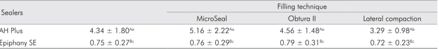
Regardless of the filling technique used, AH Plus presented higher push-out bond strength values (4.34 ± 1.80 MPa) than Epiphany SE (0.75 ± 0.27 MPa; p < 0.05).
Analysis of the interaction between filling technique and sealer showed that Obtura II and
MicroSeal illed with AH Plus presented the highest adhesion values (4.46 ± 1.48 and 5.16 ± 2.22 MPa, respectively) and lateral-compaction filled with
Epiphany SE presented the lowest adhesion values
(0.72 ± 0.23 MPa; p < 0.05). On using Epiphany SE, no statistically signiicant difference (p > 0.05) was observed between the illing techniques; in addition,
it also presented the lowest adhesion values.
There were no statistically signiicant differences (p > 0.05) between the root-thirds (cervical: 2.44 ± 2.03, middle: 2.50 ± 2.27, and apical: 2.70 ± 2.34 MPa).
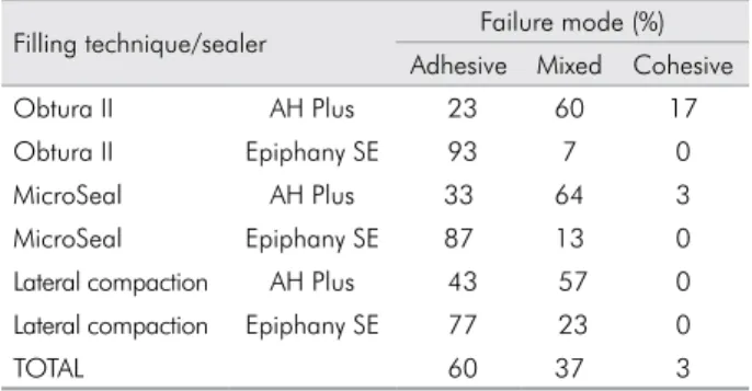
Analysis of the mode of failure showed a
predominance of adhesive failures (60%) in all groups,
particularly at the sealer/dentin interface. There
were a many mixed failures in the specimens illed
with AH Plus compared with those with Epiphany
SE. Cohesive failures were observed in 17% of the specimens illed with Obtura II and AH Plus. The
distribution of failure modes is presented in Table 2.
Discussion
The selection and use of appropriate illing techniques is essential to completely ill the root canal and prevent bacterial infection, the main cause of apical periodontitis. The quality of root canal illings is generally evaluated
using two-dimensional radiographs,19 leakage models,1,5
and cross-sectional methods.3,12 These techniques
enable visualization of the distribution of root illing materials, such as gutta-percha and sealer, inside the
canal.3 However, the sealer bond strength is related to its ability to remain adhered to the root dentin
walls,20 and this property is important in static and
dynamic situations. In static circumstances, the adhesion eliminates spaces that allow iniltration of luids into the sealer/dentin interface. In dynamic situations, the
adhesion is necessary to prevent sealer dislodgment during prosthetic procedures.21 The push-out test has been shown to be effective and reliable in the assessment
of sealers bond strength to dentin, as it evaluate the
material dislocation resistance.22 Furthermore, the push-out test causes failure parallel to the dentin/material
interface, which is similar to that observed in clinical
circumstances.23,24 In this study, the push-out test was used to compare the bond strength of two different
thermoplastic filling techniques using epoxy- and methacrylate-based sealers. Differences in bond strength
were observed between the techniques as well as the
sealer types used, thereby rejecting the null hypothesis. The indings of this study showed that, regardless of the illing technique used, AH Plus presented higher push-out bond strength values than Epiphany SE. According to Carneiro et al.,24 epoxy-based sealers (AH Plus) penetrate deeper into the microirregularities
because of its lowability and long polymerization time,
which in turn enhances the mechanical interlocking
between sealer and dentin. In addition, the cohesion
between the sealer molecules increases the resistance
Table 1. Bond strength mean and standard deviation values (in MPa) for the displacement of the filling material from the specimens in each group during the push-out test.
Sealers Filling technique
MicroSeal Obtura II Lateral compaction
AH Plus 4.34 ± 1.80Aa 5.16 ± 2.22Aa 4.56 ± 1.48Aa 3.29 ± 0.98Ab
Epiphany SE 0.75 ± 0.27Bc 0.76 ± 0.29Bc 0.79 ± 0.31Bc 0.72 ± 0.23Bc
to displacement of the material from dentin surfaces,
which translates into greater adhesion.25 Based on
previous studies,23,25,26 the outcome of Epiphany may
be a result of incomplete sealer photoactivation caused by decreased light exposure in the deepest regions of
the root canal or by oxygen inhibition. Consequently, there is a reduced degree of monomer conversion,
resulting in lower bond strengths.
With respect to the illing techniques, the highest
mean values were obtained when MicroSeal and
Obtura II were used with AH Plus, and this group was different from the lateral-compaction groups (p < 0.05),
which showed statistical similarities with different
types of sealers (p > 0.05). These results could be due
to the improved low of warm gutta-percha2,27 that,
along with vertical compaction (performed to inish the illing procedure), resulted in high volume of a compact and homogeneous mass of gutta-percha and
minimal volume of sealer. This is generally associated with higher material retention.28 Nevertheless, the cold lateral-compaction technique does not allow complete illing of the root canal system irregularities, resulting in absence of sealer and/or gutta-percha in
some canal areas and reduced illing bond strength,10
as observed in the present study.
In addition, it is important to indicate that although both systems thermoplasticize the gutta-percha,
MicroSeal has an intermediate procedure using a
taper. 04 condenser at 5000 rpm. This is used to ensure a suficient volume of gutta-percha in the canal and
therefore higher bond strength may be expected
with this technique. However, in the present study, no statistically signiicant differences were observed
in the bond strength produced by MicroSeal and Obtura II techniques.
The analysis of failure type conirmed the bond
strength results of the sealers and/or techniques used.
Mixed failures were predominant with AH Plus,
regardless of the technique used. In the Epiphany
SE and lateral-compaction groups, the prevalence
of adhesive failures can be explained by the weak
adhesion of illing material to the root dentin.18,21
Here we also investigated the effect of root-thirds on
sealer bond strength. According to Tao and Pashley,29
there are some structural differences in the root dentin, such as higher tubular density/diameter, and collagen iber density in the cervical-third. Therefore, sealer
bond strength must be higher in these parts of the root.
However, it was observed that despite the structural differences, the sealer bond strength values between the root-thirds, for different illing techniques and/or sealer types, were statistically similar. These results could be due to the crown-down instrumentation performed before root illing. Because the cervical-third dentin presents higher tubular/collagen density, the
instrumentation causes greater formation of smear
layer in this area, which could negatively affect the
illing material bond strength.29
Overall, in the present study, better results were observed with thermoplastic filling techniques, but the lateral-compaction bond strength values
obtained were in accordance with previous studies which confirmed their clinical acceptability.5,10,30
Therefore, clinical implications of illing techniques
and/or endodontic sealers should not be based on
bond strength results alone; assessment of the sealing ability using different iniltration methods as well as 3-D micro-CT evaluations could provide different
perspectives and demonstrate the actual effects of the materials/techniques used for endodontic treatment.
Conclusion
Within the limitations of the present study, it can
be concluded that MicroSeal and Obtura II produce
illings with higher bond strengths within the root canal wall than lateral-compaction. AH Plus sealers
produced higher bond strengths when compared with
Epiphany SE, regardless of the illing technique used.
Table 2. Distribution of the mode of failure after the displacement of the filling material from the specimens in each group during the push-out test.
Filling technique/sealer Failure mode (%) Adhesive Mixed Cohesive
Obtura II AH Plus 23 60 17
Obtura II Epiphany SE 93 7 0
MicroSeal AH Plus 33 64 3
MicroSeal Epiphany SE 87 13 0
Lateral compaction AH Plus 43 57 0 Lateral compaction Epiphany SE 77 23 0
1. Leonardo MR, Cervi DA, Tanomaru JMG, Silva LAB. Effect of different rotary instrumentation techniques and
thermoplastic filling on apical sealing. J Appl Oral Sci. 2004;12(1):89-92. doi:10.1590/S1678-77572004000100016
2. Venturi M. Evaluation of canal filling after using two
warm vertical gutta-percha compaction techniques in vivo: a preliminary study. Int Endod J. 2006;39(7):538-46. doi:10.1111/j.1365-2591.2006.01106.x
3. Ordinola-Zapata R, Bramante CM, Moraes IG, Bernardineli N, Garcia RB, Gutmann JL. Analysis of the gutta-percha filled area in C-shaped mandibular molars obtured with a modified MicroSeal techniques. Int Endod J. 2009;42(3):186-97. doi:10.1111/j.1365-2591.2008.01495.x
4. Ünal GÇ, Kaia BU, Taç AG, Keçece AD. A comparison
of the efficacy of conventional and new retreatment
instruments to remove gutta-percha in curved root canals: an ex vivo study. Int Endod J. 2009;42(4):344-50. doi:10.1111/j.1365-2591.2008.01518.x
5. De-Deus G, Reis C, Beznos D, Abranches AMG,
Coutinho-Filho T, Paciornik S. Limited ability of three commonly used thermoplasticized gutta-percha techniques in filling oval-shaped canals. J Endod. 2008;34(11):1401-5. doi:10.1016/j.joen.2008.08.015
6. Gulsahi K, Cehreli ZC, Onay EO, Tasman-Dagli F, Ungor
M. Comparison of the area of resin-based sealer and voids in roots obturated with Resilon and gutta-percha. J Endod. 2007;33(11):1338-41. doi:10.1016/j.joen.2007.06.015
7. Davalou S, Gutmann JL, Nunn MH. Assessment of apical and coronal root canal seals using contemporary endodontic obturation and restorative materials and techniques. Int
Endod J. 1999;32(5):388-96. doi:10.1046/j.1365-2591.1999.00246.x
8. Goldberg F, Artaza LP, Silvio A. Effectiveness of different obturation tech niques in the filling of
simulated lateral canals. J Endod. 2001;27(5):362-4. doi:10.1097/00004770-200105000-00015
9. Tagger M, Tagger E, Tjan AHL, Bakland LK. Measurement
of adhesion of endodontic sealers to dentin. J Endod. 2002;28(5):351-54. doi:10.1097/00004770-200205000-00001 10. Brosco VH, Bernardineli N, Moraes IG. In vitro evaluation
of the apical sealing of root canals obturated with
different techniques. J Appl Oral Sci. 2003;11(3):181-85. doi:10.1590/S1678-77572003000300005
11. Karabucak B, Kim A, Chen V, Iqbal MK. The comparison
of gutta-percha and resilon penetration into lateral canals with different thermoplastic delivery systems. J Endod. 2008;34(7):847-49. doi:10.1016/j.joen.2008.03.024
12. Cathro PR, Love RM. Comparison of Microseal and
System B/Obtura II obturation techniques. Int Endod J. 2003;36(12):876-82. doi:10.1111/j.1365-2591.2003.00741.x
13. Magg iore F. M ic ro S ea l system s a nd mod i f ied
technique. Dent Clin North Am. 2004;48(1):217-64. doi:10.1016/j.cden.2003.11.005
14. Soo WK, Thong YL, Gutmann JL. A comparison of four gutta-percha filling techniques in simulated C-shaped canals. Int Endod J. 2015;48(8):736-46. doi:10.1111/iej.12371
15. Bradshaw GB, Hall A, Edmunds DH. The sealing ability of
injection-moulded thermoplasticized gutta-percha. Int Endod J. 1989;22(1):17-20. doi:10.1111/j.1365-2591.1989.tb00500.x
16. Olson AK, Hartwell GR, Weller RN. Evaluation of the controlled
placement of injected thermoplasticized gutta-percha. J Endod. 1989;15(7):306-9. doi:10.1016/S0099-2399(89)80052-4
17. Gutmann JL, Saunders WP, Saunders EM, Nguyen L. An assessment of the plastic Thermafil obturation technique.
Part 2. Material adaptation and sealability. Int Endod J. 1993;26(3):179-83. doi:10.1111/j.1365-2591.1993.tb00790.x
18. Gonçalves L, Silva-Sousa YT, Raucci Neto W, Teixeira CS,
Sousa-Neto MD, Alfredo E. Effect of different irrigation
protocols on the radicular dentin interface and bond strength
with a metacrylate-based endodontic sealer. Microsc Res Tech. 2014;77(6):446-52. doi:10.1002/jemt.22365
19. Hörsted-Bindslev P, Andersen MA, Jensen MF, Nilsson JH,
Wenzel A. Quality of molar root canal fillings performed with the lateral compaction and the single-cone technique. J Endod. 2007;33(4):468-71. doi:10.1016/j.joen.2006.12.016 20. Sousa-Neto MD, Coelho FI, Marchesan MA, Alfredo E,
Silva-Sousa YTC. Ex vivo study of the adhesion of an epoxy-based sealer to human dentine submitted to irradiation with Er:YAG and Nd:YAG. Int Endod J. 2005;38(12):866-70. doi:10.1111/j.1365-2591.2005.01027.x
21. Rached-Junior FJ, Souza-Gabriel AE, Alfredo E, Miranda
CE, Silva-Sousa YT, Sousa-Neto MD. Bond strength of Epiphany sealer prepared with resinous solvent. J Endod. 2009;35(2):251-5. doi:10.1016/j.joen.2008.10.027
22. Moinzadeh AT, Jongsma L, Wesselink PR. Considerations
about the use of the push-out test in Endodontic research. Int Endod J. 2015;48(5):498-500. doi:10.1111/iej.12416 23. Shokouhinejad N1, Sharifian MR, Jafari M, Sabeti MA.
Push-out bond strength of Resilon/Epiphany self-etch and gutta-percha/AH26 after different irrigation protocols. Oral Surg Oral Med Oral Pathol Oral Radiol Endod. 2010;110(5):e88-92. doi:10.1016/j.tripleo.2010.05.069
24. Carneiro SM, Sousa-Neto MD, Rached Junior FA, Miranda CE, Silva SR, Silva-Sousa YT. Push-out strength of root
fillings with or without thermomechanical compaction. Int
Endod J. 2012;45(9):821-8. doi:10.1111/j.1365-2591.2012.02039.x
25. Nunes VH, Silva RG, Alfredo E, Sousa-Neto MD, Silva-Sousa
YT. Adhesion of Epiphany and AH Plus sealers to human root dentin treated with different solutions. Braz Dent J. 2008;19(1):46-50. doi:10.1590/S0103-64402008000100008
26. Alfredo E, Silva SR, Ozório JE, Sousa-Neto MD,
Brugnera-Júnior A, Silva-Sousa YT. Bond strength of AH
Plus and Epiphany sealers on root dentine irradiated
with 980 nm diode laser. Int Endod J. 2008;41(9):733-40. doi:10.1111/j.1365-2591.2008.01418.x
27. Wu M, van der Sluis LW, Wesselink PR. A preliminary
study of the percentage of gutta-percha-filled area
in the apical canal filled with vertically compacted
warm gutta-percha. Int Endod J. 2002;35(6):527-35. doi:10.1046/j.1365-2591.2002.00522.x
28. Schilder H. Filling root canals in three dimensions. J Endod.
2006;32(4):281-90. doi:10.1016/j.joen.2006.02.007
29. Tao L, Pashley D. Shear bond strengths to dentin: effect
of surface treatments, depth and position. Dent Mater. 1988;4(7):371-8. doi:10.1016/S0109-5641(88)80052-6