Braz Dent J 20(1) 2009
54 C.M. Silva and K.R.H.C. Dias
INTRODUCTION
New technologies have been continuously inves-tigated in esthetic dentistry with the aim of improving the physical, mechanical and esthetic properties of esthetic restorative materials.
Quartz-tungsten-halogen (QTH) lamps are com-posed of a quartz tungsten thread found in the bulb, involved by inert gas, ilter, refrigerating system and optic ibers for light conduction. These lamps operate on a 450-500 nm wavelength range and are popular vis-ible light sources. However, QTH bulbs have a limited effective lifetime and several factors may contribute to produce an inadequate polymerization output, such as presence of debris on the iber tip, breakage of the tungsten ilaments of the optical iber and voltage varia -tions. In addition, only little energy of the total energy
Compressive Strength of Esthetic Restorative
Materials Polymerized with Quartz-Tungsten-
Halogen Light and Blue LED
Cecy Martins SILVA1
Katia Regina Hostilio Cervantes DIAS2
1Dental School, Federal University of Pará, Belém, PA, Brazil 2Dental School, State of University of Rio de Janeiro, RJ, Brazil
This study compared the compressive strength of a composite resin and compomer photoactivated with a conventional quartz-tungsten halogen-light (XL 3000, 3M/SPE) and a blue light-emitting diode (LED) (SmartLite PS; Dentsply/De Trey). Forty disc-shaped
specimens were prepared using a split polytetraluoroethylene matrix (4.0 mm diameter x 8.0 mm hight) in which the materials were
inserted incrementally. The curing time of each increment was of 40 s with the QTH and 10 s with the LED. The specimens were ran-domly assigned to 4 groups (n=10), according to the light source and the restorative material. After storage in distilled water at 37oC
± 2oC for 24 h, the specimens was tested in compressive strength in a universal testing machine with load cell of 500 kgf running at a crosshead speed of 0.5 mm/min. Data (in MPa) were analyzed statistically by ANOVA and Student-Newman-Keuls test (p<0.05). For
the composite resin, light curing with the QTH source did not produce statistically signiicant difference (p>0.05) in the compressive
strength when compared to light curing with the LED source. However, light curing of the compomer with the QTH source resulted
in signiicantly higher compressive strength than the use of the LED unit (p>0.05). The composite resin presented signiicantly higher (p>0.05) compressive strength than the compomer, regardless of the light source. In conclusion, the compressive strength of the tested materials photoactivated with a QTH and a LED light source was inluenced by the energy density employed and the chemical
composition of the esthetic restorative materials.
Key Words: composite resin, compomers, light-curing, LED, QTH light.
input is effectively converted into light the remainder being generated as heat (1).
Blue light emitting diodes (LED) produce a stable, eficient, long-lasting output of blue light in a short-wave emission spectrum (450-490 nm), with peak at 470 nm, coinciding with the absorption peak of camphorquinone (468 nm), which is the photoinitiator present in most composites (2). LED units have some advantages over QTH lamps due to their potential lifetime of over 10,000 hours without a signiicant degradation in light output after this period, no need of cooling system or ilters, no noise production during function, operation with batteries and direct conversion of electrical energy into light with little amount of wasted energy and minimum heat generation (2). Light-curing units (LCUs) with one LED producing irradiance have recently been marketed.
Several studies have investigated the inluence
Correspondence: Profa. Dra. Cecy Martins Silva, Rua dos Mundurucus, 822/903, Jurunas, 66025-660 Belém, PA, Brasil. Tel: +55-91-3252-1269. Fax: +55-91-3201-7563. e-mail: [email protected]
ISSN 0103-6440
Braz Dent J 20(1) 2009
Compressive strength of dental materials 55
of halogen and LED light-curing on different properties of light-cured composites such as degree of conversion (3-5), depth of cure (6-8), hardness (6,8-13), diametral tensile strength (14), lexural strength (15), abrasion resistance (16) and compressive strength (17). There-fore, research studies investigating the effectiveness of resin-based materials photoactivated with different light sources by compressive strength testing will con-tribute to elucidate the actual participation of LCUs on the longevity of esthetic restorations in the oral cavity. The purpose of this study was to evaluate the curing eficacy of a high-irradiance blue LED LCU and a QTH lamp by assessing the compressive strength of a composite resin and a compomer photoactivated with both light sources. The null hypothesis tested is that there is no signiicant difference in the compressive strength when the materials are photoactivated with the light sources.
MATERIAL AND METHODS
The following materials and LCUs were used in this study: Dyract Ap compomer (Dentsply/Caulk, Milford, DE, USA; batch number 0201001349), TPH Spectrum composite resin (Dentsply/Caulk; batch num -ber 55596). XL 3000 QTH light source (3M/ESPE, St. Paul, MN, USA; light intensity = 470 mW/cm2; 400-510 nm wavelength range), and Smart Lite PS blue LED light source (Dentsply/DeTrey, Konstanz, Germany; intensity = 950 mW/cm2; 450-490 nm wavelength range). Before preparation of specimens, the intensity of the QTH and LED sources was measured with curing radiometers, Demetron L.E.D. Radiometer (Demetron/Kerr Corp., Orange, CA. USA; batch number 79302331, and Cure Rite Radiometer (EFOS, Mississauga, Ontario, Canada; batch number 9152), respectively.
Twenty disc-shaped specimens of each material were prepared using a split white polytetraluoroeth -ylene matrix (4.0 mm diameter x 8.0 mm height). The matrix was placed onto a 2.0-mm-thick glass plate and was illed in 4 approximately 2.0-mm-thick increments. Ten specimens of material were light cured with one of light sources. Each increment was exposed to the QTH and LED LCUs for 40 s and 10 s, respectively. After insertion of the last increment, a transparent polyester strip and a 2.0-mm-thick glass plate were placed onto the matrix and light curing was done through this set. After storage in distilled water at 37oC ± 2oC protected from
light during 24 h, the specimens were tested in compres-sive strength in a universal testing machine (EMIC DL 10.000, São José dos Pinhais, PR, Brazil) with a load cell of 500 kgf and a crosshead speed of 0.5 mm/min. Data were tabulated and analyzed statistically using ANOVA and the Student-Newman-Keuls test (SNK) for pairwise comparisons. Signiicance level was set at 5%.
RESULTS
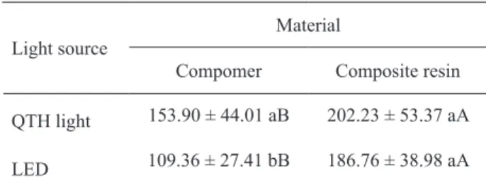
Table 1 shows the mean compressive strength values (in MPa) and standard deviations of the resin materials photoactivated with the QTH and LED sources. The SNK test revealed statistically signiicant interaction when individual comparison was done between the light sources (p<0.05) and between the materials (p<0.05) (Table 1). For the composite resin, light curing with the QTH source did not produce sig-niicant difference (p>0.05) in the compressive strength when compared to light curing with the LED source. However, light curing of the compomer with the QTH source resulted in signiicantly higher compressive strength than the use of the LED unit (p>0.05). The composite resin presented signiicantly higher (p>0.05) compressive strength than the compomer regardless of the light source.
DISCUSSION
Compressive strength has a particularly impor-tant role in the mastication process since several of the masticatory forces are of compressive nature. The
Table 1. Compressive strength means (in MPa) and standard deviations of the resin materials light cured with the QTH and LED sources.
Light source
Material
Compomer Composite resin
QTH light 153.90 ± 44.01 aB 202.23 ± 53.37 aA
LED 109.36 ± 27.41 bB 186.76 ± 38.98 aA
Means followed by same lowercase letters in columns and uppercase
Braz Dent J 20(1) 2009
56 C.M. Silva and K.R.H.C. Dias
maximum resistance to compression is calculated by the original cross-sectional area of the test specimens and the maximum force applied. The compression forces applied on each side of the test specimens are dissipated into shear forces along the cuneiform area on each side. As a result of the action of the two cones on the cylinder, traction forces arise in the central portion of the mass. Due to this tensile dissipation in the specimen, a matrix that reproduced a cylinder that was twice as long as the diameter (4.0 mm diameter and 8.0 mm length) had to be used in order to have satisfactory results (3).
Signiicant interaction was noted between the light source and the tested material. The compomer cured with the QTH lamp showed signiicantly higher mean compressive strength values than those obtained when the material was cured with the LED source. The composite resin light-cured with the QTH lamp also showed higher mean compressive strength values than those obtained with the LED, however without statistical signiicance. This result suggests that the compressive strength of the composite resin was not affected by the type of light curing unit, which has been found elsewhere (17) (Table 1). Mills et al. (9) found no statistically signiicant differences in the compressive strength of dental composites photoactivated with two experimental high-power LED prototypes and a commercial QTH lamp. However, light curing with a commercial LED LCU resulted in signiicantly lower values.
In the present study, the type of material (com-pomer vs. composite resin) had a signiicant role in light curing. The composite resin presented signii -cantly higher mean compressive strength values than the compomer (Table 1). The literature has shown the chemical composition of dental composites interfere in their mechanical properties (7,8,10,14).
The use of high irradiance with short irradiation time has been recommended to achieve satisfactory depth of cure and improved mechanical properties. However, high irradiance has also been shown to cause high polymerization shrinkage and increased marginal microleakage around composite restorations (18).
Determination of curing time was an important factor in the results obtained in this study, which are consistent with those of previous investigations (6,13). The curing time used for composite photoactivation with both units followed the manufacturer’s recommenda-tions. The use of the LED unit allowed shortening the curing time to 10 s due to the high irradiance delivered
by this light source. However, the use of high irradia-tion in a short period of time provides a rapid reacirradia-tion of polymerization, shortening the pre-gel phase, pass-ing quickly to a rigid state (post-gel phase), reducpass-ing the number of chemical reactions for the conversion of monomers into polymers, which may interfere with the mechanical properties of the material.
Light intensity or output power density or irradi-ance is expressed in W/cm² and represents the number of photons emitted per second by a light source per
unit area of the light-cured point. The energy density for light curing is calculated by multiplying the light intensity by the curing time and is expressed in J/cm² (19,20). The eficiency of LCUs depends on the total energy concept, according to which both intensity and photo-initiation time are important for an eficient light curing of dental composites.
For both materials, the light intensity of the QTH LCU was 470 mW/cm² and the exposure duration was 40 s, while the LED source had intensity of 950 mW/cm² with exposure duration of 10 s. Therefore, the energy density used by the QTH and LED LCU were 18 J/cm² and 9.5 J/cm², respectively. The higher energy output was used for the QTH LCU could help explaining the higher compressive strength obtained when the materials were light activated with the halogen lamp.
The conversion rate presents a direct relationship with the amount of energy applied, which means that higher energy intensity will promote greater monomer conversion with consequent improved mechanical pro-prieties of the material (4). It may be speculated that the high radiation emitted by the LED LCU Smart Lite PS in short exposure duration, accelerates the curing reac -tion, reducing the polymer lowability, increasing the modulus of elasticity and shortening the pre-gel phase, which could interfere with the results of resistance to compression. In order to obtain an effective light cur-ing, which means 50 to 60% of monomer conversion, a radiant energy of approximately 16 J/cm² for a 2-mm-thick resin layer is needed. The increase of output power density accelerates reaction speed.
Braz Dent J 20(1) 2009
Compressive strength of dental materials 57
compomer when photoactivated with each of the light sources. The composite resin presented better results than the compomer. In summary, the inluence of the chemi -cal composition of the materials, light intensity, curing time and output power density of the LCUs should be considered on the inal outcomes. Further research must be carried out to clarify the mechanical proprieties of composites photoactivated with LED units, addressing the factors mentioned above. In conclusion, the compres-sive strength of the tested materials photoactivated with a QTH and a LED light source was inluenced by the energy density employed and the chemical composition of esthetic restorative materials. Within the limitations of this in vitro study, it may be suggested that the superior-ity of halogen lamps over LEDs is questionable when different resin-based materials are evaluated.
RESUMO
Este estudo comparou a resistência à compressão de uma resina composta e de um compômero, fotoativados com luz halógena convencional de quarto-tungstênio (QTH) (XL 300, 3M/SPE) e LED azul (SmartLite PS; Dentsply/De Trey). Foram
confec-cionados 40 espécimes em forma de disco usando uma matriz bipartida de politetraluoretileno (4,0 mm de diâmetro x 8,0 mm
de altura) em que o material foi inserido incrementalmente. O tempo de polimerização de cada incremento foi de 40 s para a
luz halógena convencional e de 10 s para o LED. Os espécimes
foram aleatoriamente alocados em 4 grupos (n=10), de acordo com a fonte de luz e com o material restaurador. Depois de arma-zenadas em água destilada a 37°C ± 2°C por 24 h, a resistência à
compressão dos espécimes foi testada em uma máquina universal de ensaios com célula de carga de 500 kgf a uma velocidade de car -regamento de 0,5 mm/min. Os dados (em MPa) foram analisados estatisticamente por ANOVA e teste de Student-Newman-Keuls (p<0,05). Para a resina composta, a fotopolimerização com luz
halógena não produziu diferença estatisticamente signiicante (p>0,05) em sua resistência à compressão quando comparada à
fotopolimerização com LED. Contudo, a fotopolimerização do compômero com a luz halógena resultou em uma resistência à
compressão signiicativamente maior que a feita o LED (p>0,05). A resina composta apresentou resistência à compressão signii -cativamente maior que a do compômero, independente da fonte de luz. Concluiu-se que a resistência à compressão dos materiais
fotopolimerizados com luz halógena e LED foi inluenciada pela
densidade de energia empregada e pela composição química dos
materiais restauradores estéticos.
REFERENCES
1. Rueggeberg FA. Contemporary issues in photocuring. Compend Cont Educ Dent 1999;20:4-15.
2. Nomoto R. Effect of light wavelength on polymerization of
light-cured resins. Dent Mater J 1997;16:60-73.
3. Rueggeberg FA, Caughman WF, Curtis JW. Effect of light inten
-sity and exposure duration on cure of composite resin. Oper Dent
1994;19:26-32.
4. Halvorson RH, Erickson RL, Davidson CL. Energy dependent po
-lymerization of resin-based composite. Dent Mater
2002;18:463-469.
5. Arrais CAG, Pontes FM, Santos LPS, Leite ER, Giannini M. Degree of conversion of adhesive systems light-cured by LED an
halogen light. Braz Dent J 2007;18:54-59.
6. Uhl A, Mills RW, Jandt KD. Photoinitiator dependent composite
depth of cure and Knoop hardness with halogen and LED
light-curing units. Biomaterials 2003;24:1787-1795.
7. Koupis NS, Vercruysse CWJ, Marks LAM, Martens LC, Ver
-beech RMH. Curing depth of (polyacid-modiied) composite
resins determined by scraping and a penetrometer. Dent Mater
2004;20:908-914.
8. Obici AC, Sinhoreti MAC, Correr Sobrinho L, Góes MF, Consani
S. Evaluation of depth of cure and Knoop hardness in a dental composite photoactivated using different methods. Braz Dent J 2004;15:199-203.
9. Mills RW, Uhl A, Blackwell GB, Jandt KD. High power light
emitting diode (LED) arrays versus halogen light polymerization of oral biomaterials: Barcol hardness, compressive strength and radiometric properties. Biomaterials 2002;23:2955-2963.
10. Soh MS, Yap AUJ, Siow KS. The effectiveness of cure of LED
and halogen curing lights at varying cavity depths. Oper Dent
2003;28:707-715.
11. Cefaly DFG, Ferrarezi GAO, Tapety CMC, Lauris JRP, Navarro
MFLN. Microhardness of resin-based materials polymerized with
LED and halogen curing units. Braz Dent J 2005;16:98-102.
12. Correr AB, Sinhoretti MAC, Sobrinho LC, Tango RN, Schneider
LFJ, Consani S. Effect of the increase of energy density on Knoop
hardness of dental composites light-cured by conventional QTH,
LED e Xenon Plasma Arc. Braz Dent J 2005;16:218-224. 13. Price RBT, Felix CA, Andreou P. Knoop hardness of ten composite
resins irradiated with high-power LED and quartz-tungsten-halogen lights. Biomaterials 2005;26:2631-2641.
14. Tolosa MCCG, Paulillo LAMS, Giannini M, Santos AJS, Dias CTS. Inluence of composite restorative materials and light- curing
units on diametrical tensile strength. Braz Oral Res 2005;19:123-126.
15. Miyazaki M, Oshida Y, Moore BK, Onose H. Effect of light ex
-posure on fracture toughness and lexural strength of light-cured composites. Dent Mater 1996;12:328-332.
16. Martinelli J, Pires-de-Souza FCP, Casemiro LA, Tirapelli C,
Panzeri H. Abrasion resistance of composites polymerized by
light-emitting (LED) and halogen light-curing units. Braz Dent J
2006;17:29-33.
17. Jandt KD, Mills RW, Blackell GB, Ashworth SH. Depth of cure
compressive strength of dental composites cured with blue emit-ting diodes (LEDs). Dent Mater 2000;16:41-47.
18. Unterbrink GL, Muessner R. Inluence of intensity on two restor
-ative systems. J Dent 1995;23:183-189.
19. Nomoto R, McCabe JF, Hirano S. Effect of aperture size on irradi
-ance of LED curing units. Dent Mater 2004;20:687-692.
20. Nomoto R, Uchida K, Hirasawa T. Effect of light intensity on
polymerisation of light-cured composite resins. Dent Mater J 1994;13:198-205.