Body disabling chronic hemiparesis does not appear
to determine differences in the body mass index
when compared to able-bodied controls
Paulo Henrique Ferreira de Araujo-Barbosa,ILidiane Teles de Menezes,IAbraa˜o Souza Costa,I Anderson Castro Mundim,IIOsmair Gomes de Macedo,IEmerson Fachin-MartinsII
IDepartment of Physical Therapy, Faculty of Ceilandia, University of Brasilia, Campus of Ceilandia, Ceilandia Sul, Brasilia, DF, BrazilIIGraduate Program (Po´s-Graduaca˜o) in Health Sciences and Technologies, Faculty of Ceilandia, University of Brasilia, Campus of Ceilandia, Ceilandia Sul, Brasilia, Federal District, Brazil
OBJECTIVE:(a) To compare the Body Mass Index between hemiparetic (disabled-bodied) patients vs. matched able-bodied controls, establishing relationships between Body Mass Index and the anthropometric variables (weight and height) used to calculate it; (b) to determine whether the Body Mass Index could be used as a disability indicator.
METHODS:Twenty hemiparetic patients were matched by gender and age to twenty control subjects. All of them were included in procedures to measure the anthropometric parameters used to calculate Body Mass Index. The data were submitted to statistical procedures.
RESULTS:The hemiparetic group did not present significant difference in the Body Mass Index when compared with controls, despite the fact that controls were 5.5 cm taller. From the variables used to calculate Body Mass Index, only weight presented a significant correlation to the index for both groups.
CONCLUSION:Significant differences in the Body Mass Index were not observed between groups. Moreover, for both groups, the index was only associated with body mass. Our results show that Body Mass Index is not a good parameter to establish relationships with disability indicators for disable-bodied patients.
KEYWORDS: stroke; anthropometry; body composition.
de Araujo-Barbosa PHF, de Menezes LT, Costa Abraa˜o Souza, Mundim AC, de Macedo OG, Fachin-Martins E. Body disabling chronic hemiparesis does not appear to determine differences in the body mass index when compared to able-bodied controls. MEDICALEXPRESS. 2014 June;1 (3):135-139.
Received for publication onMarch 27 2014;First review completed onApril 02 2014;Accepted for publication onApril 14 2014
E-mail: [email protected]
B INTRODUCTION
Many studies have reported Body Mass Index (BMI) as a risk predictor for diseases, but few studies have hypothe-sized this index as a predictor for exacerbation, comparing BMI observed in disable-bodied patients with able-bodied controls1 – 3, in order to verify if BMI could be used to prevent exacerbation of individuals already suffering from outcomes of chronic diseases as observed after cerebrovascular diseases.
A study carried out over a 10 year span showed that the BMI was not a good risk predictor for cerebrovascular diseases4,
although associations between BMI or weight relative to incidence of cerebrovascular accident and mortality had been described5in a previous study. This evidence suggested that BMI might not be the most informative measure for relating
obesity to cerebrovascular diseases, requiring further clarifica-tion to verify the usefulness of this index.
For subjects with diseases such as diabetes mellitus, some studies6 – 8have sought to identify associations of anthropo-metric characteristics with aggravation indicators that may contribute to the monitoring of these individuals. However, to the best of our knowledge no such information has been reported for stroke survivors with acquired motor disabil-ities, such as hemiparesis.
Stroke is a major public health problem worldwide; it has a high degree of incidence that can lead to death or to a disability when the subject survives; this results in motor, cognitive and/or sensory disorders that depend on the severity of the case and the extent of the injuries1 – 3,9 – 11.
In the light of these facts, this study aimed to compare the BMI of subjects with chronic spastic hemiparesis (disable-bodied) with able-bodied controls matched by gender and age, establishing relationships between the body mass index and
DOI:10.5935/MedicalExpress.2014.03.07
the anthropometric variables used to calculate it; we also determined whether BMI could be used as disability indicator.
B MATERIALS AND METHODS Subjects
Twenty subjects with spastic hemiparesis classified as 1.3^0.2 (mean^SEM) on theAshworthmodified scale, with a chronicity span of 41.5^12.5 months and ages varying from 29 to 81 years participated in this study. Subjects with hemiparesis (disable-bodied) were included who: (a) survived a stroke for at least six months; (b) manifested spastic hemiparesis resulting from alteration in the irrigation territory of the middle cerebral artery confirmed by a physician; (c) presented with no pathology other than the cerebral ischemic event that produced the hemiparesis and (d) showed appropriate understanding of the instructions required to effectively participate in the study, as confirmed by the Mini-Mental Test. Subjects who presented other neurological aggravations besides those determined by the stroke were excluded. Twenty able-bodied volunteer subjects without hemiparesis were matched by gender and age to the hemiparetic subjects to compose a control group.
Patients and volunteers were verbally informed about the procedures to which they would be submitted and agreed to participate in the study; all subjects signed the Informed Consent Form (ICF) approved by the Research Ethics Committee of the Health Sciences Faculty of University Brasilia (UnB) – protocol number 052/2011.
Anthropometric measurements
The anthropometric measurements recorded in the study were height (cm) and body mass (kg). Body mass was measured using a Plennaw digital weighing scale with
maximum capacity of 150 kg and a precision of 100 g. Height was obtained by means of a tape measurement which quantified the vertical distance between the floor and a pencil mark on the wall at the individual’s highest point, inscribed while subjects had their backs to the wall. Based on these measurements, the BMI was then calculated, as the ratio of body mass (kg) by the square of the height (m).
A record was also made of age and chronicity, defined as the period between the cerebrovascular event that caused the hemiparesis and the assessment time in complete months.
All subjects underwent a short preliminary interview where they were questioned about their lifestyle habits, including: (a) whether they had been smokers, or still were, for more than one year, (b) whether they had drunk or continued to drink alcohol more than 3 times per week and (c) whether they practiced any type of regular physical activity more than 3 times per week. During this interview, the degree of spasticity presented by subjects with hemiparesis was evaluated by the Ashworth modified scale.12
Processing and statistical analysis of data
Initially, data were processed through descriptive statistics to take position and dispersion measurements of the quantitative variables and to distribute qualitative variable occurrence frequency. Next, the Kolmogorov-Smirnov normality test was applied for quantitative variables to verify if these showed a Gaussian distribution, which would determine the use of parametric or non-parametric tests for inference or analytical statistics. With the exception of the
chronicity variable, all other variables showed Gaussian distribution.
The Fisher’s exact test was used to identify differences between the frequency distribution proportions calculated for the qualitative variables. Those proportions in which the test demonstrated a p value less than 0.05 were considered significantly different for comparisons between the pro-portions noted in the hemiparesis group, compared to the proportions expected for the control group.
To detect significant mean differences between the set of variables obtained in the hemiparesis when compared to the control group, the matched pair student t test was used, except for chronicity comparisons in which theWilcoxontest was used, once a Gaussian distribution was not identified for this variable. A p value, 0.05 was considered a significant difference.
Associations between different variables were detected by the Spearman correlation test for tests that involved the chronicity variable (non-parametric test) and by the Pearson correlation test for the other variables (parametric test).
To classify the intensity of the correlations detected as significant by the tests (p,0.05), the limits established by Menz and collaborators13were used, defining as strong the
coefficient association of correlation greater or equal to 0.75; moderate for coefficients varying between 0.40 and 0.75 and weak for coefficients less than or equal to 0.40.
B RESULTS
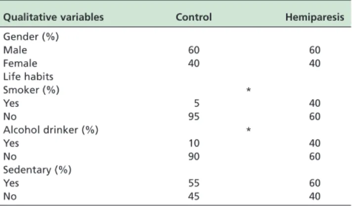
The sample used in this study was 60 % male (Table 1). Among the subjects from both groups, non-smokers and non-drinkers predominated; however, significant differences in the proportion between the groups were detected by Fisher’s exact test, showing that the hemiparesis group had more smokers (40 %) and alcohol drinkers (40 %) than the control group (5 % and 10 %, respectively). There was a small majority of sedentary subjects in both groups, without significant differences between proportions.
Because the groups were paired by gender and age, they had similar variables, whereby the same proportion was noted for each gender (Table 1); the mean age was 59.40^3.04 for the hemiparesis group and 58.55^3.11 for
Table 1 -Frequency distribution of qualitative variables of the sample for the control (able-bodies) and hemiparesis (disable-bodies) groups
Qualitative variables Control Hemiparesis
Gender (%)
Male 60 60
Female 40 40
Life habits
Smoker (%) *
Yes 5 40
No 95 60
Alcohol drinker (%) *
Yes 10 40
No 90 60
Sedentary (%)
Yes 55 60
No 45 40
the control group, with no significant difference (p¼0.3157) between them.
For the variables used for BMI calculation, height was significantly different between groups (158^2 cm for hemi-paretic subjects, 163^2 cm for control subjects (p¼0.0269). Mass for control and hemiparesis groups was 73.72^3.16 kg and 68.68^3.93 kg respectively (Figure 1), with no significant difference between the means (p¼0.2534).
Although there was a significant height difference noted between the groups as Figure 1 shows, this did not reflect a significant difference in BMI (p¼0.6765), which was 28.04^0.92 kg/m2 for the control subjects and 27.38^1.22 kg/m2 for hemiparetic subjects; thus, in both groups the subjects were overweight.
The temporal variables (age and chronicity) did not show significant correlation and demonstrated weak association with BMI, presenting p values of 0.945 and 0.411 and correlation coefficient of 0.017 and 0.195 respectively for each temporal variable (not shown in the figure).
Of the anthropometric variables measured (weight and height), only weight correlated significantly with the BMI for both groups, showing a set of values rising from 49.0 to 98.0 kg that associated themselves with BMI values, which increased from 24.0 to 39.8 kg/m2 in the control group (Figure 2A) and a set of values rising from 46.9 to 107.7 kg that associated themselves with BMI values, which increased from 18.5 to 38.2 kg/m2in the hemiparesis group (Figure 2B). Moderate and strong associations were found between the variables for the control and hemiparesis groups, respect-ively (Figure 2). No such correlation was detected between the height and BMI as also indicated in Figure 1 for both groups.
B DISCUSSION
In the last several decades, a number of studies1 – 3,9,14have identified risk factors that can or cannot be modified by any form of intervention for ischemic and hemorrhagic stroke; however, few studies described risk factors for aggravations after an individual has suffered this cerebrovascular event.
As observed in the majority of reports, this investigation also identified the usual modifiable risk factors for cerebrovascular diseases in greater proportion in the hemiparesis vs. the control group (Table 1), confirming the negative effects of life habits such as smoking, alcohol use and sedentary behavior.
In the same manner that these life habits constitute modifiable risk factors, obesity also represents a risk factor that can be modified. However, this study investigated information about obesity that could help to understand its predictive value forexacerbationin disabled-bodied stroke survivors, and did so in a way different from studies that
Figure 2 -Dispersion graphics of the anthropometric variables measured: height (white balls) and mass (grey balls), for the control group (A) and hemiparesis group (B) indicating the associations of these variables with the BMI (Body Mass Index) calculated. For each variable measured, the correlation coefficient (cc) is indicated and the p value detected by the Pearson correlation test. The white stars indicate significant
correlations (p,0.05).
Figure 1 -Bar chart showing mean and standard error of mean
(MEAN^SEM) for the anthropometric variables measured
(height and mass) and for calculated variable (Body Mass Index, BMI) both in the control group (white) and the hemiparesis group
(grey). The white star indicates significant difference (p,0.05)
described evidence of obesity as a risk factor for cerebrovascular disease15 – 17. Few studies described this
feature in literature.
As already described, one method of detecting overweight and obesity is the BMI1,3,6,11,17, because of the simplicity of its application; however, both favorable17 and unfavorable4 evidence for its use as predictors have been reported.
Our results add to the unfavorable evidence for the use of BMI as a predictor of future aggravation, because no significant differences in this index were found between groups of able and disable-bodied subjects. This, of course, shows that the motor disability resulting from spastic hemiparesis in survivors of cerebrovascular diseases would not be a determining factor for BMI alterations.
However the evidence of no difference between BMI in the two groups could never be used to affirm that subjects with hemiparesis do not suffer alterations in their body composition, seeing as studies using other forms of body composition for stroke survivors show concrete evidence that hemiparesis is capable of promoting considerable osteopenia and sarcopenia18 – 20.
Therefore, the fact that our sample of subjects with spastic hemiparesis is composed of patients in chronic condition with BMI equal to that of control subjects paired by age and gender, does not mean they do not have body composition alteration. The motor disability brought on by spastic hemiparesis results in numerous changes to daily life activities that can lead to disuse and to the subsequent process of sarcopenia and osteopenia21,22.
How then could alterations in body composition be present, even in subjects with hemiparesis who do not show BMI modifications when compared to the control subjects? For many years, there have been assertions and evidence that the isolated measurements of body mass or of mass and body height are insufficient to determine both obesity and malnutrition. Such measures do not evaluate body constitu-ents and therefore could not establish if individuals classified as fat or thin have an adequate percentage of fat in relation to total body mass23 – 25.
By the evidences and affirmations available, it is important to consider the possibility that individuals do in fact have alterations in body composition, while maintaining body mass and consequently BMI: this condition could happen, if lean mass were replaced by fat mass. The need to measure skin folds in people with physical deficiencies should be borne in mind, as a manner of identifying where the body fat may be most concentrated in this population26.
Other important information about fat mass (adipose tissue) is related to the report that muscular deficits may alter the deposition of fat in the affected body segments27. Evidence from adults with spastic hemiparesis following cerebral palsy showed that different values were found for the biceps, triceps, chest, supra-iliac area, thigh and medial calf skin fold thickness between the affected and the unaffected hemibody. Similarly, the percentage of body fat in these individuals also showed significant difference between the hemibodies28.
A limitation of this study lies in the lack of evaluation of fat deposition and of bone (osteopenia) and muscle loss (sarcopenia). It would then have been possible to affirm that the lean mass was substituted by fat mass even when differences in body mass and BMI are not noted.
Furthermore, it should be noted that the BMI of both groups is associated with the body mass, but not with the
height. This association shows that BMI modifications are mainly determined by body mass. This is reinforced by the fact that differences were noted in the height of groups that did not determine BMI differences. An analysis of the relationship between lean and fat mass, in this case, could be very enlightening in future investigations.
The results of this study corroborate the reports that BMI does not reflect the real estimate of the individual’s lean mass and fat mass, therefore being unsuitable in epidemio-logical investigations to identify risk factors in this population.
B CONCLUSION
Although the control subjects show greater height than the subjects with hemiparesis, a significant difference in BMI was not found between these groups. Apart from this, both for disabled-bodied hemiparetic subjects and their matched able-bodied controls, BMI was only significantly associated between groups with body mass in strong and moderate correlations.
B ACKNOWLEDGEMENTS
The author reports no conflicts of interest in this work. This work was supported by the grant for research from Programa Institucional de Bolsas de Iniciaca˜o Cientı´fica – PIBIC/CNPq (2011/2012) and from Conselho Nacional de Desenvolvimento Cientı´fico e Tecnolo´gicos – CNPq processo 473816/2010-5.
B RESUMO
OBJETIVO: (a) Comparar o I´ndice de Massa Corporal de pacientes hemipare´ticos com deficieˆncias motoras versus controles sa˜os, estabelecendo relaco˜es entre I´ndice de Massa Corporal e as varia´veis antropome´tricas (peso e altura) usados para calcula´-lo; (b) determinar se o ı´ndice de massa corporal pode ser usado como um indicador de deficieˆncia.
ME´TODO:Vinte pacientes hemipare´ticos foram pareados por sexo e idade com vinte controles. Todos eles foram incluı´dos nos procedimentos para medica˜o dos paraˆmetros antropome´tricos utilizados para calcular o I´ndice de Massa Corporal. Os dados foram submetidos a procedimentos estatı´sticos. RESULTADOS:O grupo hemipare´tico na˜o apresentou diferenca significativa no ı´ndice de massa corporal, quando comparado com os controles, apesar do fato de que os controles eram 5,5 centı´metros mais altos. A partir das varia´veis usadas para calcular o I´ndice de Massa Corporal, apenas o peso apresentou uma correlaca˜o significativa com o ı´ndice para os dois grupos.
CONCLUSA˜ O: Na˜o foram observadas diferencas significativas entre os grupos em relaca˜o ao ı´ndice de massa corporal. Ale´m disso, para ambos os grupos, o ı´ndice foi associado apenas com a massa corporal. Nossos resultados mostram que o ı´ndice de massa corporal na˜o e´ um bom paraˆmetro para estabelecer relaco˜es com os indicadores de incapacidade fı´sica para pacientes portadores de deficieˆncias.
B REFERENCES
1. Bazzano LA, Gu D, Whelton MR, Wu X, Chen C-S, Duan X, et al., Body mass index and risk of stroke among Chinese men and women. Ann Neurol. 2010;67(1):11-20.
2. Berg K. Heart-rate telemetry for evaluation of the energy expenditure of children with cerebral palsy. Am J Clin Nutr. 1971;24(12):1438-45. 3. Berg K. Remarks on physical training of children with cerebral palsy. Acta
Pediatr Scand. Suppl. 1971;217:106-7.
4. Brashear A, Zafonte R, Corcoran M, Galvez-Jimenez N, Gracies JM, Gordon MF, et al., Inter- and intrarater reliability of the ashworth scale and the disability assessment scale in patients with upper-limb poststroke spasticity. Arch Phys Med Rehabil. 2002;83:1349-54.
5. Chambless LE, Heiss G, Shahar E, Earp MJ, Toole J. Prediction of ischemic stroke risk in the atherosclerosis risk in communities study. Am J Epidemiol. 2004;160(3):259-69.
6. Chatzikonstantinou A, Wolf ME, Hennerici MG. Ischemic stroke in young adults: classification and risk factors. J Neurol. 2012;259(4):653-9. 7. Chaves MLF. Acidente vascular encefa´lico: conceituaca˜o e fatores de
8. Chen Z, Yang G, Offer A, Zhou M, Smith M, Peto R, et al., Body mass index and mortality in China: a 15-year prospective study of 220 000 men. Int J Epidemiol. 2012;41(2):472-81.
9. Elkind MS. Epidemiology and risk factors. Continuum (Minneapolis Minn). 2011;17(6 2ndary Stroke Prevention):1213-32.
10. Hildreth HG, Johnson RK, Goran MI, Contompasis SH. Body composition in adults with cerebral palsy by dual-energy X-ray absorptiometry, bioelectrical impedance analysis, and skin fold anthropometry compared with the 18 O isotope- dilution technique. Am J Clin Nutr. 1997; 66(6):1436-42.
11. Leite PF. Obesidade: Diagno´stico e classificaca˜o atrave´s do me´todo antropome´trico. In: Leite PF, editor. Aptida˜o fı´sica: esporte e sau´de. Sa˜o Paulo: Robe Livraria e Editora; 1990; p. 189-204.
12. Liu M, Tsuji T, Hase K, Hara Y, Fujiwara T. Physical fitness in persons with hemiparetic stroke. Keio J Med. 2003;52(4):211-9.
13. Macedo OG, Carazzato JG, Meirelles ES, Paula A, Santos CA, Bolliger-Neto R. Comparative study of skin folding of dominant and nondominant hemibodies in spastic hemiplegic cerebral palsy. Clinics. 2008;63(5):601-6. 14. Matos LN, Giorelli GdV, Dias CB. Correlation of anthropometric indicators for identifying insulin sensitivity and resistance. Sao Paulo Med J. 2011;129(1):30-5.
15. Menz HB, Latt MD, Tiedemann A, Kwan MMS, Lord SR. Reliability of the GAITRite (R) walkway system for the quantification of temporo-spatial parameters of gait in young and older people. Gait & Posture. 2004; 20(1):20-5.
16. Minelli C, Fen LF, Minelli DP, RN C. Stroke incidence, prognosis, 30-day, and 1-year case fatality rates in Mata˜o, Brazil: a population-based prospective study. Stroke. 2007;38(11):2906-11.
17. Okawa Y, Nakamura S, Kudo M, Ueda S. An evidence-based construction of the models of decline of functioning. Part 1: two major models of decline of functioning. Int J Rehabil Res. 2009;32(3):189-92.
18. Paker N, Bugdayci D, Tekdos D, Dere C, Kaya B. Relationship between bone turnover and bone density at the proximal femur in stroke patients. J Stroke Cerebrovasc Dis. 2009;18(2):139-43.
19. Pelegrini A. Diabetes mellitus auto-referido e sua associaca˜o com excesso de peso em idosos. Revista Brasileira de Cineantropometria & Desempenho Humano. 2011;13(6):442-47.
20. Ribeiro SML, Miyamoto MV, Melo CMd, Kehayias J. Ana´lise vetorial de bioimpedaˆncia e estado nutricional de idosas de acordo com o ı´ndice de massa corporal. Revista Brasileira de Cineantropometria & Desempenho Humano. 2011;13(6):415-21.
21. Scherbakov N, Doehner W. Sarcopenia in stroke-facts and numbers on muscle loss accounting for disability after stroke. J Cachexia Sarcopenia Muscle. 2011;2(1):5-8.
22. Shara NM, Wang H, Mete M, Al-Balha YR, Azalddin N, Lee ET. Estimated GFR and incident cardiovascular disease events in American Indians: the strong heart study. Am J Kidney Dis. 2012;60(5):795-803.
23. Shinton R, Shipley M, Rose G. Overweight and stroke in the Whitehall study. J Epidemiol Community Health. 1991;45(2):138-42.
24. Shiue I. Does body mass index increase risk of hemorrhagic stroke? Ann Neurol. 2010;68(1):115-6.
25. Teixeira-Salmela LF, Oliveira ESG, Santana EGS, Resende GP. Fortaleci-mento muscular e condicionaFortaleci-mento fı´sico em hemiple´gicos. Acta Fisia´trica. 2000;7(3):108-18.
26. Towfighi A, Ovbiagele B. The impact of body mass index on mortality after stroke. 2009;40(8):2704-8.
27. Walker SP, Rimm EB, Ascherio A, Kawachi I, Stampfer MJ, Willett WC. Body size and fat distribution as predictors of stroke among US men. Am J Epidemiol. 1996;144(12):1143-50.