Arterial hypertension and metabolic profile in
patients with polycystic ovary syndrome
Hipertensão arterial e perfil metabólico em pacientes com síndrome
dos ovários policísticos
GiSele HaRt ZieHe1 veRa aleta manSuR2 Flávia lúcia conceição3
Abstract
PURPOSE: To evaluate parameters related with arterial pressure and metabolic proile in women with polycystic ovary syndrome (POS). METHODS: This monocentric study at the University Hospital Endocrinology Section included 60 women aged 18-45 years, 42 being diagnosed with POS and acting as 18 controls. All women were subjected to transvaginal ultrasound and monitored for arterial pressure for 24 h in the ambulatory (MAP). Venous blood samples were taken between 07.00 and 09.00, after 12 h fasting. Basal (BG) and fasting glucose concentrations, total cholesterol and its fractions, triglycerides and insulin (to calculate the homeostatic assay insulin-resistance, HOMA-IR) were measured. Collected data were the mean arterial blood pressure (24-h awake/sleep cycle), arterial pressure nocturnal descensus, glycemia and fasting glucose for HOMA-IR, and lipid proile. The Student’s t test was used to compare homogeneous variables; the Mann-Whitney test was used to compare non-homogeneous variables; the Pearson’s correlation coeficient was used to search for correlation between the variables. The χ2 test was used for comparison of the absence of nocturnal descensus. Signiicance was taken as p<0.05. RESULTS: The mean age of the patients with POS was 27.4±5.5 (18–45 years, n=42) and the body mass index (BMI) was 30.2±6.5 kg/m2 (18.3–54.9). In the Control Group, the mean age was 31.4±6.1 (18–45 years) and the BMI was 27.1±6.2 kg/m2 (18.3–54.9, n=18). No difference in the metabolic parameters and insulin resistance was observed between the two groups. Comparison between these parameters and MAP showed that the only parameter with a correlation was the BMI, independent of the POS diagnosis. This was not seen in nocturnal descensus, which was uncorrelated with POS and any of the other studied parameters. CONCLUSION: POS women do not show higher arterial blood pressure, glycemia, HDL-col, TG, HOMA-IR and BMI compared to non-POS women. However, POS patients showed correlation between arterial pressure and BMI, suggesting that obesity is a primary factor involved in arterial pressure changes in these patients.
Resumo
OBJETIVO: Avaliar os parâmetros relacionados com a pressão arterial e o peril metabólico em portadoras de SOP. MÉTODOS: Estudo monocêntrico aberto no qual foram avaliadas 60 mulheres em idade fértil, entre 18 e 45 anos, sendo que 42 mulheres preenchiam os critérios diagnósticos para SOP, e 18 que não preenchiam critérios formaram o Grupo Controle. Todas as mulheres foram submetidas a ultrassonograia transvaginal e a monitorização ambulatorial da pressão arterial por 24 horas (MAPA). Amostras de sangue venoso foram coletadas entre 7h00min e 9h00min, após jejum prévio de 12 horas, sendo medidos glicose de jejum ou basal (GB), colesterol total e frações, triglicerídeos e insulina (para cálculo do HOMA-IR). Dados coletados: valores médios de pressão arterial-PA (no período de vigília, sono de 24hs), descenso noturno de PA; glicemia e insulina de jejum para cálculo do HOMA-IR; peril lipídico. Para comparar as variáveis com distribuição homogênea foi utilizado o teste t de Student e para as variáveis não homogêneas foi utilizado o teste não-paramétrico de Mann-Whitney; e para correlacionar as variáveis foi avaliado o coeiciente de correlação de Pearson. Para comparação das proporções da ausência de descenso noturno foi realizado o teste do χ2. Em todas as análises, foi considerado o nível de signiicância de 5% (p<0,05). RESULTADOS: A média de idade das 42 pacientes com diagnóstico de SOP foi de 27,4±5,5 (18–45 anos) e do IMC de 30,2±6,5 kg/m2 (18,3–54,9), e a média de idade das 18 mulheres controle foi de 31,4±6,1 (18–45
Study carried out at Department of Endocrinology at the Hospital Universitário Clementino Fraga Filho, Universidade Federal do Rio de Janeiro – UFRJ – Rio de Janeiro (RJ), Brazil.
1Endocrinology Section – Faculdade de Medicina e Hospital Universitário Clementino Fraga Filho, Universidade Federal do Rio de Janeiro – UFRJ – Rio de Janeiro (RJ), Brazil.
2Hospital dos Servidores do Estado – Rio de Janeiro (RJ), Brazil.
3Faculdade de Medicina e Hospital Universitário Clementino Fraga Filho, Universidade Federal do Rio de Janeiro – UFRJ – Rio de Janeiro (RJ), Brazil.
Conlict of interest: none.
Keywords
Cardiovascular diseases/etiology Cardiovascular diseases/metabolism Metabolic diseases/complications
Metabolic diseases/metabolism Polycystic ovary syndrome/complications Polycystic ovary syndrome/metabolism Obesity/complications
Palavras-chave
Doenças cardiovasculares/etiologia Doenças cardiovasculares/metabolismo Doenças metabólicas/complicações Doenças metabólicas/metabolismo Síndrome do ovário policístico/complicações Síndrome do ovário policístico/metabolismo Obesidade/complicações
Correspondence Hospital Universitario Clementino Fraga Filho – Rua Rodolfo Paulo Rocco, 255 – Department of Endocrinology, 9º andar – Ilha do Fundão CEP: 95010-003 Rio de Janeiro (RJ), Brazil
Received 08/22/2012
Accepted with modiications 11/26/2012
Introduction
Polycystic ovary syndrome (POS) is a heterogeneous clinical condition affecting 1 in 15 women in the world1,
and is the most common endocrine disorder at a reproduc-tive age2,3, varying from 6–10%, according to diagnosing
criteria4,5. In spite of its physiopathology being not fully
understood, it is of multifactorial genesis, with genetical, metabolic and neuroendocrine inluences6,7, and a
funda-mental insulin-resistance background8.
In addition to classic chronic anovulation and hyperan-drogenism, POS leads to higher risk of cardiovascular prob-lems related to insulin resistance9, such as systemic arterial
hypertension (SAH), glucose intolerance and type 2 diabetes mellitus, dyslipidemia, obesity and metabolic syndrome10-13.
One of the risk factors for cardiovascular diseases is SAH, diagnosed in one third of the population world-wide14,15. Given its high prevalence16 and high
morbid-mortality rates, it is of socio-economic importance. An association between SAH and POS is not completely established9. Insulin resistance and hyperinsulinemia are
predisposing factors for SAH in POS women17, but no
study has shown a direct correlation between the arterial pressure and the insulin resistance parameters in these patients. In spite of the comorbidity related to insulin resistance in POS patients inluencing arterial pressure18,
it is not known if the POS patients are characterized by differential arterial pressure (AP) compared to other women, and whether its parameters would be correlated with other cardiovascular risk factors. Mean arterial pres-sure (MAP) studies to evaluate arterial prespres-sure in POS patients are scarce.
Thus we decided to study the changes in arterial pressure and metabolic proile in POS patients.
Methods
Sixty women who attended the Hospital Clementino Fraga Filho (HUCFF) Endocrinology Section voluntarily between 2010 and 2012 were enrolled in this study. The study was approved by the Hospital’s Ethics and Research Committee under the number 085/09. All volunteers agreeing to participate in the study signed an informed consent form, according to the National Health Council (Resolution 196/96).
Women aged 18 to 45 included in the study presented symptoms related to hyperandrogenism (hirsutism, acne, alopecia), anovulation (amenorrhea, oligomenorrhea, infertility) and insulin resistance (obesity, acantose nigri-cans). The POS diagnosing criteria were those established by the Rotterdam consensus19, and included two of the
three following criteria: oligomenorrhea or anovulation; clinical or biochemical signs of hyperandrogenism, poly-cystic ovaries conirmed by ultrasonography (presence of 12 or more 2–9 mm diameter follicles in each ovary and/or ovarian volume >10 mm3). Women included in the
Control Group were in the same age range, had similar complaints as the ones from the POS group, but did not fulill the diagnostic criteria.
Women showing other correlated diseases result-ing in hyperandrogenism and/or anovulation, such as thyroid disfunction, hyperprolactinemia, classical and non-classical congenital adrenal hyperplasia, Cushing syndrome and androgen-producing tumors of the adrenal gland and ovaries, were not included in the study. Women administered with medicine contributing to false-positive results (corticoids, anticoagulants, oral contraceptives, anti-lipidemic or hypoglycemic drugs etc.) were also excluded. Some patients interrupted contraceptive treat-ment to take part in the study.
Evaluated parameters were age, symptoms associated with hyperandrogenism and/or anovulation, comorbidities, medications in use, physiological and familiar risk factors. Patients were given a clinical examination to determine body mass (kg), height (m), waist circumference (WC), and arterial pressure. WC was measured at the mean point between the costal margin and the anterosuperior iliac crest. BMI was determined by the Quetelet’s20 rate between
body mass (kg) divided by height in m2. A diagnosis of
overweight/obesity was determined according to the WHO criteria20: normal (18.5–24.9 kg/m2), overweight
(25–29.9 kg/m2), and obese (>30 kg/m2). In addition,
insulin resistance signs, such as acantose nigricans in skin folding region and signs of hyperandrogenism, such as hirsutism, acne and alopecia. Hirsutism was diagnosed according to the modiied semiquantitative Ferriman-Gallwey score, using a cut off of 821.
All women were submitted to transvaginal ultra-sound examination to determine the ovarian topography to determine ultrasound criteria established by the Rotterdam consensus.
MAP was measured for 24 h, using a Dynamapa monitor that involved the oscilometric method. MAP along the awake and sleep cycle and nocturnal descensus were determined. The results were interpreted according to the V Brazilian Directions for Ambulatory Monitoring of Arterial Pressure22.
Criteria established by the American Diabetes Association23 were to determine glucose value and those
from NCEP, ATP III24, for lipid proiles. Venous blood
samples were collected between 07.00 and 09.00 after 12 h fasting. Fasting and basal (BG) glucose levels, total cholesterol and fractions, triglycerides and insulin (to calculate HOMA-IR) levels were measured. Serum glu-cose was measured enzymatically, with a level lower than 99 mg/dL as reference. Total cholesterol was determined by a colorimetric enzymatic method, with a reference level <200 mg/dL; LDL-col was measured by the Friedwald formula; triglycerides were determined by a colorimetric enzymatic method, using a reference level lower than 150 mg/dL; insulin was determined by a electrochemolu-minescence method, using reference levels between 2 and 13 mcU/mL (for BMI up to 25), 2 and 19 mcU/mL (for BMIs of 25–30), and 2 and 23 mcU/mL (for BMI>30).
Roche modular equipment P was used for glucose (sensibility 2 mg/dL, intra essay variation coeficient (IaVC) 1% and inter essay variation (IeVC) 1.7%), choles-terol (sensibility 3 mg/dL, IaVC 0.8 % and IeVC 1.7%), and triglycerides (sensibility 4 mg/dL, IaVC 1.5% and IeVC 1.8%). Roche modular equipment E was used to quantify insulin
(sensibility 2 mcU/mL, IaVC 0.7–1.5% and IeVC 2.4–4.9%).
Student’s t-test was used to compare the mean of the variables in arterial pressure, glucose, HDL-col, triglycerides and HOMA-IR between the obese and non-obese POS patients. To compare the distribution of the means of variables arterial pressure, glucose, HDL-col, triglycerides, and HOMA-IR between the control and POS groups, and within the POS group those with and without nocturnal descensus, the non-parametric Mann Whitney test. To establish any correlation between the metabolic paramenters and insulin resistance with the mean arterial pressure within the POS group, the Pearson correlation coeficient was used (p>0=positive correlation; p<0=negative correlation; p=0: no correla-tion; p: 0–3.0=weak correlacorrela-tion; p: 3.0–7.0=moderate correlation; p>7,0=strong correlation). For those param-eters showing some correlation, a simple linear regression helped to quantify the inluence of these parameters in MAP. To compare the proportion of the absence of noc-turnal descensus between the control and POS groups, and between the obese and non-obese POS, the χ2 test
was employed, the values being expressed as percentages.
The results have been expressed as the mean ± stan-dard deviation, and p<0.05 was taken as statistically signiicant. Statistical Analysis System (SAS) software for Windows, version 9.1, was used throughout this work.
Results
Clinical and biochemical characteristics of the groups are presented in Table 1. The mean age of the 42 POS patients was 27.4±5.5 (18–45 years) and the BMI was 30.2±6.5 kg/m2 (18.3–54.9).
Fifty-two percent (22/42) were obese (IMC>30) and forty-eight percent (20/42) were non-obese (IMC<29.9). The mean age of the 18 women in the Control Groups was 31.4±6.1 (18–45 years), and the BMI was 27.1±6.2 kg/m2 (18.3–54.9). Thirty-nine percent of
them (7/18) were obese (IMC>30) and sixty-one percent (11/18) were non-obese (IMC<29.9).
The mean values of systolic and diastolic arterial pres-sures in the 24 h awake/sleep cycle are given in Table 2. Sixteen POS patients (36.8%) and six controls (33.3%) did not show nocturnal descensus, with no difference between the two groups (p=0.95). There was no signii-cant difference between POS and control with respect to the metabolic parameters, insulin resistence and MAP parameters (mean values for systolic and diastolic arterial pressure, and nocturnal descensus). This was also observed when the obese women in the two groups were compared.
Table 1. Clinical and biochemical characteristics of groups
HDL-col: high density lipoprotein; TG: triglyceride; HOMA-IR: homeostasis mode assessment-insulin resistance.
Note:*p signiicant <0.05 using the the non-parametric Mann Whitney test (Reference values: glucose <100 mg/dL; HDL-col>40 mg/d; TG<150 mg/dL).
Parameters
POS (n=42)
Control
(n=18) p-value
Mean DP Mean DP
Age (years) 27.4 5.5 31.4 6.1
BMI (kg/m2) 30.2 6.5 27.1 6.2 0.1
Glucose (mg/dL) 86.8 17.4 81.8 8.4 0.5
HDL-col (mg/dL) 50.3 14.8 57.5 13.5 0.08
TG (mg/dL) 116.8 80.6 116.5 66 0.8
HOMA-IR 4.4 6.8 2.1 1.1 0.2
Table 2. Mean values of systolic and diastolic arterial pressures in the 24 h/ awake/sleep cycle
AP: arterial pressure; ASP: arterial systolic pressure; ADP: arterial diastolic pressure. Note: *p signiicant <0.05 using the the non-parametric Mann Whitney test.
AP (mmHg)
POS (n=42)
Control
(n=18) p-value
Mean DP Mean DP
ASP 24h 111.8 12.0 107.3 15.0 0.3
ASP awake 117.5 10.9 115.5 11.7 0.5
ASP sleep 103.8 11.1 100.8 11.4 0.5
ADP 24h 70.2 9.0 71 13.5 0.8
ADP awake 73.6 8.4 73 9.2 0.7
Discussion
There was no difference between the metabolic pa-rameters (RI, glucose, HDL and triglycerides) between the women diagnosed with POS and Control Group. However, the prevalence of this metabolic alteration was signiicantly higher in obese and non-obese POS women, revealing the relevant role of obesity in the metabolic proile of POS patients.
In spite of the prevalence of metabolic disorders in POS patients25,26, no difference was seen in the glucose, HDL,
triglycerides, HOMA-IR and BMI levels between the POS and control women. This might be correlated with the fact that both groups showed metabolic syndrome symptoms, hyperandrogenism and anovulation. This similarity between the clinical presentations in the two groups could perhaps result from equivalent metabolic characteristics. However, when obese and non-obese POS patients were compared, signiicantly different levels of glucose, triglycerides and HOMA-IR were found. Studies on the BMI as a risk fac-tor for POS are conlicting. Some authors stress that BMI inluences the prevalence of metabolic disorders in POS patients, while other claim that a poorer metabolic proile of POS patients is independent of BMI27,28.
SAH is a risk factor for different diseases and, as such, it is of high socio-economic concerns. The associa-tion between POS and SAH is not well established. The actual mechanism responsible for SAH in POS patients is a matter of great controversy. In addition to a crucial role in SAH in POS patients, other mechanisms may be involved, such as the characteristic pro-inlammatory state in the syndrome, hyperandrogenism, and the inluence of both the angiotensin-aldosterone and sympathetic system10,29,30.
By comparing the metabolic parameters and the HOMA-IR with mean arterial pressure, the only vari-able with a positive correlation was with BMI. In POS patients, a positive correlation between BMI and MAP was apparent. A moderate correlation existed between the BMI and the systolic arterial pressure, and a weak correlation existed between BMI and diastolic arterial pressure (Table 3).
Within the POS group, there were signiicant dif-ferences between the non-obese (BMI<29.9) and obese women (BMI>30) with respect to glycemia, triglycerides, HOMA-IR, and the mean arterial pressure (Table 4).
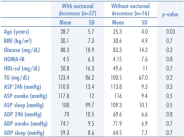
There was no difference with respect to the metabolic proile, HOMA-IR, and mean arterial pressure when the POS patients with and without nocturnal descensus were compared to each other (Table 5).
Finally, the presence or absence of nocturnal de-scensus did not show statistically signiicant differences with respect to POS (p=0.9). The same was observed for the BMI, meaning that the nocturnal descensus was not correlated with obesity (p=0.2).
Table 3. Pearson correlation coeficient between Body Mass Index with the mean arterial pressures
AP: arterial pressure; ASP: arterial systolic pressure; ADP: arterial diastolic pressure. The Pearson correlation coeficient was used (p>0= positive correlation; p<0= negative correlation; p=0: no correlation; p: 0–3.0=weak correlation; p: 3.0–7.0=moderate correlation; p>7.0= strong correlation).
AP (mmHg) Correlation (P)
POS Control
ASP 24h 0.61 0.4
ASP awake 0.63 0.61
ASP sleep 0.59 0.50
ADP 24h 0.37 0.45
ADP awake 0.47 0.7
ADP sleep 0.42 0.45
Table 4. Mean arterial pressures, glucose, triglycerides and HOMA-IR in obese and non-obese POS patients
Parameters
Non-obese (n=20)
Obese
(n=22) p-value
Mean DP Mean DP
ASP 24h (mmHg) 106 11.7 117 8.9 0.0001
ASP awake (mmHg) 111 9.2 120.5 10.1 0.001
ASP sleep (mmHg) 96 8.6 109.5 9.8 0.00
ADP 24h (mmHg) 64 10.4 70.5 6.9 0.03
ADP awake (mmHg) 69.5 7.3 74.5 8.4 0.006
ADP sleep (mmHg) 55.5 6.5 64 8.2 0.0008
GLUCOSE (mg/dL) 79.6 6.9 90 22.4 0.02
TG (mg/dL) 70 55.4 118 91.4 0.01
HOMA-IR 1.8 1.2 3.42 9.1 0.05
ASP: arterial systolic pressure; ADP: arterial diastolic pressure; TG: triglyceride; HOMA-IR: homeostasis model assessment- insulin resistance.
Note:*p signiicant <0.05 using the Student’s t-test.
With nocturnal descensus (n=27)
Without nocturnal
descensus (n=16) p-value
Mean SD Mean SD
Age (years) 28.7 5.7 25.3 4.0 0.03
BMI (kg/m²) 30.1 7.3 30.6 4.9 0.7
Glucose (mg/dL) 88.3 18.9 83.3 14.3 0.2
HOMA-IR 4.5 6.3 4.15 7.6 0.8
HDL-col (mg/dL) 50.8 16.3 49.6 11 0.7
TG (mg/dL) 123.4 86.2 100.5 67.0 0.2
ASP 24h (mmHg) 110.0 13.4 113.8 9.3 0.3
ASP awake (mmHg) 117.8 12 116 9.4 0.5
ASP sleep (mmHg) 100 99.7 109.3 10.1 0.5
ADP 24h (mmHg) 70 10.5 69.6 6.6 0.8
ADP awake (mmHg) 74.1 9.5 71.9 6.9 0.7
ADP sleep (mmHg) 59.3 8.6 64.5 7.7 0.7 Table 5. Clinical and biochemical characteristic of the polycystic ovary syndrome patients with and without nocturnal descensus
MAP is an appropriate way to inspect cardiovascular status in these patients22. By keeping indirect and
inter-mittent registers of arterial pressure for 24 h, MAP could detect circadian cycle variations in arterial pressure, which had considerable prognostic implications. Among the parameters studied, the most indicative were the mean arterial pressure and the absence of nocturnal descensus. The mean arterial systolic pressure (ASP) and the mean arterial diastolic pressure (ADP) obtained in a 24 h period, including the awake/sleep cycle, show consistent correla-tions with lesions in the target organs and cardiovascular morbidity and mortality31,32. When comparing the mean
arterial pressure detected by MAP in POS and control patients, no difference was found. These results are dificult to consider, since most of the results in the literature are controversial and use casual ambulatorial arterial pressure measurements, and MAP studies are scarce.
The only cardiovascular risk factor analysed in our investigation that showed a correlation with the arterial systolic and diastolic pressures was BMI. Two interesting results were a moderate correlation between the mean arterial systolic pressure, and a weak correlation between the mean arterial diastolic pressure, and POS. The same inluence of BMI occurred between the mean arterial pressure and obesity in POS patients. A 5% signiicance was found in the comparison between these parameters, conirming previous reports indicating an association be-tween obesity and arterial pressure33,34. Nonetheless, the
differences between the levels of glycemia, triglycerides and HOMA-IR in the obese and non-obese groups was signiicant, conirming an interaction between the adipose tissue and the comorbidities related to the insulin resistance.
The adipose tissue is recognized as a complex en-docrine organ, capable of inluencing the sympathetic system and producing chemical signal that, together with insulin resistance, inluence arterial pressure in POS women. Overstimulation of sympathetic system in obesity promotes an expansion of the extracellular vol-ume and regional lux, causing increase cardiac debit35.
Obese patients show higher plasma rennin activity, in-creasing the plasma levels of angiotensinogen, increased activity of tissue converting enzyme and higher plasma
levels of aldosterone, implicating the renin-angiotensin-aldosterone system in ovary physiology and pathology36.
Another important aspect of adipose tissue physiology is hyperinsulinemia that compensates in the insulin re-sistance, which exercises a trophic effect on blood vessel smooth muscle, causing increased vascular resistance and arterial pressure37. POS might share a common genetic
background with arterial hypertension and obesity; ind-ing this trait could elucidate the mechanisms involved in these diseases and other comorbidities due to insulin resistance38. However, it is not possible to conclude that
POS patients are at greater risk of cardiovascular diseases, and the validity of this association needs testing.
The correlation between nocturnal descensus and risk factors for cardiovascular disease remains debatable since the results are controversial22,31,39. In the present
study, the risk of presenting nocturnal descensus was similar between the POS, the controls and their BMIs. No correlation existed between nocturnal descensus and the mean values of glycemia, HDL-col, triglycerides and HOMA-IR. Similarly, no correlation was found between MAP.These indings, in general, contradict those in the literature. This might be attributed to the fact that sleep disorders, such as obstructive dyspnea, were not excluded and could inluence the prevalence of nocturnal descen-sus and present a relevant bias in our study40.
One advantage of our study was the uniform proile of the patients seeking treatment in our institution by presenting suspected POS, all of whom had the same diagnosis. In both groups — POS and Control — the complaints were related to hyperandrogenism, anovulation and obesity. POS diagnosis was careful and followed the Rotterdam consensus; other causes of hyperandrogenism were excluded. These criteria were fundamental to the reliability of the study. However, the small number of patients in the Control Group might have introduced some bias to the analyses.
Acknowledgements
To Clínica Prev Total Laboratório de Imagem LTDA for the MAP measurements and Laboratório Sérgio Franco for the laboratorial tests.
References
1. Norman RJ, Dewailly D, Legro RS, Hickey TE. Polycystic ovary syndrome. Lancet. 2007;370(9588):685-97.
2. Elghblawi E. Polycystic ovary syndrome and female reproduction. Br J Nurs. 2007;16(18):1118-21.
3. March WA, Moore VM, Wilson KJ, Phillips DI, Norman RJ, Davies MJ. The prevalence of polycystic ovary syndrome in a community sample assessed under contrasting diagnostic criteria. Hum Reprod. 2010;25(2):544-51.
4. Ehrmann DA. Polycystic ovary syndrome. N Engl J Med. 2005;352(12):1223-36.
5. Marcondes JAM, Barcellos CRG, Rocha MP. Diiculdades e armadilhas no diagnóstico da síndrome dos ovários policísticos. Arq Bras Endocrinol Metab. 2011;55(1):6-15.
7. Burt Solorzano CM, Beller JP, Abshire MY, Collins JS, McCartney CR, Marshall JC. Neuroendocrine dysfunction in polycystic ovary syndrome. Steroids. 2012;77(4):332-7.
8. de Paula Martins W, Santana LF, Nastri CO, Ferriani FA, de Sa MF, Dos Reis RM. Agreement among insulin sensitivity indexes on the diagnosis of insulin resistance in polycystic ovary syndrome and ovulatory women. Eur J Obstet Gynecol Reprod Biol. 2007;133(2):203-7.
9. Iftikhar S, Collazo-Clavell ML, Roger VL, St Sauver J, Brown RD Jr, Cha S, et al. Risk of cardiovascular events in patients with polycystic ovary syndrome. Neth J Med. 2012;70(2):74-80.
10. Azevedo MF, Costa EC, Oliveira AIN, Silva IBO, Marinho JCDB, Rodrigues JAM, et al. Elevated blood pressure in women with polycystic ovary syndrome: prevalence and associated risk factors. Rev Bras Ginecol Obstet. 2011;33(1):31-6.
11. El-Mazny A, Abou-Salem N, El-Sherbiny W, El-Mazny A. Insulin resistance, dyslipidemia, and metabolic syndrome in women with polycystic ovary syndrome. Int J Gynaecol Obstet. 2010;109(3):239-41.
12. Kandaraki E, Christakou C, Diamanti-Kandarakis E. Metabolic syndrome and polycystic ovary syndrome... and vice versa. Arq Bras Endocrinol Metab. 2009;53(2):227-37.
13. Moran LJ, Misso ML, Wild RA, Norman RJ. Impaired glucose intolerance, type 2 diabetes and metabolic syndrome in polycystic ovary syndrome: a systematic review and meta-analysis. Hum Reprod Update. 2010;16(4):347-63.
14. Baracat EC, Soares-Junior JM. Polycystic ovaries: insulin resistance and metabolic syndrome. Rev Bras Ginecol Obstet. 2007;29(3):117-9. 15. Sathyapalan T, Atkin SL. Recent advances in cardiovascular aspects of
polycystic ovary syndrome. Eur J Endocrinol. 2012;166(4):575-83. 16. Magalhães MEC, Brandão AA, Pozzan R, Campana EMG, Fonseca
FL, Pizzi OL, et al. Prevention of arterial hypertension: when to start and with whom? Rev Bras Hipertens. 2010;17(2):93-7. 17. Williams B. The year in hipertension. J Am Coll Cardiol. 2009;55(1):
65-73.
18. Kargili A, Karakurt F, Kasapoglu B, Derbent A, Koca C, Selcoki Y. Association of polycystic ovary syndrome and a non-dipping blood pressure pattern in young women. Clinics (Sao Paulo). 2010;65(5):475-9.
19. Rotterdam ESHRE/ASRM-Sponsored PCOS Consensus Workshop Group. Revised 2003 consensus on diagnostic criteria and long-term health risks related to polycystic ovary syndrome. Fertil Steril. 2004;81(1):19-25.
20. World Health Organization. Obesity: preventing and managing the global epidemic. Report of a WHO Consultation on Obesity. Geneva: WHO; 1997.
21. Hatch R, Rosenield RL, Kim MH, Tredway D. Hirsutism: implications, etiology, and management. Am J Obstet Gynecol. 1981;140(7):815-30. 22. Sociedades Brasileira de Cardiologia, de Hipertensão e de Nefrologia.
V Diretrizes Brasileiras de Monitorização Ambulatorial de Hipertensão Arterial (MAPA). Rev Bras Hipertens. 2011;18(1):7-17.
23. Expert Commitee on the Diagnosis and Classiication of Diabetes Mellitus. Report of the Expert Commitee on the Diagnosis and Classiication of Diabetes Mellitus. Diabetes Care. 1997;20(7):1183-97.
24. Expert Panel on Detection, Evaluation,
and Treatment of High Blood Cholesterol in Adults. Executive Summary of the Third Report of the National Cholesterol Education
Program (NCEP) Expert Panel on Detection, Evaluation, and Treatment of High Blood Cholesterol in Adults (Adult Treatment Panel III). JAMA. 2001;285(19):2486-97.
25. RomanoLGM, Bedoschi G, Melo AS, Albuquerque FO, Rosa e
SilvaACJ, Ferriani RA, et al. Metabolic abnormalities in polycystic
ovary syndrome women: obese and non obese. Rev Bras Ginecol Obstet. 2011;33(6):310-6.
26. Melo AS, Macedo CSV, Romano LGM, Ferriani RA, Navarro PAAS. Women with polycystic ovary syndrome have a higher frequency of metabolic syndrome regardless of body mass index. Rev Bras Ginecol Obstet. 2012;34(1):4-10.
27. Costa EC, Soares EMM, Lemos TMA, Maranhão TMO, Azevedo GD. Índices de obesidade central e fatores de risco cardiovascular na síndrome dos ovários policísticos. Arq Bras Cardiol. 2010;94(5):633-8.
28. de Groot PC, Dekkers M, Romijn JA, Dieben SW, Helmerhorst FM. PCOS, coronary heart disease, stroke and the inluence of obesity: a systematic review and meta-analysis. Hum Reprod Update. 2011;17(4):495-500.
29. Lansdown A, Rees DA. The Sympathetic nervous system in polycystic ovary syndrome: a novel therapeutic target? Clin Endocrinol (Oxf). 2012;77(6):791-801.
30. Reckelhoff JF. Polycystic ovary syndrome: androgens and hypertension. Hypertension. 2007;49(6):1220-1.
31. Vaz-de-Melo RO, Toledo JCY, Loureiro AAC, Cipullo JP, Moreno
Júnior H, Martin JFV.Ausência de descenso noturno se associa
a acidente vascular cerebral e infarto do miocárdio. Arq Bras Cardiol. 2010;94(1):79-85.
32. Conen D, Bamberg F. Noinvasive 24-h ambulatory blood pressure
and cardiovascular disease: a systematic review and meta-analysis. J Hypertens. 2008;26(7):1290-9.
33. Luque-Ramírez M, Alvarez-Blasco F, Mendieta-Azcona C, Botella-Carretero JI, Escobar-Morreale HF. Obesity is the major determinant of the abnormalities in blood pressure found in young women with the polycystic ovary syndrome. J Clin Endocrinol Metab. 2007;92(6):2141-8.
34. Kotchen T. Obesity-related hypertension: epidemiology, pathophysiology, and clinical management. Am J Hypertens. 2010;23(11):1170-8.
35. Landsberg L. Obesity, metabolism, and hypertension. Yale J Biol Med. 1989;62(5):511-9.
36. Raposo-Costa AP, Reis AM. O sistema renina-angiotensina em ovário. Arq Bras Endocrinol Metab. 2000;44(4):306-13. 37. Martins WP, Soares GM, Vieira CS, dos Reis RM, Silva de Sá
MF, Ferriani RA. Cardiovascular risk markers in polycystic ovary syndrome in women with and without insulin resistance. Rev Bras Ginecol Obstet. 2009;31(3):111-6.
38. Leibel NI, Baumann EE, Kocherginsky M, Rosenield RL. Relationship of adolescent polycystic ovary syndrome to parental metabolic syndrome. J Clin Endocrinol Metab. 2006;91(4):1275-83. 39. Ben-Dov IZ, Kark JD, Ben-Ishay D, Mekler J, Ben-Arie L, Bursztyn M.
Predictors of all-cause mortality in clinical ambulatory monitoring: unique aspects of blood pressure during sleep. Hypertension. 2007;49(6):1235-41.