Evaluating the Clinical and Physiological
Effects of Long Term Ultraviolet B
Radiation on Guinea Pigs (Cavia porcellus)
Megan K. Watson1, Adam W. Stern2, Amber L. Labelle1, Stephen Joslyn1, Timothy M. Fan1, Katie Leister1, Micah Kohles3, Kemba Marshall4, Mark A. Mitchell1*
1.Department of Veterinary Clinical Medicine, University of Illinois, Urbana, Illinois, United States of America,
2.Department of Pathobiology, University of Illinois, Urbana, Illinois, United States of America,3.School of Veterinary Medicine and Biomedical Sciences, University of Nebraska-Lincoln, Lincoln, Nebraska, United States of America; Oxbow Animal Health, Murdock, Nebraska, United States of America,4.PetSmart, Phoenix, Arizona, United States of America
Abstract
Vitamin D is an important hormone in vertebrates. Most animals acquire this hormone through their diet, secondary to exposure to ultraviolet B (UVB) radiation, or a combination thereof. The objectives for this research were to evaluate the clinical and physiologic effects of artificial UVB light supplementation on guinea pigs (Cavia porcellus) and to evaluate the long-term safety of artificial UVB light supplementation over the course of six months. Twelve juvenile acromelanic Hartley guinea pigs were randomly assigned to one of two treatment groups: Group A was exposed to 12 hours of artificial UVB radiation daily and Group B received only ambient fluorescent light for 12 hours daily. Animals in both groups were offered the same diet and housed under the same conditions. Blood samples were collected every three weeks to measure blood chemistry values, parathyroid hormone, ionized calcium, and serum 25-hydroxyvitamin D3 (25-OHD3) levels.
Serial ophthalmologic examinations, computed tomography scans, and dual energy x-ray absorptiometry scans were performed during the course of the study. At the end of the study the animals were euthanized and necropsied. Mean¡SD serum 25-OHD3 concentrations differed significantly in the guinea pigs (p,0.0001)
between the UVB supplementation group (101.49¡21.81 nmol/L) and the control group (36.33¡24.42 nmol/L). An increased corneal thickness in both eyes was also found in the UVB supplementation compared to the control group (right eye [OD]: p,0.0001; left eye [OS]: p,0.0001). There were no apparent negative clinical or pathologic side effects noted between the groups. This study found that exposing guinea pigs to UVB radiation long term significantly increased their circulating serum 25-OHD3levels, and that this increase was sustainable over time. Providing
OPEN ACCESS
Citation:Watson MK, Stern AW, Labelle AL, Joslyn S, Fan TM, et al. (2014) Evaluating the Clinical and Physiological Effects of Long Term Ultraviolet B Radiation on Guinea Pigs (Cavia porcellus). PLoS ONE 9(12): e114413. doi:10. 1371/journal.pone.0114413
Editor:Silvana Allodi, Federal University of Rio de Janeiro, Brazil
Received:August 27, 2014
Accepted:November 10, 2014
Published:December 17, 2014
Copyright:ß2014 Watson et al. This is an open-access article distributed under the terms of the
Creative Commons Attribution License, which permits unrestricted use, distribution, and repro-duction in any medium, provided the original author and source are credited.
Data Availability:The authors confirm that all data underlying the findings are fully available without restriction. All relevant data are within the paper.
Funding:PetSmart Charities provided funding for this project. Dr. Kemba Marshall was included as a co-author as a member of the team that designed the study, provided additional resources, and helped with the preparation of the manuscript. Oxbow Animal Health provided funding for this project. Dr. Micah Kohles was included as a co-author as a member of the team that designed the study, provided additional resources, and helped with the preparation of the manuscript. Fluker Farms (Port Allen, LA) was a sponsor that contributed funding but played no role in study design, data collection and analysis, decision to publish or preparation of the manuscript.
guinea pigs exposure to UVB may be an important husbandry consideration that is not currently recommended.
Introduction
Guinea pigs (Cavia porcellus) are members of the order Rodentia and family Caviidae. They were native to the Andes and other mountainous regions of South America, where they were predominately used for food in countries such as Peru, Colombia, Venezuela, and Brazil [1]. At this time they are considered extinct in the wild [2]. In the United States, guinea pigs are popular pets. In a survey performed by the AVMA in 2006, there were over 1.3 million guinea pigs being kept as pets in the United States [3]. Guinea pigs are docile, responsive animals with a lively personality, and therefore make appealing pets for most households. Guinea pigs have also been used in research for over 400 years [4].
Current husbandry recommendations for guinea pigs are made with the goal of improving their health and longevity. However, at this time there are no specific lighting recommendations for guinea pigs other than providing a photoperiod of 12 hours to mimic the natural diurnal cycle [5]. This is interesting to note as these diurnal animals have evolved in a high altitude environment and would be expected to have increased exposure to ultraviolet B (UVB) radiation compared to animals at lower altitudes. Vertebrates can utilize UVB exposure as a method of generating endogenous vitamin D, an essential hormone. Whether exposure to UVB radiation is important for these animals relative to this function is not known.
Vitamin D is a circulating hormone that is important to homeostasis and normal physiology, including bone development, growth, neuromuscular function, reproduction, cardiovascular health, and immune function [6,7]. Vitamin D deficiencies in humans and other vertebrates have been shown to cause rickets, osteomalacia, and reproductive failure [6,7]. Not only do deficiencies directly lead to abnormal calcium metabolism, it is also becoming more apparent that vitamin D levels are important to overall health. Vitamin D receptors are found in numerous tissues throughout the body [8,9]. In humans, adequate levels of vitamin D have been shown to decrease the risk of developing many different conditions, including diabetes, muscular dystrophy, hypertension, and inflam-matory bowel disease [8]. When considering the many different physiological functions in vertebrates that are intimately related to vitamin D and calcium metabolism, it should be considered that disease processes in captive species could be related to hypovitaminosis D.
housed indoors and not exposed to natural or artificial UVB light, it is possible that these animals may experience chronic vitamin D deficiencies. Inappropriate levels of this essential nutrient and hormone could contribute to common disease processes in these animals such as dental disease, cardiovascular disease, or impaired immune function [4,5].
While the benefits of UVB exposure for captive animals appear to be strong, it is not without risk. UVB radiation has been associated with the development of skin neoplasia, with the groups at greatest risk including humans with fair complexions and albino or white animals [10,11]. Direct UVB radiation also has the potential to cause structural damage to the eye at the level of the cornea (keratitis, corneal edema), lens (cataracts), or retina (blindness) [11]. Direct UVB radiation can also cause short-term damage such as photodermatitis and
erythema. For these reasons, the safety of UVB supplementation in small mammals should be investigated.
The objectives for this research were to evaluate the clinical and physiologic effects of artificial UVB light supplementation on guinea pigs and to evaluate the long-term safety of artificial UVB light supplementation over the course of six months in these species. The clinical effects being evaluated between the UVB exposed and non-exposed guinea pigs included whether there would be an increased likelihood of ocular disease associated with the cornea, lens, or retina; gross or histologic skin changes; or differences in bone mineralization. The physiologic changes being assessed were related to the 25-hydroxyvitamin D levels, biochemistries, and blood cells. The hypotheses for this study were: 1) animals supplemented with UVB light would produce higher levels of vitamin D than those without supplementation, and that 2) there would be no significant detrimental side effects associated with UVB light supplementation.
Materials and Methods
Animals
Twelve juvenile (14-16 weeks), purpose bred, female intact Hartley guinea pigs were used in this study. Hartley guinea pigs are acromelanic albino animals. The pigmentation for acromelanic animals is genetically determined, with slightly variable amounts of pigmentation on the extremities. The animals were obtained from a commercial breeding facility (Charles River Laboratories, Wilmington, MA). Prior to their arrival at the study site, the animals received a standard commercial pelleted feed (Lab Diet, St. Louis, MO) with no additional
supplemental vitamin D or UVB light. The animals were allowed to acclimate to their surroundings for 7 days prior to the onset of data collection. This project was approved and performed in accordance with the regulations established by the Institutional Animal Care and Use Committee at the University of Illinois (protocol #13-024).
The animals were housed in 183 cm long6137 cm wide pens. Three animals
to the ceiling, and the animals were housed on the floor. Room temperature was measured weekly and was kept consistent throughout the study with an average range of 23 to 27 degrees Celsius. Paper shavings (Harlan Laboratories,
Indianapolis, IN) were provided as substrate lining the floor of the pen. General room lighting was provided by non-UVB producing ambient fluorescent lighting. The animals were provided with water ad libitum through sipper bottles placed on the front of the pens. All animals were all fed the same weight based diet consisting of Oxbow timothy grass hay (Oxbow Animal Health, Murdoch, NE) and Oxbow Essentials adult guinea pig pellets (Oxbow Animal Health). The vitamin D content of this particular pelleted diet is 900 IU/kg.
Biochemistry
Population and Animal Health, East Lansing, MI) for measurement of serum 25-hydroxyvitamin D (25-OHD3, radioimmunoassay), ionized calcium (iCa,
ion-selective electrode/pH electrode at pH 7.4) and parathyroid hormone (PTH, radioimmunoassay).
Ophthalmology
Complete ophthalmic examinations under manual restraint were also performed at the initiation of the study. One drop of Tropicamide 1% (Bausch & Lomb Inc., Tampa, FL) was administered into each eye prior to the onset of the exam. The examinations included a neuro-ophthalmic examination, fundic examination, tonometry, slit lamp biomicroscopy (Kowa-SL2, Kowa, Tokyo, Japan), and corneal pachymetry. Tonometry was performed with a rebound tonometer (Tonovet, Icare Finland Oy, Espoo, Finland). The rebound tonometer has three available calibration settings: canine, equine, and ‘‘other’’: other is used for species for which a calibration table has not been established. The calibration setting for ‘‘other’’ was used in this study. A corneal pachymeter (Compuscan P ultrasonic pachymeter, 20 MHz, Strorz Instrument Company, St. Louis, Missouri) was used in the exam as a gauge of corneal thickness. Pachymetry readings were taken in triplicate for each eye during the exam. Serial ophthalmic examinations were performed at two-month intervals, and at the end of the study period, with the same study procedure repeated for each examination period. A single, blinded investigator (ALL) performed all of the ophthalmic exams.
Imaging
Computed tomography (CT) scans were performed under sedation at the onset of the study. Computed tomography (CT) of the skull was performed on all subjects using a 16-slice helical CT scanner (GE Lightspeed 16 slice CT Milwaukee, WI, USA) using the following parameters: kVp of 80, mA of 180, slice width of 0.625 mm, pitch of 0.5625, scan field of view 9.6 cm, 5126512 matrix, a rotation
time of 0.8 s and using a bone convolution kernel. Each guinea pig was
administered a combination of 0.05 mg/kg buprenorphine hydrochloride (Reckitt Benckiser Pharmaceuticals Inc., Richmond, VA) and 0.5 mg/kg midazolam (Hospira Inc., Lake Forest, IL) via intramuscular injection. Once the animal became sedate, it was positioned in sternal recumbency and perpendicular to the gantry with forelimbs positioned caudally. After the scan was complete and the animal recovered from sedation, it was returned to laboratory housing.
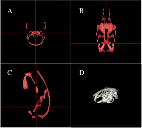
plug-in (Mialite, Center for Medical Image Science and Visualization (CMIV), Linko¨ping University, Sweden) (Fig. 1, Fig. 2). The first step was removing the soft tissue surrounding the skull and neuro tissue within the calvarium and proximal cervical spinal cord. This was achieved by segmentation using a threshold minimum of -100HU, a maximum of 150HU and a smoothing factor of 0.1 [12]. The resulting soft tissue region of interest (ROI) was removed by changing all contained pixels to a value of -2000HU. The segmentation plugin was used again to isolate the skull using a threshold minimum of 0HU, no maximum HU constrains, smoothing factor of 0.1, and a seed (starting point) within the temporal bone. An additional blocker point was used to prevent segmentation of the cervical vertebral bodies. The parameters of the resulting three-dimensional (3D) ROI were recorded for each specimen, such as volume, average attenuation (HU), and attenuation standard deviation.
UVB Supplementation
After initial sample collection, guinea pigs were assigned to two treatment groups of six animals each using a random number generator (www.random.org). Each
Fig. 1. Multiplanar reconstruction view of the bone threshold process using a range of 0 Hounsfield units (HU) – max HU, following initial soft tissue removal.Images A, B and C are the transverse, dorsal and sagittal plane sample images, respectively. Image D is a 3-dimensional volume rendered image of the included bone.
group was then randomly divided into two pens housing three animals each. Of the treatment groups, the supplemented (treatment) group was exposed to 12 hours of artificial UVB radiation daily that was controlled by a timer; non-supplemented (control) groups received ambient fluorescent light with no UVB supplementation for 12 hours daily. To provide direct UVB radiation, nine dome lights (Fluker Farms, Port Allen, LA) were suspended above the animals at equidistant intervals, comprising three rows of three lights each. The 5.5 inch domes were anchored to a shelving unit several feet off the ground and suspended directly above the guinea pigs, with no physical barrier present between the animals and light. UVB light was provided using one Oxbow bulb (Oxbow Animal Health, Murdock, NE) in each dome. The domes were suspended 12–14 inches above the ground, in order to maximize exposure to the animals without impeding movement, as well as attempting to mimic the distance that would be used in a typical laboratory or captive cage setting. The amount of UVB radiation and irradiance was measured in microwatts per centimeter squared (mW/cm2) by
placing a digital UVB meter (Solartech, Inc., Harrison Township, MI) and an irradiance meter (Zoo Med Laboratories Inc., San Luis Obispo, CA) on the ground directly under each bulb, as well as in between the bulbs. The height of the
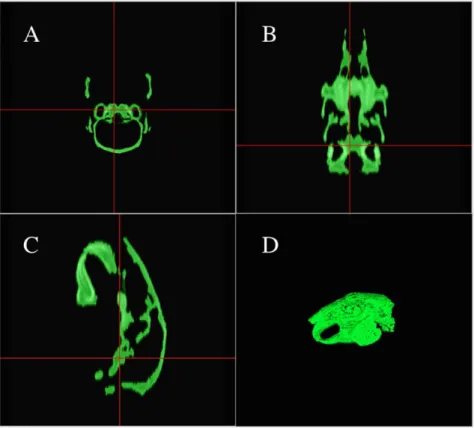
Fig. 2. Final window showing the segmented bone and summary of 3D volume parameters: Volume (mL), Mean HU, STD HU, Max HU and Min HU.A-C are the multiplanar reconstruction sample images and the final 3D volume region of interest (D).
meter was approximately at eye level of the guinea pigs. Measurements were collected weekly, approximately 3 hours after the bulbs were turned on and approximately 3 hours prior to the bulbs being turned off. To correct for UVB degradation over time and provide consistent levels of supplementation, bulbs were replaced if at any time the UVB output from a bulb measured less than 20 mW/cm2. The UVB radiation levels measured in the UVB treatment groups
ranged from 21–70 mwatts/cm2 directly under the lights and 2–19mwatts/cm2in
between the lights. UVB radiation levels in the non-UVB groups were,1 mwatts/
cm2. The maximum UVB irradiance level in the pens was 3.5, which falls under IV in the UV index spectrum. This value is considered high and equivalent to mid day baskers; however it is well within the safe region, with greater than 7 considered dangerous.
The study was conducted over 6 months. After the UVB light supplementation was initiated, blood samples were collected every three weeks to measure blood chemistries, serum PTH, ionized calcium, and 25-hydroxyvitamin D3 levels, for a
total of 9 sample collections. Weights were collected during each sample
collection. Collection, processing and testing of the blood samples were similar to the techniques described for the samples obtained on day 0. Serial ophthalmologic examinations were performed at two month intervals, and at the end of the study period, for a total of four examinations.
Histopathologic Evaluation
At the end of the six month period, anesthesia was induced as previously described for initial sample collection. Each animal was placed in dorsal recumbency and a final 3 ml blood sample was collected from the cranial vena cava. While being maintained on 5% isoflurane and 1 L oxygen, 2–3 ml of pentobarbital (Vortech Pharmaceuticals, Dearborn, MI) was administered to the animal via intracardiac injection. Euthanasia was confirmed via auscultation and negative corneal reflex.
CT scans were performed immediately following euthanasia using the identical scanning and positioning parameters described at the beginning of the study. Segmentation of the skull, mandible and teeth was also acquired using the techniques described previously. ROI volume, mean and SD of the attenuations were recorded for each specimen.
A postmortem examination was performed on each animal. A select tissue set was collected from all animals except one animal, which experienced an anesthetic death in which a complete set of tissues was collected. The select tissue set included lung, heart, haired skin- head, haired skin-ear, haired skin-dorsum, haired skin-ventrum, liver, pulmonary artery, aorta, adrenal gland, kidney, duodenum, pancreas, thyroid glands, spleen, esophagus, reproductive tract, trachea, skeletal muscle, eye, and femur. Tissues were fixed in 10% neutral buffered formalin. Following fixation, tissues were embedded in paraffin wax, sectioned at 4 mm, and stained with hematoxylin and eosin. Additionally,
lesions were noted and scored using the following categories: no lesions, mild lesions, moderate lesions, and severe lesions. Attempts to characterize apoptosis in the skin, the skin associated with the head, the skin associated with the ears, and the eyes were made to determine if there were any UVB specific injuries caused in these tissues. A single, blinded pathologist (AWS) performed all of the
microscopic examinations.
Bone mineral densities (BMD) of guinea pig skulls were measured by dual energy x-ray absorptiometry (Hologic QDR 2000, Marlborough, MA). Prior to analysis, all skulls were preserved and stored at 220 degrees Celsius. For BMD assessment, all skulls were positioned dorsoventrally, and a standardized region of interest (R1) of a constant area (cm2) was created to encompass the entire skull size of each specimen. Bone mineral density was measured and expressed as grams of bone per area (gm/cm2). The analyst of the bone concentrations was blinded to the study groups (TMF).
Statistical Analysis
The sample size selected for this study (n512; 6 per group) was based on the following assumptions: an expected mean difference in serum 25-OHD3
concentrations of 35 nmol/L between treatment and control groups with a SD of 20 nmol/L [13,14], an alpha of 0.05, and a power of 0.8 (MedCalc 11.3.2.0). Distribution of the data was evaluated by use of the Shapiro-Wilk test. Mean, SD, and minimum-maximum (min-max) values were reported for data that had a normal distribution, whereas the median, 10th to 90th percentiles (%), and min-max values were reported for data that did not have a normal distribution. Data that were not normally distributed were logarithmically transformed in order to perform parametric analysis. If the data was not normally distributed after transformation, appropriate nonparametric statistics were performed.
A linear mixed model was used to evaluate the normally distributed and correlated blood chemistry values, iCa, and 25-OHD3 collected in the repeated
measures design of the study. This statistical method was selected because one animal died over the course of the study and this type of model can account for missing data. Group and time represented the fixed and random factors,
t- test was used to determine if bone mineral density differed between groups. Chi square one-tailed tests with a Yates correction or Fisher exact tests were performed to determine if there was any difference in the pathologic findings documented at necropsy between groups. A value of P#0.05 was used to determine significance. Commercial statistical software (SPSS, version 22.0, SPSS Inc, Chicago, IL) and graphpad.com were used to analyze the data.
Results
Twelve guinea pigs (6 not exposed to UVB, and 6 exposed to UVB) were used in this study. At the initiation of the study the average body weight of the guinea pigs was 497.2 grams (SD: 15.6, min-max: 465.4-520.2 grams), while at the conclusion of the study the mean body weight of the guinea pigs was 946.1 g (SD: 15.6, Min-Max 831-1070); there was no significant difference in body weight noted between groups (F50.347, p50.557). The mean difference in weight gain from initiation to completion of the study was 449.38 g (SD: 82.1, Min-Max: 349–556) for all animals. Over the study period, weight gain among guinea pigs exposed to supplemental UVB lighting and those not exposed did not differ (t50.079, p50.938).
Complete blood counts were performed at the beginning and completion of the study to assess the general health of the guinea pigs. Unfortunately, a number of the samples collected at both the beginning and end of the study were clotted and could not be evaluated. Because of the small sample set, statistical comparisons were not performed. However, at least one complete blood count result was available for each guinea pig at either the beginning or conclusion of the study, and all results were within published reference limits [5].
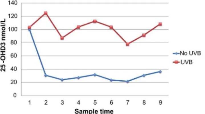
Serum 25-OHD3 values differed significantly by group over time (F5576.126,
p,0.0001) (Fig. 3, Fig. 4) with higher values seen in the UVB supplemented group. Since the animals arrived at the University of Illinois facility having been fed a different diet than the study diet, mean, SD, and min-max values including initial baseline data and excluding the baseline data are shown in Table 1. There was no significant difference in the 25-OHD3 concentrations between the two
pens of guinea pigs exposed to the UVB lights (F50.022, p50.884) or between the two pens of guinea pigs not exposed to the UVB lights (F51.082, p50.357). This comparison was done to rule out potential differences in light exposure between treatment groups or quantity of food consumed between control groups.
There was a significant difference in corneal thickness between groups for each eye (right eye [OD]: F5149.527, p,0.0001; left eye [OS]: F530.525,
p5,0.0001), with the UVB supplemented group having thicker corneas (OS and OD). Average corneal pachymetry measurements by group are shown in Table 3. There was no significant difference in intraocular pressure noted between groups for either eye (right eye [OD]: F50.196, p50.66; left eye [OS]: F51.1, p5 0.3). No other abnormalities were noted upon examination of the eyes, with minor exceptions. One animal developed a corneal foreign body (OS) from what appeared to be organic material (i.e., bedding or food). Proparacaine was applied to the eye and the foreign body was easily removed with sterile eye wash using gentle lavage. Fluorescein stain was applied, and the cornea was then observed via slit lamp to have a mild superficial ulcer and keratitis, secondary to the corneal foreign body. The animal was administered one treatment of triple antibiotic ointment in that eye and rechecked in 48 hours. At that time the keratitis had completely resolved and no further treatment was necessary. Another animal developed serous ocular discharge OS with no associated conjunctivitis, which resolved within 48 hours without treatment. Both of these animals were in the UVB supplementation group.
For the initial CT exams, the mean Hounsfield units were 1076.478 (SD: 32.8606, Min-Max1015.18-1126.14); for the final exams the mean HU was 1208.003 (SD: 44.360, Min-Max 1152.64-1293.49). There was a significant increase in Hounsfield units noted on the CT images over time (F5173.414, p5,0.0001) but not between control and UVB supplemented groups (F50.559, p50.472). There was no evidence of dental disease on the CT scans. There also was no significant difference noted in the bone mineral density between the two groups obtained via DEXA scan (t 5 20.713, p50.494).
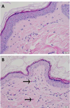
No significant differences were found when comparing necropsy lesions between groups (all p.0.05) (Table 4). Mild dermatologic changes were noted equally across groups consisting of rare suprabasilar apoptotic keratinocytes, small numbers of lymphocytes, plasma cells, and eosinophils scattered in the superficial dermis, and minimal to mild epidermal hyperplasia on skin from the ears (Fig. 5)
Fig. 3. Serum 25-OHD3in guinea pigs between groups over time (sample number).
Fig. 4. A) Guinea pig serum 25-OHD3 levels with no UVB supplementation.B) Guinea pig serum 25-OHD3 levels for UVB supplemented group. (Time5sample number).
doi:10.1371/journal.pone.0114413.g004
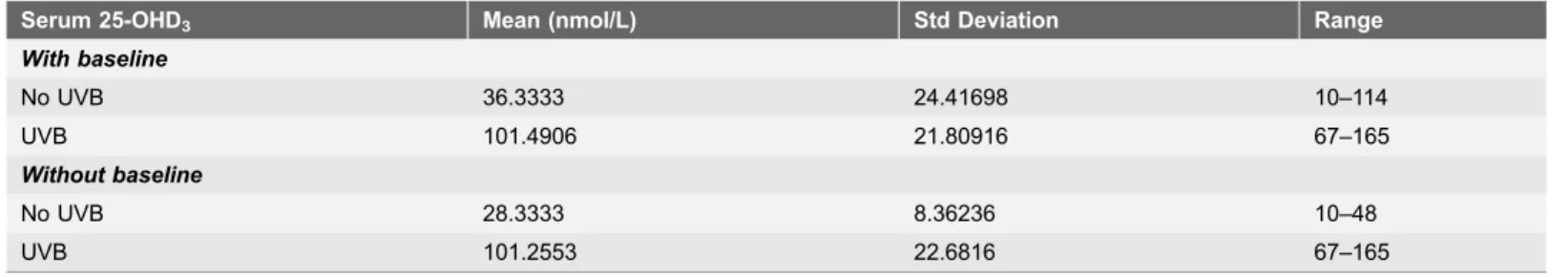
Table 1.Guinea pig serum 25-OHD3values with and without baseline values included by group.
Serum 25-OHD3 Mean (nmol/L) Std Deviation Range
With baseline
No UVB 36.3333 24.41698 10–114
UVB 101.4906 21.80916 67–165
Without baseline
No UVB 28.3333 8.36236 10–48
UVB 101.2553 22.6816 67–165
or head (Fig. 6). No elastin abnormalities were noted on Verhoeff-Van Gieson staining (Fig. 7). Multifocal and mild vacuolar hepatopathy (glycogen) were also noted equally across groups. The only mineralization documented was multifocal and mild mineralization within renal cortical tubules in a guinea pig with no UVB supplementation, and a single mineralized muscle myofiber in the skeletal muscle of a guinea pig with UVB supplementation.
Discussion
Findings from the present study demonstrated that guinea pigs provided artificial supplementation of UVB light produced and maintained significantly higher serum 25-OHD3levels compared to those without UVB supplementation. In
many vertebrates, vitamin D3 (cholecalciferol) can be synthesized after exposing
the skin to UVB radiation. This initiates the photobiochemical conversion of the precursor 7-dehydrocholesterol in the dermis to previtamin D3, which undergoes
isomerization into vitamin D3 and is hydroxylated to 25-OHD3in the liver [15–
17]. 25-OHD3 is the storage form of vitamin D, and therefore is considered the
Table 2.Guinea pig biochemical values by group in which statistical significance was found (p,0.0001).
Variable Mean SD Range
Ionized Calcium(mmol/L)
No UVB 1.52 0.07 1.35–1.65
UVB 1.58 0.09 1.29–1.74
Sodium(mmol/L)
No UVB 144.32* 0.38 138–150
UVB 140.19* 0.35 137–148
Albumin(g/dL)
No UVB 4.40 0.28 4.0–5.5
UVB 4.22 0.26 3.7–4.8
Total protein(g/dL)
No UVB 6.08* 0.35 5.0–6.5
UVB 5.79* 0.36 4.6–6.3
*Denotes estimated marginal mean
doi:10.1371/journal.pone.0114413.t002
Table 3.Guinea pig corneal pachymetry values for each eye by group (p,0.0001).
Eye Mean (mm) Std Deviation Range Right (OD)
No UVB 245.72 14.16 212–269
UVB 257.62 21.35 213–288
Left (OS)
No UVB 242.64 13.40 216–268
UVB 262.77 21.95 213–295
Table 4.Guinea pig lesions found on necropsy by organ.
Organ No UVB UVB
n percentage n percentage
Liver 3* 50% 4# 66.7%
Kidney 1 16.7% 0
Haired Skin- Ear 2 33.3% 1# 16.7%
Haired skin- Head 1* 16.7% 0
Haired skin- Ventrum 1 16.7% 0
Esophagus 1* 16.7% 0
Trachea 1* 16.7% 2# 33.3%
Femur 1* 16.7% 0
Lung 0 2# 33.3%
Spleen 0 1# 16.7%
Eye 0 1# 16.7%
Muscle 0 1 16.7%
*Denotes all lesions were documented in the same animal.
#All lesions documented in the same animal. This animal died post-anesthetic recovery.
doi:10.1371/journal.pone.0114413.t004
Fig. 5. A. Haired skin, ear.Microscopically normal skin from the ear. B. Haired skin, ear. Rare keratinocyte apoptosis within the stratum spinosum. Apoptotic keratinocyte denoted with arrow. Hematoxylin and eosin stain.
Fig. 6. A. Haired skin, head.Microscopically normal skin from the head. B. Haired skin, head. Increased cellularity of the dermis by small numbers of lymphocytes and eosinophils. Arrows denote eosinophils. Hematoxylin and eosin stain.
doi:10.1371/journal.pone.0114413.g006
Fig. 7. Haired skin, ear.Microscopically normal skin from the ear. Note the lack of positive (black) staining within the dermis. Verhoeff Van-Giessen stain.
best measure of vitamin D status in the body [18,19]. When vitamin D is needed systemically, the kidneys are responsible for conversion of 25-OHD3 to
1,25(OH)2D3, although conversion has been demonstrated in other tissues as
well.. The findings in this study, along with a previous short term pilot study, reinforce that guinea pigs have the ability to generate significant quantities of vitamin D through UVB exposure, despite adequate levels being provided in the diet, and thus may be an important conserved evolutionary mechanism not currently utilized in captive guinea pig husbandry practices.
While a significant difference was noted between groups over time, reference ranges for optimum serum 25-OHD3in wild or captive guinea pigs have not been
established. In humans, the mammal in which vitamin D deficiency is most commonly diagnosed and treated, a serum 25-OHD3concentration of,50 nmol/
L is considered deficient [6]. However, it should be noted that this minimum value is a general recommendation by most experts, and a general consensus on optimum levels of 25-OHD3 in humans does not exist. Since guinea pigs and
humans share similar methods of vitamin D acquisition, both utilizing UVB exposure and diet, it might be tempting to extrapolate the guinea pig values to human reference ranges. Based on the current results, the group not
supplemented with UVB light (28.33¡8.36 nmol/L, range 10–48) would be considered chronically deficient in vitamin D. However, such a direct comparison is difficult because of differences in physiology between our two species. Humans are omnivores that are capable of deriving vitamin D from animal and fortified sources in their diet, in addition to the vitamin D they can obtain from the plant material in their diet, as in the case of guinea pigs. On the other hand, guinea pigs have evolved in a high altitude, low latitude environment where exposure to UVB radiation would be greater than that which humans evolved. While it may appear as though both species are adapted to synthesize and maintain vitamin D levels under these different conditions, evaluating the two different methods for which they derive the vitamin D is important to consider. Dietary sources of vitamin D tend to persist in the body, while vitamin D produced via photoconversion can be turned on or off based on need. In theory, guinea pigs may be adapted to lower levels than humans, but the results of the current study suggest otherwise since animals exposed to UVB maintained 25-OHD3 levels well over 50 nmol/L.
An important finding in this study was that, over a six-month period, prolonged exposure of guinea pigs to artificial UVB light sources led to a statistically significant increase in serum 25-OHD3 levels that were sustainable
over time. This ability to sustain substantially higher levels of 25-OHD3from UVB
exposure compared to animals provided vitamin D in diet alone reinforces that these animals evolved with this mechanism to procure and maintain adequate levels of vitamin D. Guinea pigs are naturally a high altitude, low latitude species, with ample access to sunlight and natural UVB radiation in their home range. A recent pilot study also demonstrated that chinchillas (Chinchilla lanigera), which are also native to the Andes Mountains, have the ability to produce 25-OHD3
and alpacas (Vicugna pacos), have been shown to develop significant disease secondary to a seasonally dependent hypovitaminosis D [20]. When comparing the vitamin D levels of the guinea pigs in this study to rabbits maintained in a similar study, the average 25-OHD3level in guinea pigs was higher, despite similar
exposure. This could suggest these animals have a higher vitamin D requirement in relation to their natural habitat being at a higher altitude and lower latitude.
An interesting observation in this study was that the baseline serum 25-OHD3
values were most similar to the group supplemented with UVB light, and contrary to what was expected, actually decreased after initial baseline collection in the group not supplemented with UVB. Unfortunately, the initial baseline sample collection after arrival at the University of Illinois took place three months prior to the first sample collection post-UVB exposure because of logistics, so it is not known how long it took for the decline in serum 25-OHD3values to occur.
During this three-month period, the guinea pigs were fed the same diet and housed under the conditions described previously. After contacting the breeding facility it was determined that, prior to acquisition at the University of Illinois, the animals were fed a pelleted guinea pig feed (LabDiet, St. Louis, MO) that contained a higher concentration of vitamin D (3400 IU/kg), in comparison to the diet fed throughout the time they were housed for the study which contained 900 IU/kg of vitamin D. The 3.8 times higher dietary concentration seen in the diet offered at the breeding facility is almost in direct agreement with the higher mean levels of serum 25-OHD3 demonstrated between groups after the initial baseline
collection (3.6 times). This finding reinforces that guinea pigs indeed have the capability of increasing their serum 25-OHD3levels via either dietary ingestion or
photobiochemical conversion, and also may hint towards optimum serum 25-OHD3 maintenance levels in guinea pigs. However, how they acquire vitamin D
could be important, especially as it relates to the potential development of hypervitaminosis D. The dietary vitamin D requirements for guinea pigs are not known, but it is suggested to be 1000 IU/kg diet [21]. Under these guidelines, the commercial diet fed throughout the study adheres to those recommended requirements. The formula used at the breeding facility was 3.8 times higher than this and could, long term, lead to problems as dietary sources are more likely to be attributed to cases of hypervitaminosis D.
When evaluating circulating levels of 25-OHD3, one important factor to
consider is that systemic mineralization has been observed due to hypervitami-nosis D secondary to increased levels of vitamin D in the diet via clinical or experimental report [22]. Toxicity was described in a cohort of guinea pigs fed 150 times the normal concentration of vitamin D in their diet, and subsequently they developed systemic disease with lesions consisting of renal interstitial fibrosis with tubular mineralization, soft tissue mineralization in multiple organs, hepatic lipidosis, and pneumonia [22]. Serum 25-OHD3 levels were obtained in two
is tightly regulated within the body. In humans, mechanisms have been elucidated which have shown sunlight actually destroys excess previtamin D3or vitamin D3
when exposure is prolonged or excessive [23]. Previtamin D3 can revert to the
parent compound, 7-DHC, or other biologically inert metabolites, to avoid potentially harmful accumulation of circulating vitamin D in the body [24]. Since other pathways involving photoconversion in guinea pigs are similar to the human, it is highly likely that these control mechanisms exist in the guinea pig as well; however, specific investigation in guinea pigs is warranted.
Ionized calcium values were found to be significantly increased in the group supplemented with UVB radiation, while total protein and albumin were
significantly higher in the non-UVB supplemented group. Vitamin D3is essential
for active calcium absorption in the intestine, as well as maintaining serum calcium levels in most vertebrates. In the blood, calcium can be free (iCa), bound to proteins, or complexed with other molecules [25]. Ionized calcium is the most important indicator of calcium status, since it is the major active form in the body. It is also typically tightly regulated, as small increases can lead to cell dysfunction. In women, intestinal calcium transport increased by 45–65% when 25-OHD3levels were increased from 50 to 72 nmol/L [26]. Although biologically
within normal limits, this statistically significant increase in the guinea pigs represents a trend in available active calcium, secondary to increased circulating vitamin D. In the non-UVB supplemented group, the higher albumin and total protein in combination with the lower iCa and 25-OHD3 suggest that these
animals have lower availability to active calcium. These findings are important because they reinforce that vitamin D plays an important role in calcium homeostasis for guinea pigs. While the levels obtained were within reference ranges, the dynamic nature of ionized calcium suggests that interpreting its meaning from single reference points could underestimate its true value. Follow-up longitudinal studies evaluating the relationship of iCa and 25-OHD3 in
clinically normal and diseased guinea pigs are needed.
In children, chronic vitamin D deficiency can lead to developmental
noted during the growth phase of these animals, regardless of UVB exposure. Because these animals were juvenile animals that were only observed until approximately one year of age, it is not likely we would witness any effects secondary to chronic vitamin D deficiency. Dental disease, which has been hypothesized to be due to chronic vitamin D deficiency, often involves resorption of alveolar bone and also often occurs progressively over time [30,31]. The results of this study do confirm that a dietary source of vitamin D at 900 IU/kg during development is sufficient for normal skeletal growth. Future studies that follow guinea pigs longitudinally will be important to determine whether animals exposed to UVB, and subsequently maintain higher levels of circulating 25-OHD3,
do so to minimize the potential for developing specific pathologies. Corneal thickness was found to be significantly increased in guinea pigs exposed to UVB radiation; this group also had higher circulating levels of 25-OHD3. Although increased corneal thickness may be a normal aging change, a
difference was identified between groups over time. The phenotype, diet, and environmental factors were all the same between groups with the exception of the UVB supplementation and there was no significant difference in weight between groups over time. Due to these factors, we can attribute the increase in corneal thickness to the UVB radiation. No evidence of keratitis that could be associated with UV toxicity was identified during slit lamp examinations, or on
histopathology. Corneal thickness in both groups was slightly increased in comparison to another study, which found an average thickness of
227.85¡14.09 mm [32]. Since no ocular pathology was identified in any of the
eyes, it is possible that the increase in corneal thickness may be a protective mechanism to limit the potential for damage to internal structures of the eye such as the lens and retina. Cataracts are the most common pathology reported in the eyes of vertebrates exposed to unhealthy levels of ultraviolet radiation (UVA) [33]. However, it has been demonstrated that the cornea provides a significant filter to the amount of UV radiation that reaches the lens [34]. A study that placed UV protective contacts on guinea pig eyes found increased damage to corneas without UV protective contacts [35]. In this high altitude species, it is possible the cornea has developed protective mechanisms over time. The cornea has also been shown to have the ability to synthesize vitamin D in a similar fashion to that of the skin [36]. It has been demonstrated that increased vitamin D levels enhance the epithelial barrier function of the cornea [37].
No significant differences were noted in the gross necropsy or histopathologic findings between study groups. However, there were lesions recorded in
[10]. In this study, albino animals were used because it was expected that they would be most sensitive to UVB exposure and at the greatest risk for potential adverse side effects secondary to UVB exposure. Presence of rare suprabasilar apoptotic keratinocytes were noted in three cases, with more noted in animals without UVB supplementation than with UVB. Typically, apoptotic keratinocytes are associated with UV exposure. In humans, it is well documented that UV radiation induces DNA damage, oxidative stress, and inflammatory processes in keratinocytes, which can result in photocarcinogenesis [38]. When apoptotic keratosis is noted in small amounts, it is likely an indication of a defense mechanism network that has been developed to control potentially harmful environmental contaminants. Damage to skin collagen and elastin is a hallmark of long term exposure to solar UV radiation in humans [39]. Verhoeff-Van Gieson staining was performed to detect elastin abnormalities that may be present in conjunction with solar elastosis [40]; however, no elastin abnormalities were noted in either group. This finding, in addition to the lack of ocular pathology, suggests that the UVB levels and irradiance used in this study are not toxic in guinea pigs over an extended period (6 months).
Conclusions
The results of this study indicate that guinea pigs have the ability to increase their 25-OHD3levels via photobiochemical production, and that the utilization of this
pathway is sustainable over a six month period. Ionized calcium had a positive association with increased serum vitamin D levels, while albumin and total protein had an inverse relationship. Corneal thickness increased in animals exposed to UVB light; with no ocular pathology noted, this may be indicative of a protective mechanism. This study also showed that there were no apparent detrimental health effects associated with artificial UVB exposure as characterized by complete blood counts, serum biochemistry analyses, ophthalmologic
examinations, CT scans, a DEXA scan, and necropsy and histopathology. While it is known that serum 25-OHD3 levels can also be increased via dietary
Acknowledgments
The authors thank Melissa Cavaretta for assistance with sample collection, as well as the Department of Animal Resources staff for their care of the study animals.
Author Contributions
Conceived and designed the experiments: MKW MAM MK KM. Performed the experiments: MKW MAM AWS ALL SJ TMF KL. Analyzed the data: MKW MAM. Contributed reagents/materials/analysis tools: MKW MAM AWS ALL SJ TMF MK KM. Wrote the paper: MKW MAM AWS ALL SJ TMF KL MK KS.
References
1. Pigie`re F, Van Neer W, Ansieau C, Denis M (2012) New archaeozoological evidence for the introduction of the guinea pig to Europe. J Archaeol Sci 39: 1020–1024.
2. Kraus C, Ro¨ del HG(2004) Where have all the cavies gone? Causes and consequences of predation by the minor grison on a wild cavy population. Oikos 105: 489–500.
3. Shepherd AJ(2008) Results of the 2006 AVMA survey of companion animal ownership in US pet-owning households. J Am Vet Med Assoc 232: 695–696.
4. Quesenberry K, Donnelly TM, Mans C(2012) Biology, Husbandry, and Clinical Techniques of Guinea pigs and Chinchillas. In: Quesenberry KE and Carpenter JW, editors. Ferrets, rabbits and rodents: clinical medicine and surgery. St. Louis, MO, Saunders Elsevier: 279–294.
5. Riggs S(2009) Guinea Pigs. In: Mitchell MA, Tully T, editors. Manual of Exotic Pet Practice. St. Louis, MO: Saunders Elsevier. pp. 456–473.
6. Holick MF(2007) Vitamin D deficiency. N Engl J Med 357: 266–281.
7. Stumpf WE, Sar M, Reid FA, Tanaka Y, DeLuca HF(1979) Target cells for 1, 25-dihydroxyvitamin D3 in intestinal tract, stomach, kidney, skin, pituitary, and parathyroid. Science 206: 1188–1190.
8. Holick MF(2014) Sunlight, UV-radiation, vitamin D and skin cancer: how much sunlight do we need? Sunlight, Vitamin D and Skin Cancer. Springer. pp.1–15. Available:http://link.springer.com/chapter/10. 1007/978-0-387-77574-6_1. Accessed 4 June 2014.
9. Nagpal S, Na S, Rathnachalam R(2005) Noncalcemic actions of vitamin D receptor ligands. Endocr Rev 26: 662–687.
10. Dorn CR, Taylor DON, Schneider R(1971) Sunlight exposure and risk of developing cutaneous and oral squamous cell carcinomas in white cats. J Natl Cancer Inst 46: 1073–1078.
11. Gallagher RP, Lee TK(2006) Adverse effects of ultraviolet radiation: A brief review. Prog Biophys Mol Biol 92: 119–131.
12. Wang C, Frimmel H, Smedby O¨ (2011) Level-set based vessel segmentation accelerated with periodic monotonic speed function. In: Dawant BM, Haynor DR, editors. p. 79621M–79621M–7. Available:http:// proceedings.spiedigitallibrary.org/proceeding.aspx?articleid51349869. Accessed 4 June 2014.
13. Emerson JA, Whittington JK, Allender MC, Mitchell MA (2014) Effects of ultraviolet radiation produced from artificial lights on serum 25-hydroxyvitamin D concentration in captive domestic rabbits (Oryctolagus cuniculi). Am J Vet Res 75: 380–384.
14. Rivas AE, Mitchell MA, Flower J, Welle KR, Whittington JK(2014) Effects of Ultraviolet Radiation on Serum 25-Hydroxyvitamin D Concentrations in Captive Chinchillas (Chinchilla lanigera). J Exot Pet Med 23: 270–276.
16. How KL, Hazewinkel HAW, Mol JA (1994) Dietary vitamin D dependence of cat and dog due to inadequate cutaneous synthesis of vitamin D. Gen Comp Endocrinol 96: 12–18.
17. Webb AR, Holick MF(1988) The role of sunlight in the cutaneous production of vitamin D3. Annu Rev Nutr 8: 375–399.
18. Holick MF(1990) The use and interpretation of assays for vitamin D and its metabolites. J Nutr 120: 1464–1469.
19. Schmidt-Gayk H, Bouillon R, Roth HJ(1997) Measurement of vitamin D and its metabolites (calcidiol and calcitriol) and their clinical significance. Scand J Clin Lab Invest 57: 35–45.
20. Van Saun RJ(2009) Nutritional Diseases of Llamas and Alpacas. Vet Clin North Am Food Anim Pract 25: 797–810.
21. Nutrient Requirements of Laboratory Animals, Fourth Revised Edition, 1995 (n.d.). Available:http:// www.nap.edu/openbook.php?isbn50309051266. Accessed 6 June 2014.
22. Jensen JA, Brice AK, Bagel JH, Mexas AM, Yoon SY, et al.(2013) Hypervitaminosis D in Guinea Pigs witha-Mannosidosis. Comp Med 63: 156.
23. Holick MF, Garabedian M (2006) Vitamin D: photobiology, metabolism, mechanism of action, and clinical applications. In: Favus MJ, editor. Primer on the metabolic bone diseases and disorders of mineral metabolism. Washington, D.C.: American Society for Bone and Mineral Research. pp.129–137.
24. Holick MF, MacLaughlin JA, Doppelt SH (1981) Regulation of cutaneous previtamin D3 photosynthesis in man: skin pigment is not an essential regulator. Science 211: 590–593.
25. Stockham SL, Scott MA (2008) Calcium, phosphorus, magnesium, and regulatory hormones. In: Stockham SL, Scott MA, editors. Fundamentals of Veterinary Clinical Pathology. Ames, IA, Blackwell publishing. pp 593–638.
26. Heaney RP, Dowell MS, Hale CA, Bendich A(2003) Calcium absorption varies within the reference range for serum 25-hydroxyvitamin D. J Am Coll Nutr 22: 142–146.
27. Hazewinkel HAW, Tryfonidou MA(2002) Vitamin D3 metabolism in dogs. Mol Cell Endocrinol 197: 23– 33.
28. Lebas F(2010) Vitamins in rabbit nutrition: Literature review and recommendations. World Rabbit Sci 8: p–185.
29. Brommage R, Miller SC, Langman CB, Bouillon R, Smith R, et al.(1988) The effects of chronic vitamin D deficiency on the skeleton in the adult rabbit. Bone 9: 131–139.
30. Harcourt-Brown FM(2007) The Progressive Syndrome of Acquired Dental Disease in Rabbits. J Exot Pet Med 16: 146–157.
31. Jekl V, Redrobe S(2013) Rabbit dental disease and calcium metabolism - the science behind divided opinions. J Small Anim Pract 54: 481–490.
32. Cafaro TA, Ortiz SG, Maldonado C, Espo´sito FA, Croxatto JO, et al.(2009) The cornea of Guinea pig: structural and functional studies. Vet Ophthalmol 12: 234–241.
33. Balasubramanian D(2000) Ultraviolet radiation and cataract. J Ocul Pharmacol Ther 16: 285–297.
34. Zigman S(1995) Environmental near-UV radiation and cataracts. Optom Vis Sci 72: 899–901.
35. Bergbauer KL, Kuck Jr JF, Su KC, Yu N-T(1991) Use of a UV-blocking contact lens in evaluation of UV-induced damage to the guinea pig lens. Int Contact Lens Clin 18: 182–187.
36. Lin Y, Ubels JL, Schotanus MP, Yin Z, Pintea V, et al.(2012) Enhancement of Vitamin D Metabolites in the Eye Following Vitamin D3 Supplementation and UV-B Irradiation. Curr Eye Res 37: 871–878.
37. Yin Z, Pintea V, Lin Y, Hammock BD, Watsky MA(2011) Vitamin D Enhances Corneal Epithelial Barrier Function. Invest Ophthalmol Vis Sci 52: 7359–7364.
38. Afaq F, Mukhtar H(2001) Effects of solar radiation on cutaneous detoxification pathways. J Photochem Photobiol B 63: 61–69.