Schmallenberg Virus in Belgian Field Samples and
Identifies Genetic Variation in Europe
Toon Rosseel1, Matthias Scheuch2, Dirk Ho¨per2, Nick De Regge3, Ann Brigitte Caij3,
Frank Vandenbussche1, Steven Van Borm1*
1Virological Platform Unit (VIRPLAT), Operational Direction Viral Diseases, Veterinary and Agrochemical Research Center, Brussels, Belgium,2Institute of Diagnostic Virology, Friedrich-Loeffler-Institut, Greifswald-Insel Riems, Germany,3Unit of Enzootic and (Re)emerging Viral Diseases (ENZOREM), Operational Direction Viral Diseases, Veterinary and Agrochemical Research Center, Brussels, Belgium
Abstract
In 2011, a novel Orthobunyavirus was identified in cattle and sheep in Germany and the Netherlands. This virus was named Schmallenberg virus (SBV). Later, presence of the virus was confirmed using real time RT-PCR in cases of congenital malformations of bovines and ovines in several European countries, including Belgium. In the absence of specific sequencing protocols for this novel virus we confirmed its presence in RT-qPCR positive field samples using DNase SISPA-next generation sequencing (NGS), a virus discovery method based on random amplification and SISPA-next generation sequencing. An in vitro transcribed RNA was used to construct a standard curve allowing the quantification of viral RNA in the field samples. Two field samples of aborted lambs containing 7.66 and 7.64 log10RNA copies permL total RNA allowed
unambiguous identification of SBV. One sample yielded 192 SBV reads covering about 81% of the L segment, 56% of the M segment and 13% of the S segment. The other sample resulted in 8 reads distributed over the L and M segments. Three weak positive field samples (one from an aborted calf, two from aborted lambs) containing virus quantities equivalent to 4.27–4.89 log10RNA copies permL did not allow identification using DNase SISPA-NGS. This partial sequence information
was compared to the whole genome sequence of SBV isolated from bovines in Germany, identifying several sequence differences. The applied viral discovery method allowed the confirmation of SBV in RT-qPCR positive brain samples. However, the failure to confirm SBV in weak PCR-positive samples illustrates the importance of the selection of properly targeted and fresh field samples in any virus discovery method. The partial sequences derived from the field samples showed several differences compared to the sequences from bovines in Germany, indicating sequence divergence within the epidemic.
Citation:Rosseel T, Scheuch M, Ho¨per D, De Regge N, Caij AB, et al. (2012) DNase SISPA-Next Generation Sequencing Confirms Schmallenberg Virus in Belgian Field Samples and Identifies Genetic Variation in Europe. PLoS ONE 7(7): e41967. doi:10.1371/journal.pone.0041967
Editor:Houssam Attoui, Institute for Animal Health, United Kingdom ReceivedMay 8, 2012;AcceptedJune 28, 2012;PublishedJuly 27, 2012
Copyright:ß2012 Rosseel et al. This is an open-access article distributed under the terms of the Creative Commons Attribution License, which permits unrestricted use, distribution, and reproduction in any medium, provided the original author and source are credited.
Funding:This study was supported by internal research and development grant RandSeq11 of the Veterinary and Agrochemical Research Center, European Union FP7 project RAPIDIA-FIELD (FP7-289364), and by the Network of Competence of Agricultural and Nutritional Research ‘‘PHENOMICS’’ of the German Federal Ministry of Education and Research. The funders had no role in study design, data collection and analysis, decision to publish, or preparation of the manuscript. Competing Interests:The authors have declared that no competing interests exist.
* E-mail: [email protected]
Introduction
During the summer and autumn of 2011, a novel disease with symptoms including fever, decreased milk production and diarrhea, was identified in dairy cattle in Germany and The Netherlands [1,2]. Using a metagenomic analysis on next generation sequence data produced from the blood of symptom-atic animals, a novel Orthobunyavirus was shown to be associated with the disease [1]. The virus was preliminary named Schmallen-berg virus (SBV) according to the geographical location of the index case. A specific real time RT-PCR test was developed, confirming the presence of the virus in diseased bovines. Animal experiments with the isolated virus further supported a causal relationship between the virus and the disease [1]. In addition, the virus proved to be associated with an outbreak of congenital malformations and abortions in both ovine and bovine [1,3,4]. The dissemination of real time RT-PCR protocols to laboratories throughout Europe allowed the detection of Schmallenberg virus
in six additional countries, including Belgium, France, Luxem-bourg, United Kingdom, Italy, and Spain [5] and provided evidence for the involvement of Culicoidessp. midges as possible vectors [6].
combination with quantitative real time RT-PCR results from the field tissue samples allowed a first estimate of the sensitivity of this approach using field samples infected with an emerging disease.
Results and Discussion
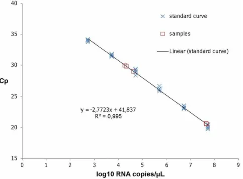
To test the feasibility of virus identification using DNase SISPA-next generation sequencing (NGS) and to get a first estimate of its sensitivity on field samples, we selected both strong and weak positive SBV infected field samples. For viral RNA quantification, in vitro transcribed RNA was used as a standard curve in the previously described L gene real time RT-PCR. The curve showed a linear range at least from 2.75 to 7.75 log10RNA copies permL,
and a sensitivity of less than 2.75 log10 RNA copies per mL
(Figure 1).
Two field samples of aborted lambs with Cp values of 20.59 and 20.65 corresponding to 7.66 and 7.64 log10RNA copies permL
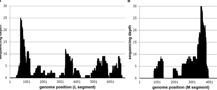
(Table 1) allowed unambiguous identification of Schmallenberg virus. One sample (BE/12-2478) yielded 2 S segment sequences (covering about 13% of the S segment), 81 M segment sequences (covering about 56% of the M segment) and 109 L segment sequences (covering about 81% of the L segment) (Table 1, Figure 2). The other strong positive sample (BE/12-2068) resulted in a total of 8 SBV specific sequence reads distributed over the L and M genomic segments. This difference in sequence coverage for two samples with a comparable SBV RNA load is most likely due to the difference in the amount of raw sequence data (Table 1). While the sequencing of BE/12-2478 yielded about 95000 reads, BE/12-2068 only resulted in circa 25000 reads probably due to DNA library quantification issues at the sequencing facility. Moreover, more tissue sample was available for DNase SISPA protocol from sample BE/12-2478 (Table 1), although the ratio of viral reads to total raw reads was superior (0.01) in sample BE/12-2068 compared to sample BE/12-2478 (0.002 ; Table 2).
Three weak positive field samples (one from an aborted calf, two from aborted lambs) containing virus quantities equivalent to 4.27–4.63 log10 RNA copies permL did not allow identification
using DNase SISPA-NGS (Table 1). This is consistent with an approximate sensitivity of 104–106 virions per ml estimated in previous studies using in vitro virus dilutions [11] or tissue biopsy samples [12]. However, it should be noted that the exact ratio between viral RNA quantities and intact virion quantities in field samples (the functional unit being detected in this method) remains to be determined. Our preliminary data indicate that RNA extracted from an SBV isolate containing 105TCID50/ml may contain up to 8.46 log10 RNA copies per mL (Table 1). This
indicates that precaution should be taken in interpreting RNA quantities in terms of DNase SISPA-NGS sensitivity, which is determined by the amount of viral nucleic acids that remain protected in intact virions during nuclease treatment. Moreover, a comparison of approximate sensitivity with other viral discovery methods is almost impossible, as the utilized sequencing effort varies from a few 100 Sanger sequencing reads [11,13] to about 30 million Illumina GAII reads [12]; and different sample types (targeted tissue selection, freshness of the sample) may result in different levels of host and contaminating nucleic acids. Given the limited approximate sensitivity of DNase SISPA-NGS, as any virus discovery method, careful selection of properly targeted and fresh field samples is necessary.
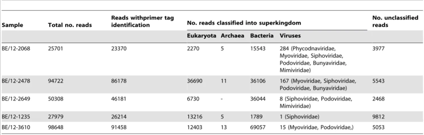
As expected in any metagenomic approach, a considerable part of the sequence reads represented diverse bacterial species and host nucleic acids (Table 2). It should be noted that the field samples were stored for a considerable time before the pretreat-ment and RNA extraction, during which opportunistic bacteria probably grew in the samples. Although we use a 0.22mM filter to
remove remaining cell fragments and bacteria, nucleic acids from disrupted cells can pass through the filter.
Figure 1. Quantification of Schmallenberg virus L segment RNA using quantitative real time RT-PCR.The viral RNA load in field samples was quantified with real time RT-PCR using a standard curve consisting of in vitro transcribed L segment RNA spanning the diagnostic RT-qPCR, which was run in five replicates (blue crosses). The linear trendline and the associated standard curve equation are displayed. The samples are indicated by red squares.
Other viral reads could be mainly identified as bacteriophages belonging to the families Myoviridae, Siphoviridae and Podoviridae (Table 2). Three reads showed partial similarity to a virus belonging to the Phycodnaviridae and two reads showed some similarity to viruses of theMimiviridaefamily. None of these viruses have relatives known to infect animals. These sequences most likely represent contamination of the tissue samples during storage until analysis.
Compared to the metagenomic approach used by Hoffmann and colleagues [1] that initially identified this novel Orthobunya-virus by shotgun sequencing of total RNA extracted from clinical samples, our virus discovery protocol attempts an enrichment in viral nucleic acids by selective filtration and nuclease treatment. A direct comparison between both data sets is impossible as we treated limited amounts of tissue samples representing a different host species. Moreover, the sequencing effort per library was not identical. Both studies indicate the need of high sequence throughput and proper sample selection as critical factors for successful virus discovery using metagenomics.
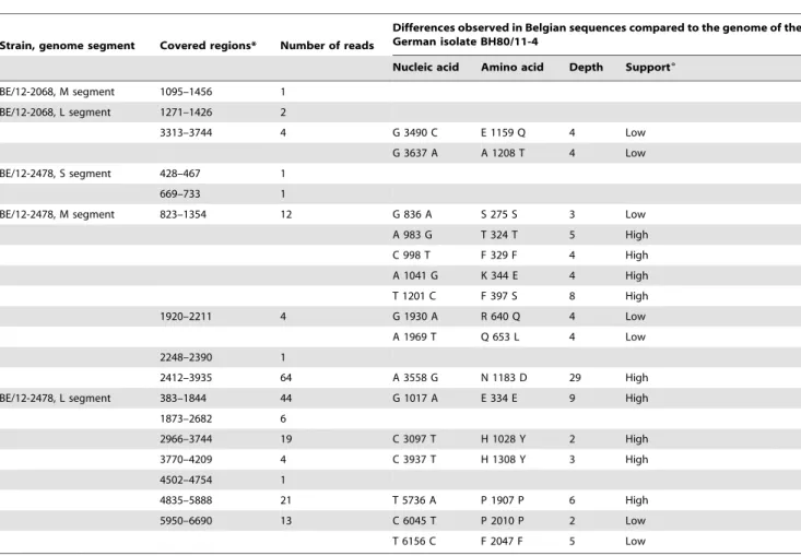
The partial SBV sequence info we obtained was compared to the whole genome sequence that was determined from the virus originally isolated from diseased bovines in Germany. Several coding and noncoding mutations could be observed (Table 3). The partial data from the two Belgian ovine field samples showed together 16 nucleotide differences (of which 9 well-supported by the sequence data, Table 3) corresponding to 9 amino acid differences (of which 5 well-supported). Although this can be expected for an RNA virus that has now shown a distribution throughout a large part of Western Europe, this is to our knowledge the first documentation of genetic diversity within the Schmallenberg virus outbreak. Based upon the 8134 nucleotides in common between our partial sequence (BE/12/2478) and the genome of the virus isolated from diseased bovine in Germany (accession codes HE649912-HE649914), a mutation frequency of 1.7 1023mutations per site was observed. The time frame between the two samples was about three months and the geographical distance between the sampling sites roughly 325 km. Although only based on a three month period, the observed mutation
Figure 2. Mapping of Schmallenberg virus specific reads of sample BE/12-2479 against the German isolate BH80/11-4.Positional sequence coverage (number of sequence reads for given nucleotide position) of the M and L segments of sample BE/12-2478, based on reference assembly to HE649912 and HE649913.
doi:10.1371/journal.pone.0041967.g002
Table 1.SBV virus quantification and confirmation by DNase SISPA-NGS in selected field samples from Belgium.
Sample (location, date) Description
SBV log10 RNA copies/ml (Cp value)
Total no. reads
No. SBV reads per segment (% of RNA segment covered)
S M L
BE/12-2068 (Ghoy, 13.01.2012) Brain tissue, 180 mg, aborted lamb 7,664 (20.59) 25701 - 1 (8.59) 6 (8.69) BE/12-2478 (Deinze, 18.01.2012) Brain tissue, 1000 mg, aborted lamb 7,642 (20.65) 94722 2 (13.68) 81(59.1) 109 (81.9) BE/12-2649 (Reningelst, 22.01.2012) Brain tissue, 1130 mg, aborted lamb 4,331 (29.83) 50308 - - -BE/12-1235 (Sivry, 06.01.2012) Brain tissue homogenate, 1.8 ml,
aborted calf
4,630 (29.00) 27979 - -
-BE/12-3610 (Izenberge, 28.01.2012) Brain tissue, 620 mg, aborted lamb 4,270 (30.00) 98648 - - -Isolate BE/12-2068 (Ghoy, 13.01.2012) Tissue culture supernatant. 5 log10
TCID50/ml
8,418 (18.50) nd nd nd nd
frequency is within the documented range of Bunyavirus variability (1022 to 1024 mutations/site/year documented for
Hantavirus ; [14]) and within the documented range of RNA virus variability (1023to 1024mutations/site/year ; [15,16]). It should be noted that, while the samples in Germany were taken from acutely infected bovines, the ovine samples from Belgium present aborted lambs, making an estimation of the infection time of the maternal animal impossible. Future targeted molecular epidemi-ological studies including samples from the complete geographic range of the virus may shed light on the origin and time of introduction of this novel virus in Europe.
Our data show that DNase SISPA-NGS viral discovery technology can be used on limited amounts of field tissue samples to identify emerging diseases. However, the sensitivity of the method seems to limit its applicability to samples containing about 104to 106virions per ml. Consequently, when applying this methodology to a cluster of cases of an undiagnosed disease, it is important to select properly targeted and fresh samples as well as to test multiple diseased animals to allow correct identification of an associated virus.
Materials and Methods
Samples
Diagnostic field samples from suspected cases of SBV related congenital malformations in lambs and calves were selected based on their geographical location (Table 1) and on the Cp values of the RT-qPCR detecting the L-segment of the virus that was used for diagnosis [1]. This study was conducted under the authoriza-tion and supervision of the Bioethics Committee at the Veterinary and Agrochemical Research Center (VAR), following national and European regulations.
Viral RNA quantification
Briefly, approximately 0.5 cm3of brain tissue was added to 1 ml PBS and homogenized (2 min, 25 Hz) in a TissueLyser (Qiagen, Venlo, The Netherlands). The RNA was extracted using the RNeasy minikit (Qiagen) following manufacturer instructions and eluted in 50ml. 2ml of this RNA mixture was further analyzed by a one-step PCR using the LightCycler 480 RNA Master Hydrolysis Probes kit (Roche Diagnostics, Vilvoorde, Belgium) on a LightCycler 480 Real-time PCR system following manufac-turer instructions. Primers and probe sequences for the SBV L segment detection were kindly provided by Dr. B. Hoffmann (FLI, Germany, available on request) and used at a final concentration of 1 and 0.1875mM respectively. Two highly positive brain tissue
samples (Cp,21) from aborted lambs were selected for confirma-tion by DNase SISPA-NGS. In addiconfirma-tion, two weak positive brain tissue samples from lambs and one weak positive sample form a calf were selected (Cp.28). The viral RNA load in RNA directly extracted from these samples was quantified in the above described real time RT-PCR using a standard curve consisting of in vitro transcribed L segment RNA spanning the diagnostic RT-qPCR, which was run in five replicates. The in vitro transcribed RNA was independently quantified using three different approaches: NanoDrop Spectrophotometer (Nanodrop Technologies, Wilmington DE, USA), QubitHRNA assay kit on QubitH fluorometer (Invitrogen-Life Technologies, Gent, Bel-gium), and RNA 6000 Pico chip on the Agilent 2100 Bioanalyser (Agilent Technologies, Diegem, Belgium).
DNase SISPA and 454 sequencing
The maximum available quantity of the limited tissue samples (,1,5 g; Table 1) was homogenized in about 1500mL PBS per g of
tissue using gentle homogenization in a TissueLyser (Qiagen). Sample pretreatment and SISPA was largely performed as previously described [8,9]. Briefly, after a centrifugation and filtration step using 0.22mM filters, the eluate was subjected to
DNase I treatment (100 U/200ml sample). The resulting
virion-enriched samples were subjected to a viral RNA extraction using the QIAamp Viral RNA Mini Kit (Qiagen). Forty units of Protector RNase inhibitor (Roche) were added to the eluted RNA, and RNA quality was checked using a Bioanalyzer 2100 (Agilent Technolo-gies). The random first- and second strand cDNA synthesis was performed with the primer FR26RV-N (59GCC GGA GCT CTG CAG ATA TCN NNN NN 39) and primer FR20RV (59-GCC GGA GCT CTG CAG ATA TC-39) was used in subsequent PCR to amplify the resulting cDNA. After visualisation of the random amplified DNA fragments on a 1% agarose gel, the fragments between 200 and 1000 bp were excised, purified and quantified using a Nanodrop spectrophotometer (Nanodrop Technologies). Five micrograms of each size selected (200–1000 bp) and purified random amplified sample were sequenced on a GS FLX+(Roche, Mannheim, Germany) by the Genomics Core of the University Hospital (University of Leuven, Belgium) using multiplex identifier (MID) identification during library preparation and their standard procedures using GS FLX Titanium series reagents (Roche, Mannheim, Germany). The DNA fragmentation step by nebuliza-tion was skipped and the intennebuliza-tion was to obtain 30000–40000 reads per library.
Table 2.Output of the metagenomic analysis on raw sequence data from the sequencing libraries from SBV-positive samples.
Sample Total no. reads
Reads withprimer tag
identification No. reads classified into superkingdom
No. unclassified reads
Eukaryota Archaea Bacteria Viruses
BE/12-2068 25701 23370 2270 5 15543 284 (Phycodnaviridae,
Myoviridae, Siphoviridae, Podoviridae, Bunyaviridae, Mimiviridae)
3977
BE/12-2478 94722 86178 36690 11 36106 167 (Myoviridae, Siphoviridae,
Podoviridae, Bunyaviridae)
5543
BE/12-2649 50308 46181 6730 - 36044 8 (Siphoviridae, Podoviridae,
Mimiviridae)
2468
BE/12-1235 27979 26214 13216 5 1789 1 (Siphoviridae) 9812
BE/12-3610 98648 91458 12403 13 69057 15 (Myoviridae, Podoviridae,) 5053
Metagenomic analysis
As an additional control to exclude reads originating from potential DNA contamination during the library preparation steps, only reads containing the SISPA primer sequence were included in the assembly. Subsequently, the SISPA primer sequences plus additional six bases were trimmed off the reads. By using a combination of BLAST [17] and sequence mapping with the 454 reference mapper application (version 2.6; Roche), contigs (i.e. sets of overlapping sequence reads) and reads were classified into different taxa.
Reference assembly
To map the obtained raw sequence data to the genome of Schmallenberg virus, the complete coding sequence of SBV isolate BH80/11-4 (accession codes HE649912–HE649914) was used as reference genome in the reference assemblies of our different field samples using SeqMan NGenH version 3 (DNASTAR, Madison, WI, USA). The reads were first trimmed to remove primer sequences (including the primer-encoded random N positions) as well as low quality ends. Standard assembling and filtering parameters were used, except for a reduced minimum match percentage. The partial sequence information was made accessible through GenBank accession numbers JQ861686–JQ861692, except for fragments that were
less than 200 bp in length due to the minimum fragment length requirements dictated by GenBank.
Variability analysis
The obtained partial sequence information of SBV was compared to the sequence of isolate BH80/11-4 (accession codes HE649912– HE649914). Single nucleotide polymorphisms (SNP’s) were identified using SeqMan Pro version 9 (DNASTAR, Madison, WI, USA) and are listed in Table 3. Only sequence differences where we had at least 2 sequence reads were included. Observed sequence variants at a position with more than 2 reads and/or with reads in both cDNA and complementary sense and/or with reads having different start positions were assigned a high support (Table 3).
Acknowledgments
We thank Dr. Martin Beer and Dr. Bernd Hoffmann (FLI) for providing the SBV real time RT-PCR protocol and control reagents. We are grateful to Orkun Ozhelvaci for excellent technical assistance. The professional services of the Genomics Core of the University Hospital of Leuven are appreciated.
Author Contributions
Conceived and designed the experiments: SVB TR FV. Performed the experiments: TR. Analyzed the data: DH TR MS SVB. Contributed reagents/materials/analysis tools: NDR ABC. Wrote the paper: TR SVB. Table 3.Differences observed in Belgian SBV sequences in comparison with the German genome sequence of isolate BH80/11-4.
Strain, genome segment Covered regions* Number of reads
Differences observed in Belgian sequences compared to the genome of the German isolate BH80/11-4
Nucleic acid Amino acid Depth Support6
BE/12-2068, M segment 1095–1456 1 BE/12-2068, L segment 1271–1426 2
3313–3744 4 G 3490 C E 1159 Q 4 Low
G 3637 A A 1208 T 4 Low
BE/12-2478, S segment 428–467 1
669–733 1
BE/12-2478, M segment 823–1354 12 G 836 A S 275 S 3 Low
A 983 G T 324 T 5 High
C 998 T F 329 F 4 High
A 1041 G K 344 E 4 High
T 1201 C F 397 S 8 High
1920–2211 4 G 1930 A R 640 Q 4 Low
A 1969 T Q 653 L 4 Low
2248–2390 1
2412–3935 64 A 3558 G N 1183 D 29 High
BE/12-2478, L segment 383–1844 44 G 1017 A E 334 E 9 High
1873–2682 6
2966–3744 19 C 3097 T H 1028 Y 2 High
3770–4209 4 C 3937 T H 1308 Y 3 High
4502–4754 1
4835–5888 21 T 5736 A P 1907 P 6 High
5950–6690 13 C 6045 T P 2010 P 2 Low
T 6156 C F 2047 F 5 Low
References
1. Hoffmann B, Scheuch M, Hoper D, Jungblut R, Holsteg M, et al. (2012) Novel orthobunyavirus in cattle, europe, 2011. Emerg Infect Dis 18 (3):469–472 2. Muskens J, Smolenaars AJ, van der Poel WH, Mars MH, van Wuijckhuise L,
et al. (2012) [Diarrhea and loss of production on Dutch dairy farms caused by the Schmallenberg virus]. Tijdschr Diergeneeskd 137 (2):112–115
3. van den Brom R, Luttikholt SJ, Lievaart-Peterson K, Peperkamp NH, Mars MH, et al. (2012) Epizootic of ovine congenital malformations associated with Schmallenberg virus infection. Tijdschr Diergeneeskd 137 (2):106–111 4. Bilk S, Schulze C, Fischer M, Beer M, Hlinak A, et al. (2012) Organ distribution
of Schmallenberg virus RNA in malformed newborns. Vet Microbiol. http://dx. doi.org/10.1016/j.vetmic.2012.03.35
5. ProMED-Mail (2012) Schmallenberg virus - Europe: update, international impact. http://www.promedmail.com (accessed 2012 March 29) archive no. 20120324.1079633
6. ProMED-mail (2012) Schmallenberg virus - Europe: vector, morphology. http://www.promedmail.com, archive no201203111066949 2012
7. Allander T, Emerson SU, Engle RE, Purcell RH, Bukh J (2001) A virus discovery method incorporating DNase treatment and its application to the identification of two bovine parvovirus species. Proc Natl Acad Sci U S A 98 (20):11609–11614
8. Rosseel T, Lambrecht B, Vandenbussche F, van den Berg T, Van Borm S (2011) Identification and complete genome sequencing of paramyxoviruses in mallard ducks (Anas platyrhynchos) using random access amplification and next generation sequencing technologies. Virol J 8:463
9. Van Borm S, Rosseel T, Vangeluwe D, Vandenbussche F, van den Berg T, et al. (2012) Phylogeographic analysis of avian influenza viruses isolated from
Charadriiformes in Belgium confirms intercontinental reassortment in gulls. Arch Virol. In press
10. Margulies M, Egholm M, Altman WE, Attiya S, Bader JS, et al. (2005) Genome sequencing in microfabricated high-density picolitre reactors. Nature 437 (7057):376–380
11. Djikeng A, Halpin R, Kuzmickas R, Depasse J, Feldblyum J, et al. (2008) Viral genome sequencing by random priming methods. BMC Genomics 9:5 12. Daly GM, Bexfield N, Heaney J, Stubbs S, Mayer AP, et al. (2011) A viral
discovery methodology for clinical biopsy samples utilising massively parallel next generation sequencing. PLoS One 6 (12):e28879
13. Allander T, Tammi MT, Eriksson M, Bjerkner A, Tiveljung-Lindell A, et al. (2005) Cloning of a human parvovirus by molecular screening of respiratory tract samples. Proc Natl Acad Sci U S A 102 (36):12891–12896
14. Ramsden C, Melo FL, Figueiredo LM, Holmes EC, Zanotto PM (2008) High rates of molecular evolution in hantaviruses. Mol Biol Evol 25 (7):1488–1492
15. Jenkins GM, Rambaut A, Pybus OG, Holmes EC (2002) Rates of molecular evolution in RNA viruses: a quantitative phylogenetic analysis. J Mol Evol 54 (2):156–165
16. Hanada K, Suzuki Y, Gojobori T (2004) A large variation in the rates of synonymous substitution for RNA viruses and its relationship to a diversity of viral infection and transmission modes. Mol Biol Evol 21 (6):1074–1080 17. Altschul SF, Madden TL, Schaffer AA, Zhang J, Zhang Z, et al. (1997) Gapped