online | memorias.ioc.fiocruz.br
Assessment of Epstein-Barr virus nucleic acids in gastric but not in breast
cancer by next-generation sequencing of pooled Mexican samples
Ezequiel M Fuentes-Pananá1, Violeta Larios-Serrato2, Alfonso Méndez-Tenorio2,
Abigail Morales-Sánchez1, Carlos F Arias3, Javier Torres4/+
1Hospital Infantil de México Federico Gómez, Unidad de Investigación en Virología y Cáncer, México, DF, México
2Escuela Nacional de Ciencias Biológicas, Unidad Profesional Lázaro Cárdenas, Laboratorio de Biotecnología y Bioinformática Genómica, México, DF, México 3Universidad Nacional Autónoma de Mexico, Instituto de Biotecnología,
Departamento de Genética del Desarrollo y Fisiología Molecular, Cuernavaca, Morelos, México 4Centro Médico Nacional Siglo XXI, Hospital de Pediatría, Unidad de Investigación Médica en Enfermedades Infecciosas y Parasitarias, México, DF, México
Gastric (GC) and breast (BrC) cancer are two of the most common and deadly tumours. Different lines of evi-dence suggest a possible causative role of viral infections for both GC and BrC. Wide genome sequencing (WGS) technologies allow searching for viral agents in tissues of patients with cancer. These technologies have already contributed to establish virus-cancer associations as well as to discovery new tumour viruses. The objective of this study was to document possible associations of viral infection with GC and BrC in Mexicanpatients. In order to gain idea about cost effective conditions of experimental sequencing, we first carried out an in silico simulation of WGS. The next-generation-platform IlluminaGallx was then used to sequence GC and BrC tumour samples. While we did not find viral sequences in tissues from BrC patients, multiple reads matching Epstein-Barr virus (EBV) sequences were found in GC tissues. An end-point polymerase chain reaction confirmed an enrichment of EBV sequences in one of the GC samples sequenced, validating the next-generation sequencing-bioinformatics pipeline.
Key words: gastric cancer - breast cancer - gastritis - wide genome sequencing - EBV
doi: 10.1590/0074-02760150405
Financial support: CONACyT (69450 - to J Torres), Fondo SEP CONACyT (176880 - to EMF-P), FIS/IMSS/PROT/G12/1120 (to EMF-P), Fondo de Apoyo a la Investigación HIM (2013-051 - to EMF-P) EMF-P and VL-S contributed equally to this work. + Corresponding author: [email protected] Received 22 October 2015
Accepted 12 January 2016
Globalisation, climate change, urbanisation of wild ar-eas, and other modern conditions of life are contributing to an increased exposure to a number of infectious agents. Concomitantly, worldwide populations are experiencing a demographic transition that has placed cancer as one of the leading causes of death. To date, a number of patho-gens, mostly viruses, have been classified as carcinogenic to humans by the International Agency for Research on Cancer. These viruses include: high-risk human papillo-mavirus (HPV), Epstein-Barr virus (EBV), Kaposi sar-coma herpesvirus, Merkel cell polyomavirus (MCPyV), hepatitis B virus, hepatitis C virus, and human T-lympho-tropic virus 1(Morales-Sánchez & Fuentes-Pananá 2014). Most families of oncogenic viruses are either strict DNA or viruses in which their life cycle has a DNA stage, such as retroviruses, which convert their genomic RNA into proviral DNA that is integrated into the host genome.
It is estimated that human cancers of viral aetiology comprise up to 20% of all tumours, with higher frequen-cies found in developing countries (Crawford 2005). How-ever, viral agents often defy isolation and recognition by
traditional culture or molecular methods. Next-generation sequencing (NGS) technologies are currently used for wide-genome sequencing (WGS) providing high through-put data from single genomes, making them optimal to interrogate for the presence of known and previously un-recognised viral agents. Furthermore, WGS has recently al-lowed the identification of a novel polyomavirus associated with Merkel cell carcinoma (MCC) (Feng et al. 2008).
Gastric (GC) and breast (BrC) cancerare two of the most common cancers and two of the most important problems in public health now days. GC is the fifth most frequent cancer worldwide and the third cause of death by cancer, whilst BrC is the most frequent cancer in working-age woman (Ferlay et al. 2014). GC is consid-ered primarily of infectious aetiology, with Helicobacter pylori infection recognised as the most important risk factor (Crew & Neugut 2006). Colonisation of the gas-tric mucosa by H. pylori triggers a chronic inflammato-ry response that when unregulated chronically damages the gastric mucosa, generating progressive lesions of increased severity and risk of ending in cancer (Correa et al. 2006). Lesions usually start with a nonatrophic gastritis (NAG), progress to atrophic gastritis, intestinal metaplasia and dysplasia, to finally evolve into GC (Cor-rea et al. 2006). More recently, several lines of evidence also support infection by EBV as an important causative agent for GC (Murphy et al. 2009, Camargo et al. 2011).
vi-rus (MMTV), HPV and EBV are the agents reported as probably associated with BrC (Joshi & Buehring 2012). However, data have been highly variable, with reported infection prevalence ranging from 0-100% and the viral infection-BrC association remains highly controversial.
In this study, we searched for fingerprints of viral infection in pools of GC and BrC tissues using the Illu-minaGallx NGS platform. Previously, we implemented an in silico NGS simulation assay aimed to find a man-ageable cost effective pipeline of analysis to interrogate for the presence of viral sequences in cancer samples. We did not find sequences supporting viral participa-tion in breast tumours, whereas multiple reads matching EBV sequences were found in gastric tumours. An end-point polymerase chain reaction (PCR) confirmed EBV sequences in one of the GC samples sequenced ratifying the utility of the bioinformatics pipeline of analysis. The development and implementation of specific and sensi-tive NGS together with bioinformatics strategies will be-come critical to dissect the biome associated with cancer and many other diseases of infectious origin.
SUBJECTS, MATERIALS AND METHODS
Study population - Patients with confirmed diagnosis of GC, BrC, and NAG were included in the study. Five patients formed every study group. All patients were re-cruited in Mexico City, GC and BrC patients from the Oncology Hospital and patients with a NAG diagnosis from the Specialities Hospital, both from Mexican In-stitute of Social Security (IMSS) at the XXI Century National Medical Center. Tumour and tumour-adja-cent tissues were derived from the organ resection. Tu-mour-adjacent tissues served as controls for specific-ity of tumour cell infection and these control samples
were taken ≥ 2 cm apart from the tumour mass from
the same patient in which tumour tissue was obtained. Gastric biopsies were from patients referred to the gas-troenterology unit of the Specialities Hospital because of gastric symptoms. A fragment of all tissues was fixed in formaldehyde and embedded in paraffin and a slide was stained with haematoxylin-eosin and analysed by a pathologist to confirm the diagnosis. All tumour tis-sues included in the study were carcinomas with at least 70% of tumour cells; all GCs included in the study were classified as mixed type (intestinal and diffuse) accord-ing to the Lauren’s criteria. All BrC were classified as ductal infiltrating. The BrC molecular classification was the following: three patients were luminal A, one patient was with human epidermal growth factor receptor 2 pos-itive, and one patient was triple negative. In the gastritis samples, we included cases without atrophy or pre-neo-plastic lesions with a diagnosis of NAG.
Sample preparation - Ten milligrams of each tissue sample were disrupted in a TissueLyser II (Qiagen, Ger-many) for 20 s and homogenates were subjected to DNA purification with QIAamp DNA mini kit in a QIAcube automated sample processing workstation (Qiagen). Puri-fied DNA was quantiPuri-fied using a spectrophotometer Na-noDrop 1000 (Thermo Fisher Scientific, USA) and DNA quality was determined with the 260/280 ratio of
absorb-ance, integrity by electrophoresis in agarose gels, and by
PCR of β-actin (670 bp) endogenous gene using primers
previously described (Fuentes-Pananá et al. 2004).
Sample analysis - DNA from BrC, BrC tumour-ad-jacent controls, GC, GC tumour-adtumour-ad-jacent controls, and NAG was sequenced. Pools of five patients formed every
group of study for a total of five pools. We used 1 μg of
DNA from each patient for sequencing a total of 5 mg per group. DNA from each group was loaded into separated lanes of a flow cell from a Genome Analyzer IIx (Illumi-na, USA). Sequencing was performed through 36 cycles of single base pair extensions. Fluorescent images were analysed using the Illumina base calling pipeline v.1.4 to obtain data sequences. The resulting initial sequences of the samples have a length of 36-mer. Those reads obtained for all samples (in FASTQ format) were filtered from un-desired sequences using the assembly Perl tools from the Euler-SR program (Chaisson & Pevzner 2008). Then, a collection of programs developed with the Lazarus Free Pascal programming language was used to (i) eliminate sequences of low complexity such as mononucleotide re-peats, (ii) trim end-nucleotides that did not fulfill a phred quality value < 30, (iii) change the data file format from FASTQ to FASTA, and (iv) eliminate repeated reads.
In silico preliminary analysis - Simulation of Illu-mina Sequencing was carried out in ART (Huang et al. 2012) with the ART’s parameterised quality profiles and model error specific of the platform. The inputs contained two copies of human genome (GRCh37.p13) plus one of the following options: (i) 100 copies of the HPV 16 genome (NC_001526.2) (Theelen et al. 2010), (ii) 10 copies of the MMTV genome (NC_001503.1) (Morris et al. 1977), or seven copies of the EBV genome (NC_009334.1) (Liu et al. 2011). Emulation generated synthetic Illumina sequencing reads according to differ-ent covertures (0.1X, 0.2X, 0.5X and 1X) and two dif-ferent read sizes (36 mer and 100 mer). Subtraction of poor quality and human sequences, as well as mapping of viral sequences, is described in the next section.
genomes/H_sapiens/RNA/) and Ensembl Homo sapiens
cDNA database (ftp.ensembl.org). The subtraction of hu-man reads was done with a suite of PERL developed pro-grams. The remaining non-human reads were analysed by BLASTN (v.2.2.28, word size = 9, E-value = 1 x 10⁻⁶)
to 1,520,849 viral sequences [downloaded from NCBI Nucleotide (ncbi.nlm.nih.gov/nucleotide)] using the search term “viruses” [porgn: txid10239] on 1 January 2013. The viral reads obtained were contrasted against a dataset of bacterial, protozoa, and fungi sequences to confirm their authenticity using Bowtie (parameter 1 mismatch). The dataset was a compilation of the follow-ing: 3,336 bacteria complete genomes (2012) (ftp://ftp. ncbi.nih.gov/genomes/Bacteria/), 500 human microbi-ome bacteria (ftp.ncbi.nih.gov/genmicrobi-omes/HUMAN_MI- (ftp.ncbi.nih.gov/genomes/HUMAN_MI-CROBIOM/Bacteria/), 716,562 protozoa sequences (NCBI Nucleotide database terms: “apicomplexans” [porgn: txid5794], “amoebozoa” [porgn: xid554915], diplomonadida [porgn: txid5738], “kinetoplastida” [por-gn: txid5653], “platyhelminthes” [por[por-gn:__txid6157]), and 522,571 fungi sequences (NCBI nucleotide database terms: “ascomycetes” [porgn: txid4890], “neocallimas-tigales” [porgn: txid29006], “microsporidians” [porgn: txid6029], “mucorales” [porgn: txid4827], “glomer-ales” [porgn: txid1028384], “tremellomycetes” [porgn: txid155616]). All generated sequences were deposited in SRA database with the next BioSample accessions 774161 (BrC), 795877 (BrC control), 795890 (GC), 796157 (GC control), and 796243 (gastritis). The analysis pipeline was constructed using the Perl 5.8.1 program. Data processing was carried out in a Mac Pro equipment with a Mac OS X server operating system, a 2x2.66 GHz 6-core Intel Xeon processor, and 32 GB RAM memory.
Direct search of viral sequences - To corroborate the lack of hits for viral agents previously documented in samples, 6,782 sequences of HPV [porgn: txid10566], 258 sequences of mouse mammary tumour virus [por-gn: txid11901], and 814 sequences of bovine leukaemia virus [porgn: txid11901] were downloaded and match-es were directly searched in the BrC and breast nontu-mour control databases. Sequences were aligned using Bowtie, allowing up to 1 mismatch and without remov-ing any human sequence; the only filters used in this latter search were quality phred, mononucleotides, and repeated sequences of data sets.
PCR detection of EBV - DNA samples were sub-jected to a first PCR with primers LLW1 and LLW2 (Labrecque et al. 1995) which amplify a region within the BamHI W fragment in the EBV genome. The Dau-di cell line was used as positive control. The PCR mix
(50 μL) contained 200 ng of template DNA, 200 µM of
dNTPs mix, 2.5 mM of MgCl2, 5 μL of Taq Polymerase buffer 10x with (NH4)2SO4, 200 nM of each primer, and 2.5 U of Taq Polymerase (all from Thermo Fisher Scien-tific). The PCR reaction was: an initial denaturation step of 5 min at 94ºC and then 30 cycles of 94ºC for 1.5 min, 57ºC for 45 s and 72ºC for 1 min, and a final extension of 72ºC for 7 min. Internal primers to the first PCR am-plicon were designed for a nested PCR: LLWint1 5’CT-TTGTCCAGATGTCAGGGG3’ and LLWint2
5’GCCT-GAGCCTCTACTTTTGG3’. The 50 μL PCR mixture contained 1 μL of the first PCR (1:1000 final dilution), 200 µM of dNTPs mix, 2.5 mM of MgCl2, 5 μL of Taq
Polymerase buffer 10x with (NH4)2SO4, 400 nM of each primer, and 2.5 U of Taq Polymerase (all from Thermo Fisher Scientific). The reaction was performed with an initial denaturation step at 94ºC for 5 min, followed by 15 cycles of 94ºC for 20 s, 57ºC for 20 s, and 72ºC for 30 s, and a final extension of 72ºC for 7 min.
Sanger sequencing - The identity of the EBV positive PCR product was confirmed by sequencing both for-ward and reverse strands. The PCR product was purified using QIAquick gel extraction kit (Qiagen) according to manufacturer’s instructions, and sequencing of the isolated DNA fragment was carried out in the Biology Institute, National Autonomous University of Mexico. Sequences were compared with the GenBank database using the BLAST program (Altschul et al. 1990).
Ethics - The National Commission of Scientific Re-search and the Ethical Committee on ReRe-search of the IMSS approved this project. All patients were informed on the nature of the study and those willing to participate signed a written informed consent prior to specimen collection.
RESULTS
Preliminary simulation analysis - Looking to imple-ment a bioinformatics pipeline that allowed manageable and cost effective sequencing conditions, we first carried out an NGS simulation analysis. We selected three pre-viously GC and BrC-associated viruses with different genome sizes, ranging from about 8,800 bp (MMTV) to 172,000 bp (EBV). Three different inputs containing human and viral genomes were constructed. The num-ber of human and viral genomes used in every input was adjusted mimicking real infected tumour cells. Thus, 100 copies of HPV 16 genome (Theelen et al. 2010), 10 copies of MMTV genome (Morris et al. 1977), or seven copies of EBV genome (Liu et al. 2011) were used per diploid human genome. Generation of synthetic reads was followed by several rounds of filtration (See Fig. 1 and Subjects, Materials and Methods). Multiple viral reads mapped to filtered data sets. This analysis predict-ed the minimal coverage to detect viral fingerprints. We also interrogated in silico for the most suitable size read, and a 36 mer vs. a 100 mer read size was compared (not shown). This simulation showed that using 36 mer read size was enough to detect viral hits (Fig. 1). This pre-liminary analysis guided us about adequate conditions of sequencing for viral detection in biological samples.
isolated from the same cancer patients and located at least 2 cm apart from the tumour mass served as controls for specificity of tumour cell infection, while biopsies of patients with NAG addressed the possible viral partici-pation in early inflammatory precursor lesions.
The pipeline analysis of the data generated by the Il-lumina GAIIx sequencing and the percentage of non-hu-man reads remaining after each level of filtration are shown in Fig. 2. Ten to 20% of sequences were eliminat-ed on the bases of quality and composition, 76-88% reads preferentially matched human sequences and were also eliminated, to end with an average of 2.39% non-human reads for all tissues sequenced. Table I shows the number of initial and final non-human reads after all filtration steps. Human sequences were a compendium of seven different databases, which allowed a more stringent tool for filtration and a better selection of sequences with more distant similarities to human genomes. The data-base of human sequences can be found freely at NCBI and European Molecular Biology Laboratory, and all human sequences found in pools of sequenced tissues at drive.google.com/folderview?id=0B3AZ9N8M5ZM-fVGtpbUFtQ1JBbE0&usp=sharing for readers use.
The number of non-human reads obtained consti-tuted a set of manageable information for more robust analysis; such reads were blasted against different mi-crobial databases, including a database of 1,520,849 vi-ral sequences. The vivi-ral database was based in available sequences deposited in the NCBI, and it is also freely available (drive.google.com/folderview?id=0B3AZ9N-8M5ZMfYjJCT0xlNzZSdkU&usp=sharing).
Different types of viral hits were found, most of them were of no interest since they were not from members of
a family of tumour viruses or because of they were pres-ent across all tumour and control tissues. For instance, several hits matched sequences present in phages or vectors that are commonly used as tools for molecular biology research: eight-18 hits matching enterobacteria phage sequences were found across all sequenced tissues, one-two hits matching baculovirus sequences were also found across all tissues, and one hit matching SAdV-40 was found in BrC tissue. The origin of this genetic mate-rial most likely comes from contamination with enzymes and reagents used to process tissues. Two hits were found matching the Torque teno virus, which is commonly found in cells of the immune system: one in BrC tumour-adja-cent control and one in GC. Slightly more interesting was to find hits matching retroviral sequences; although, those hits showed higher similarity with human endogenous retrovirus H and K and less to oncogenic MMTV, which has been linked to BrC. Furthermore, these retroviral hits were found in all tissues sequenced as shown in Table II.
Several hits matching members of the Herpesviri-dae family were found: six against human herpesvirus type 7 (HHV7), one in BrC tumour-adjacent, one in GC, and four in NAG; two against citomegalovirus, one in GC, and one in GC tumour-adjacent; six against herpes simplex virus type 1 (HSV1), one in BrC, one in GC tu-mour-adjacent, and four in GC. These HSV1 sequences also exhibited high similarity (> 90%) to other members of the Alphaherpesvirinae subfamily, to gallid herpesvi-rus 2 (infects birds), angullid herpesviherpesvi-rus (infects eels), and cyprinid and Koi herpesvirus (both infect fish).
Fig. 1: flowchart of next-generation sequencing (NGS) simulation. Illu-mina sequencing emulation generating 36 or 100 mer (not shown) reads was run in ART (Huang et al. 2012). Different coverages were tested. Several viral hits from human papillomavirus (HPV) 16, mouse mamma-ry tumour virus (MMTV), and Epstein-Barr virus (EBV) were mapped from human-viral inputs mimicking genomic load present in tumours as-sociated to these agents. Viral sequences were screened allowing 0 (0M) or 1 (1M) mismatch. Tens or hundreds of viral reads were found.
Hits matching HHV type 6 (HHV6) and EBV se-quences exhibited tissue specificity; HHV6 had 61 hits in breast and zero in gastric tissue. However, there was
TABLE I
Number of reads
Tissue Total reads Non-human reads
BrC 15,600,870 269,740
BrC control 30,100,894 513,073
GC 29,213,391 770,760
GC control 27,296,544 507,667 Gastritis 14,421,599 329,850
BrC: breast cancer; GC: gastric cancer.
TABLE II
Viral hits
Virus BrC BrC control GC GC control Gastritis
EBV (HHV4) 0 0 10 1 2
HHV6 16 45 0 0 0
HHV7 0 1 1 0 4
CMV (HHV5) 0 0 1 1 0
Other herpesviruses 1 0 4 1 0
HERVS 10 6 7 7 5
BrC: breast cancer; CMV: citomegalovirus; EBV: Epstein-Barr virus; GC: gastric cancer; HERVS: human endogenous retrovi-rus; HHV: human herpesvirus.
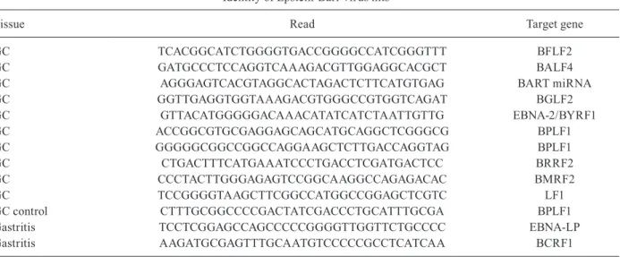
TABLE III
Identity of Epstein-Barr virus hits
Tissue Read Target gene
GC TCACGGCATCTGGGGTGACCGGGGCCATCGGGTTT BFLF2
GC GATGCCCTCCAGGTCAAAGACGTTGGAGGCACGCT BALF4
GC AGGGAGTCACGTAGGCACTAGACTCTTCATGTGAG BART miRNA
GC GGTTGAGGTGGTAAAGACGTGGGCCGTGGTCAGAT BGLF2
GC GTTACATGGGGGACAAACATATCATCTAATTGTTG EBNA-2/BYRF1
GC ACCGGCGTGCGAGGAGCAGCATGCAGGCTCGGGCG BPLF1
GC GGGGGCGGCCGGCCAGGAAGCTCTTGACCAGGTAG BPLF1
GC CTGACTTTCATGAAATCCCTGACCTCGATGACTCC BRRF2
GC CCCTACTTGGGAGAGTCCGGCAAGGCCAGAGACAC BMRF2
GC TCCGGGGTAAGCTTCGGCCATGGCCGGAGCTCGTC LF1
GC control CTTTGCGGCCCCGACTATCGACCCTGCATTTGCGA BPLF1
Gastritis TCCTCGGAGCCAGCCCCCGGGGTTGGTTCTGCCCC EBNA-LP
Gastritis AAGATGCGAGTTTGCAATGTCCCCCGCCTCATCAA BCRF1
GC: gastric cancer.
not specificity of infection for the tumour sample (16 hits in BrC and 45 in BrC tumour-adjacent control tissue); therefore, this result does not support a direct etiological role for HHV6 in BrC. On the other hand, EBV had 10 hits in GC, while only one in GC tumour-adjacent tissue and two in NAG. Table III shows the identity of the gene targets of the EBV hits. One hit overlaps to two overlap-ping coding sequences: EBNA-2 and BYRF1.
Although, the main goal of this study was to analyse the virome of BrC and GC, but since H. pylori infection is recognised as the main risk factor to develop NAG and GC, a blast of the non-human reads found in gastric lesions was performed against a database of 3,336 full bacterial genomes. These data is shown in Supplementa-ry Figure. Hits mapping to the Helicobacter genus were found in 2%, 12%, and 36% of all bacterial reads in GC, GC adjacent tissue control and NAG samples, respec-tively. Helicobacter was the most abundant genera found in gastritis, while Paracoccus and Propionobacterium
were the most represented in GC and GC controls. These data is congruent with the literature documenting that
H. pylori tents to be absent of tumour tissues, while it is highly abundant in early gastric lesion (Kokkola et al. 2003, Camorlinga-Ponce et al. 2008).
EBV detection by PCR - To confirm the presence of EBV in GC tumours and validate the results from the WGS, a PCR test was implemented using primers LLW1 and LLW2 (Labrecque et al. 1995) in a reaction set to
de-tect ≥ 40,000 viral genomes (Martínez-López et al. 2014).
Tissues from the five GC tumours and their counterpart tumour-adjacent controls and the five NAG samples were individually tested by the PCR. Furthermore, since EBV usually resides in B cells in a low number of infected cells, mononuclear cells isolated from peripheral blood of GC and NAG patients were also included in the analy-sis. Fig. 3 shows the result of the PCR test; one of the GC tissues was found positive for EBV sequences, while the other four patients were negative. The patient positive by the PCR test was negative in the tumour-adjacent control tissue and peripheral mononuclear cells, supporting an
enrichment of EBV infected cells in the tumour tissue. None of the NAG patients were positive, in agreement with the NGS data. EBV sequences were confirmed with
a more sensitive nested PCR (detects ≥ 1,500 viral par
-ticles) (Martínez-López et al. 2014) and by Sanger se -quencing of the first positive PCR-amplicon.
Taken together, these data highlight the importance of NGS technologies as a powerful and manageable tool to in-terrogate cancer tissues for the presence of viral sequences looking to better understand the aetiology of the disease.
DISCUSSION
NGS technologies have opened new perspectives for viral research and diagnostic in multiple human and veterinary diseases. In recent years, this technology has allowed the identification and characterisation of new viruses, such as the Bundiubugyo virus, a virus relat-ed to Ebola and responsible for severe haemorrhagic fe-vers in humans (Towner et al. 2008), and an arenovirus closely related to lymphocytic choriomeningitis viruses (Kim et al. 2011), associated with fatal post-transplant disease. MCC was a long suspected cancer of infectious aetiology because it develops preferentially in immu-nosuppressed individuals. However, the identity of the causative agent remained elusive until high throughput sequencing and transcriptome subtraction methodolo-gies allowed the identification of a novel polyomavirus in MCC samples (Feng et al. 2008). Now, it has been firmly documented that MCPyV is responsible for up to 100% of MCC (Agelli et al. 2010).
Here, we used a similar approach to interrogate for the presence of viral sequences in BrC and GC tissues, two of the most common cancers. However, considering that NGS-based methodologies result in an enormous set of data difficult for processing and interpretation, we imple-mented some strategies to reduce both cost and time of analysis to the experimental and bioinformatics approach. DNA samples were pooled (with DNA from 5 patients in every lane of the sequencer), thus, we reduced the cost of massive sequencing. Additionally, we carried out an in silico preliminary analysis simulating several NGS condi-tions according to the reported viral genome copies found in cancer. Through this analysis, we selected an afforda-ble but sufficient coverage to detect viral sequences.
A database of multiple human sequences was con-structed and used to digitally subtract candidate sequenc-es of viral origin, sequenc-especially those matching members of viral families with oncogenic characteristics. This strat-egy leaded us to find HHV6 sequences associated with breast tissue and EBV with gastric tumour samples. GC has been extensively associated with H. pylori infection and more recently with EBV, and we found evidence of both pathogens in gastric samples. Since EBV is a wide-spread pathogen and NGS could detect low levels of con-taminant virus, sequencing data were confirmed via two PCR tests of increased sensitivity. EBV was detected in the tumour, but not in tumour-adjacent tissue or periph-eral mononuclear cells of one GC patient. EBV usual-ly resides in B-cells in frequencies estimated between one-20 cells per million (Rickinson & Kieff 2007), a frequency of infection that was under the limit of
detec-Fig. 3: detection of Epstein-Barr virus (EBV) by polymerase chain re-action (PCR). A: one of five gastric cancer (GC) samples was positive (sample four) to EBV; by first round PCR (upper panel) and nested PCR (middle panel); B: nontumour control tissues (C) and peripheral blood (PB) from the same GC patients were negative to EBV by both PCRs; D: none of the gastritis samples (E) nor PB of these patients were pos-itive to EBV by any PCR. DNA from Daudi cells was used as pospos-itive control (C+). A reaction without DNA was used as negative control (C-); M: molecular weight marker. Lower panels shows the PCR of loading
tion of either PCR (Ryan et al. 2009, Martínez-López et
al. 2014). The enrichment of EBV infection observed in tumour tissue is in agreement with the known direct on-cogenic mechanism of EBV through expression of viral oncogenes within the transformed cell. Multiple lines of evidence now support a role for EBV in GC and a recent meta-analysis reports a 10% world-wide prevalence of EBV associated GCs (Murphy et al. 2009, Camargo et al. 2011). We found a similar frequency of EBV infection
in Mexican GC samples (Martínez-López et al. 2014). In
EBV associated lymphomas, the number of viral copies has been estimated in 50 viral episomes per tumour cell (Gulley et al. 1994), while seven viral copies were found in an EBV associated nasopharyngeal carcinoma (Liu et al. 2011), which is within the limits of the PCR test used here. It is possible that gastric carcinoma more closely resembles NPC, which highlights the power of the NGS even in pools of tissues from different patients.
Like EBV, HHV6 also belongs to the Herpesviridae family and it is also a highly common infection. HHV6 is the causative agent of roseola infantum and together with HHV7 are classified as the human roseoloviruses. HHV6 has been associated with the nodular sclerosis form of Hodgkin’s lymphoma (Siddon et al. 2012), al-though its role in other tumours is unknown. Our NGS result does not support an oncogenic role for HHV6 in BrC, since viral sequences were found in both BrC and tumour-adjacent tissues. There are evidences for a mod-ulatory role in tumour development for HHV6; for in-stance, HHV6-induced secretion of interleukin-2 causes T-cell leukaemias to progress more rapidly (Ojima et al. 2005). Contrary to EBV, HHV6 presents a wide range of tropism, infecting all types of immune cells, neurons, and fibroblasts (Yamanishi et al. 2007). HHV6 increased infection/reactivation often occurs in immunosup-pressed individuals. Since we observed HHV6 infection specific to BrC but no GC patients, cancer induced im-munosuppression would not explain the enrichment of HHV6 in breast tissue; although our observation is more in line with HHV6 infecting nontumour cells. HHV6 infection may promote inflammation and thus indirect-ly participate in tumour growth as it has been recentindirect-ly shown for b and g herpesviruses (Abate et al. 2015, Pan-dya et al. 2015). Still, our data is a preliminary observa-tion that needs to be addressed in future studies. In any case, our work also shows the importance of studying in parallel tumour and tumour-adjacent tissues to be able to identify the specificity of the viral infection and the plausibility of its association with cancer development.
Different studies also support an association of HPV with GC and HPV, EBV, and MMTV with BrC. How-ever, reports have been highly variable, with evidence in favour or against, and the issue remains controversial (Sasco et al. 1993, Wang et al. 1995, Zapata-Benavides et al. 2007, Park et al. 2011). Although, some studies support up to 80% of BrC with an infectious aetiology (Joshi et al. 2009) and even infection by multiple viruses (Glenn et al. 2012), we did not find evidence of these as-sociations. According with our results, Tang et al. (2013) analysed transcriptome sequencing reads from 810 BrC tumours finding no support for viral aetiology.
A recent study by Widschwendter et al. (2004) found that secondary BrC after invasive cervical cancer is im-portantly associated with the presence of HPV DNA, suggesting viral spread and a possible etiological role for HPV. Widschwendter et al. (2004) highlight the im-portance of knowing the previous infection history of the patients. We do not know the EBV or HPV infection status of the patients. However, both viruses are highly prevalent worldwide. In Mexico, tissue-specific HPV detection has been performed for research purposes in patients with cervical, anal, oral, and other HPV-related cancers, as well as in individuals with human immuno-deficiency virus infection (Berumen et al. 2001, Volkow et al. 2001, Anaya-Saavedra et al. 2008,
Méndez-Mar-tínez et al. 2014). In cervical cancer, HPV has been found
in close to 100% of samples supporting the high prev-alence of infection in the adult population. Similarly, EBV is a ubiquitous agent infecting approximately 95% of the adult population worldwide (Henle et al. 1969). In Mexico, there are not population-based studies of EBV seroprevalence, but in our group we analysed Mexican patients with diagnoses of NAG, GC, and premalignant lesions, finding 94.2% of EBV positive cases (Cárdenas-Mondragón et al. 2015). Therefore, it is possible that in this study all patients were infected with HPV and EBV.
We observed a significant difference in the sensitiv-ity of emulated vs. real sequencing. For example, in the case of EBV screening, the number of viral hits found
in silico was of several hundred while only 10 hits were found in the experimental sequencing (equivalent to 50 hits if we consider that the sequenced sample was a pool of 5 genomes). That means than between the number of synthetic and real reads there is roughly a log difference. Thus, virus whose genomes is short and may be present as low copy number as MMTV could have been lost in experimental sequencing. Therefore, although in silico
computation-al resources and, importantly, it may facilitate to trans-late sequencing technologies into clinical application in countries with limited resources.
REFERENCES
Abate F, Ambrosio MR, Mundo L, Laginestra MA, Fuligni F, Rossi M, Zairis S, Gazaneo S, de Falco G, Lazzi S, Bellan C, Rocca BJ, Amato T, Marasco E, Etebari M, Ogwang M, Calbi V, Ndede I, Patel K, Chumba D, Piccaluga PP, Pileri S, Leoncini L, Rabadan R 2015. Distinct viral and mutational spectrum of endemic burkitt lymphoma. PLoS Pathog11: e1005158.
Agelli M, Clegg LX, Becker JC, Rollison DE 2010. The etiology and ep-idemiology of merkel cell carcinoma. Curr Probl Cancer34: 14-37.
Altschul SF, Gish W, Miller W, Myers EW, Lipman DJ 1990. Basic local alignment search tool. J Mol Biol215: 403-410.
Anaya-Saavedra G, Ramírez-Amador V, Irigoyen-Camacho ME, García-Cuellar CM, Guido-Jiménez M, Méndez-Martínez R, García-Carrancá A 2008. High association of human papilloma -virus infection with oral cancer: a case-control study. Arch Med Res39: 189-197.
Berumen J, Ordoñez RM, Lazcano E, Salmeron J, Galvan SC, Estrada
RA, Yunes E, García-Carrancá A, González-Lira G, de la Campa
AM 2001. Asian-American variants of human papillomavirus 16 and risk for cervical cancer: a case-control study. J Natl Cancer Inst93: 1325-1330.
Camargo MC, Murphy G, Koriyama C, Pfeiffer RM, Kim WH, Herre-ra-Goepfert R, Corvalan AH, Carrascal E, Abdirad A, Anwar M, Hao Z, Kattoor J, Yoshiwara-Wakabayashi E, Eizuru Y, Rabkin CS, Akiba S 2011. Determinants of Epstein-Barr virus-positive gastric cancer: an international pooled analysis. Br J Cancer105: 38-43.
Camorlinga-Ponce M, Flores-Luna L, Lazcano-Ponce E, Herrero R,
Bernal-Sahagun F, Abdo-Francis JM, Aguirre-García J, Muñoz
N, Torres J 2008. Age and severity of mucosal lesions influence the performance of serologic markers in Helicobacter pylori -associated gastroduodenal pathologies. Cancer Epidemiol Bio-markers Prev17: 2498-2504.
Cárdenas-Mondragón MG, Torres J, Flores-Luna L, Camorlinga-Ponce M, Carreón-Talavera R, Gomez-Delgado A, Kasamatsu E, Fuentes-Pananá EM 2015. Case-control study of Epstein-Barr vi-rus and Helicobacter pylori serology in Latin American patients with gastric disease. Br J Cancer112: 1866-1873.
CGARN - Cancer Genome Atlas Research Network 2014. Compre-hensive molecular characterization of gastric adenocarcinoma.
Nature513: 202-209.
Chaisson MJ, Pevzner PA 2008. Short read fragment assembly of bac-terial genomes. Genome Res18: 324-330.
Correa P, Piazuelo MB, Camargo MC 2006. Etiopathogenesis of gas-tric cancer. Scand J Surg95: 218-224.
Crawford DH 2005. An introduction to viruses and cancer. Microbiol-ogy today5: 110-112.
Crew KD, Neugut AI 2006. Epidemiology of gastric cancer. World J Gastroenterol 12: 354-362.
Feng H, Shuda M, Chang Y, Moore PS 2008. Clonal integration of a polyomavirus in human Merkel cell carcinoma. Science319: 1096-1100.
Ferlay J, Soerjomataram I, Dikshit R, Eser S, Mathers C, Rebelo M, Parkin DM, Forman D, Bray F 2014. Cancer incidence and mor-tality worldwide: sources, methods and major patterns in GLO-BOCAN 2012. Int J Cancer 136: E359-E386.
Fuentes-Pananá EM, Bannish G, Shah N, Monroe JG 2004. Basal Igal-pha/Igbeta signals trigger the coordinated initiation of pre-B cell antigen receptor-dependent processes. J Immunol173: 1000-1011.
Glenn WK, Heng B, Delprado W, Iacopetta B, Whitaker NJ, Lawson JS 2012. Epstein-Barr virus, human papillomavirus and mouse mammary tumour virus as multiple viruses in breast cancer.
PLoS ONE7: e48788.
Gulley ML, Eagan PA, Quintanilla-Martinez L, Picado AL, Smir BN, Childs C, Dunn CD, Craig FE, Williams Jr JW, Banks PM 1994. Epstein-Barr virus DNA is abundant and monoclonal in the Reed-Sternberg cells of Hodgkin’s disease: association with mixed cellularity subtype and Hispanic American ethnicity.
Blood83: 1595-1602.
Henle G, Henle W, Clifford P, Diehl V, Kafuko GW, Kirya BG, Klein G, Morrow RH, Munube GM, Pike P, Tukei PM, Ziegler JL 1969. Antibodies to Epstein-Barr virus in Burkitt’s lymphoma and con-trol groups. J Natl Cancer Inst43: 1147-1157.
Huang W, Li L, Myers JR, Marth GT 2012. ART: a next-generation sequencing read simulator. Bioinformatics28: 593-594.
Joshi D, Buehring GC 2012. Are viruses associated with human breast cancer? Scrutinizing the molecular evidence. Breast Can-cer Res Treat135: 1-15.
Joshi D, Quadri M, Gangane N, Joshi R, Gangane N 2009. Associa-tion of Epstein-Barr virus infecAssocia-tion (EBV) with breast cancer in rural Indian women. PLoS ONE 4: e8180.
Kim HH, van den Heuvel AP, Schmidt JW, Ross SR 2011. Novel com-mon integration sites targeted by mouse mammary tumor virus insertion in mammary tumors have oncogenic activity. PLoS ONE6: e27425.
Kokkola A, Kosunen TU, Puolakkainen P, Sipponen P, Harkonen M, Laxen F, Virtamo J, Haapiainen R, Rautelin H 2003. Spontaneous disappearance of Helicobacter pylori antibodies in patients with advanced atrophic corpus gastritis. APMIS111: 619-624.
Kostic AD, Ojesina AI, Pedamallu CS, Jung J, Verhaak RG, Getz G, Mey-erson M 2011. PathSeq: software to identify or discover microbes by deep sequencing of human tissue. Nat Biotechnol 29: 393-396.
Kubista E 2001. Breast cancer: figures and facts. Wien Med Wochensch 151: 548-551.
Labrecque LG, Barnes DM, Fentiman IS, Griffin BE 1995. Epstein-Barr virus in epithelial cell tumors: a breast cancer study. Cancer Res55: 39-45.
Langmead B, Trapnell C, Pop M, Salzberg SL 2009. Ultrafast and memory-efficient alignment of short DNA sequences to the hu-man genome. Genome Biol 10: R25.
Liu P, Fang X, Feng Z, Guo YM, Peng RJ, Liu T, Huang Z, Feng Y, Sun X, Xiong Z, Guo X, Pang SS, Wang B, Lv X, Feng FT, Li DJ, Chen LZ, Feng QS, Huang WL, Zeng MS, Bei JX, Zhang Y, Zeng YX 2011. Direct sequencing and characterization of a clinical isolate of Epstein-Barr virus from nasopharyngeal carcinoma tissue by using next-generation sequencing technology. J Virol85: 11291-11299.
Martínez-López JL, Torres J, Camorlinga-Ponce M, Mantilla A, Leal
YA, Fuentes-Pananá EM 2014. Evidence of Epstein-Barr virus association with gastric cancer and non-atrophic gastritis. Vi-ruses6: 301-318.
Méndez-Martínez R, Rivera-Martínez NE, Crabtree-Ramírez B, Si
-erra-Madero JG, Caro-Vega Y, Galván SC, de León DC,
Morales-Sánchez A, Fuentes-Pananá EM 2014. Human viruses and cancer. Viruses6: 4047-4079.
Morales-Sánchez A, Molina-Muñoz T, Martínez-López JL,
Hernández-Sancen P, Mantilla A, Leal YA, Torres J, Fuentes-Pananá EM 2013. No association between Epstein-Barr virus and mouse mammary tumor virus with breast cancer in Mexican women. Sci Rep3: 2970.
Morris VL, Medeiros E, Ringold GM, Bishop JM, Varmus HE 1977. Comparison of mouse mammary tumor virus-specific DNA in inbred, wild, and Asian mice, and in tumors and normal organs from inbred mice. J Mol Biol114: 73-91.
Murphy G, Pfeiffer R, Camargo MC, Rabkin CS 2009. Meta-analysis shows that prevalence of Epstein-Barr virus-positive gastric can-cer differs based on sex and anatomic location. Gastroenterology 137: 824-833.
Ojima T, Abe K, Ohyashiki JH, Shirakata M, Yamamoto K 2005. IL-2-regulated persistent human herpesvirus-6B infection facilitates growth of adult T-cell leukemia cells. J Med Dent Sci 52: 135-141.
Pandya D, Mariani M, He S, Andreoli M, Spennato M, Dowell-Mar-tino C, Fiedler P, Ferlini C 2015. Epstein-Barr virus microRNA expression increases aggressiveness of solid malignancies. PLoS ONE10: e0136058.
Park DJ, Southey MC, Giles GG, Hopper JL 2011. No evidence of MMTV-like env sequences in specimens from the Australian breast cancer family study. Breast Cancer Res Treat 125: 229-235.
Rickinson AB, Kieff E 2007. Epstein-Barr virus. In DM Knipe, PM Howley (Eds.), Fields virology, 5th ed., Lippincott-Raven Pub-lishers, Philadelphia, p. 2655-2700.
Ryan JL, Morgan DR, Domínguez RL, Thorne LB, Elmore SH,
Mino-Kenudson M, Lauwers GY, Booker JK, Gulley ML 2009. High levels of Epstein-Barr virus DNA in latently infected gas-tric adenocarcinoma. Lab Invest89: 80-90.
Sasco AJ, Lowenfels AB, Jong PP 1993. Review article: epidemiology of male breast cancer. A meta-analysis of published case-control studies and discussion of selected aetiological factors. Int J Can-cer53: 538-549.
Siddon A, Lozovatsky L, Mohamed A, Hudnall SD 2012. Human herpesvirus 6 positive Reed-Sternberg cells in nodular sclerosis Hodgkin lymphoma. Br J Haematol 158: 635-643.
Tang KW, Alaei-Mahabadi B, Samuelsson T, Lindh M, Larsson E 2013. The landscape of viral expression and host gene fusion and adaptation in human cancer. Nat Commun4: 2513.
Theelen W, Reijans M, Simons G, Ramaekers FC, Speel EJ, Hopman AH 2010. A new multiparameter assay to assess HPV 16/18, viral load, and physical status together with gain of telomerase genes in HPV-related cancers. Int J Cancer126: 959-975.
Towner JS, Sealy TK, Khristova ML, Albarino CG, Conlan S, Reeder SA, Quan PL, Lipkin WI, Downing R, Tappero JW, Okware S, Lut-wama J, Bakamutumaho B, Kayiwa J, Comer JA, Rollin PE, Ksiazek TG, Nichol ST 2008. Newly discovered ebola virus associated with hemorrhagic fever outbreak in Uganda. PLoS Pathog 4: e1000212.
Volkow P, Rubi S, Lizano M, Carrillo A, Vilar-Compte D, García-Carrancá A, Sotelo R, García B, Sierra-Madero J, Mohar A 2001.
High prevalence of oncogenic human papillomavirus in the geni-tal tract of women with human immunodeficiency virus. Gynecol Oncol82: 27-31.
Wang Y, Holland JF, Bleiweiss IJ, Melana S, Liu X, Pelisson I, Can-tarella A, Stellrecht K, Mani S, Pogo BG 1995. Detection of mammary tumor virus env gene-like sequences in human breast cancer. Cancer Res 55: 5173-5179.
Widschwendter A, Brunhuber T, Wiedemair A, Mueller-Holzner E, Marth C 2004. Detection of human papillomavirus DNA in breast cancer of patients with cervical cancer history. J Clin Virol 31: 292-297.
Yamanishi K, Mori Y, Pellet PE 2007. Human herpesvirus 6 and 7. In DM Knipe, PM Howley (eds.), Fields virology, 5th ed., Lippin-cott-Raven Publishers, Philadelphia, p. 2819-2845.
Zapata-Benavides P, Saavedra-Alonso S, Zamora-Avila D,
Vargas-Rodarte C, Barrera-Rodríguez R, Salinas-Silva J,