Chloroquine-Enhanced Efficacy of Cisplatin in
the Treatment of Hypopharyngeal
Carcinoma in Xenograft Mice
Xing-guo Zhao1, Rui-jie Sun1, Xiao-yan Yang2, Da-yu Liu1, Da-peng Lei1, Tong Jin1, Xin-liang Pan1*
1Department of Otolaryngology, Qilu Hospital, Shandong University, Jinan, Shandong Province, China, 2Key Laboratory of Cardiovascular Remodeling and Function Research, Qilu Hospital, Shandong University, Jinan, Shandong Province, China
*pan_xinl@163.com
Abstract
Hypopharyngeal squamous cell carcinoma (HSCC) has the worst prognosis among head and neck cancers. Cisplatin (DDP)-based chemotherapy is an important part of multimodal treatments. However, resistance to DDP severely impairs the effectiveness of chemothera-py for HSCC. Chloroquine (CQ) has been reported to enhance the effectiveness of chemo-therapy and radiochemo-therapy in liver, pancreas, breast, prostate and colon tumors, but it is unclear whether CQ could increase the efficacy of DDP for treating HSCC. We inoculated BALB/c nude mice with a subcutaneous injection of human hypopharyngeal FaDu cells to generate our animal model. Mice were randomly divided into 4 groups and treated with vehi-cle control, CQ (60 mg/kg/day), DDP (5 mg/kg/6 days), or a combination of DDP and CQ. Tumor growth and survival of the mice were monitored. We found that CQ inhibited autop-hagy and increased DDP-induced apoptosis in the xenograft mouse model. CQ enhanced the efficacy of DDP, resulting in decreased tumor growth and prolonged survival of the mice. To test whether blocking autophagy enhanced the efficacy of DDP, FaDu cells were infected with lentiviral shRNA to Beclin-1 and inoculated into the flanks of nude mice. Inhibi-tion of autophagy markedly enhanced the DDP-induced antitumor effect. Our study sug-gests that the addition of CQ to DDP-based chemotherapy could be a potential therapeutic strategy for treating HSCC, and the inhibition of autophagy may contribute to chemotherapy sensitization in HSCC.
Introduction
Hypopharyngeal squamous cell carcinoma (HSCC) accounts for approximately 3 to 5% of all head and neck cancers. The prognosis of HSCC is very poor, and the 5-year overall survival rate is approximately 15 to 45% [1,2,3]. Cisplatin (DDP)-based chemotherapy is an important part of the multimodality treatment for head and neck cancers [4,5]. However, intrinsic and acquired resistance to DDP is common in HSCC treatment, and the effectiveness of chemotherapy is
a11111
OPEN ACCESS
Citation:Zhao X-g, Sun R-j, Yang X-y, Liu y, Lei D-p, Jin T, et al. (2015) Chloroquine-Enhanced Efficacy of Cisplatin in the Treatment of Hypopharyngeal Carcinoma in Xenograft Mice. PLoS ONE 10(4): e0126147. doi:10.1371/journal.pone.0126147
Academic Editor:Ying-Jan Wang, National Cheng
Kung University, TAIWAN
Received:December 8, 2014
Accepted:March 30, 2015
Published:April 29, 2015
Copyright:© 2015 Zhao et al. This is an open access article distributed under the terms of the Creative Commons Attribution License, which permits unrestricted use, distribution, and reproduction in any medium, provided the original author and source are credited.
Data Availability Statement:All relevant data are within the paper and its Supporting Information files.
Funding:This work was supported by Independent
Innovation Foundation of Shandong University (IIFSDU, #2012TS156 for DYL) and Shandong Provincial Natural Science Foundation, China (#ZR2013HM107 for XLP). XLP had a role in the study design. DYL had a role in data analysis.
Competing Interests:The authors have declared
often severely compromised [6]. Over the decades, it has remained difficult to effectively over-come DDP resistance in chemotherapy for head and neck cancers.
Chloroquine (CQ) is widely used as an anti-malarial and anti-rheumatoid drug [7]. Recent-ly, CQ has been reported to enhance the efficacy of drugs and radiation in antitumor studies, including studies on prostate cancer [8], malignant peripheral nerve sheath tumor [9], hepato-cellular cancer [10,11,12], colon cancer [13], breast cancer [14] and esophageal cancer [15]. CQ significantly suppressed the growth of pancreatic cancerin vitroandin vivoas a mono-drug therapy [16]. In contrast, CQ did not sensitize 4T1 tumors [17] or small cell lung cancers [18]. The CQ-induced enhancement of the antitumor effect seems to depend on the tumor type and context. It is unclear whether CQ could enhance the efficacy of DDP in treating HSCC.
The CQ-mediated enhancement of antitumor efficacy has mainly been attributed to its autophagy inhibition mechanism, as reported in the aforementioned literature. Autophagy is a cellular homeostatic process in which cytoplasmic components are sequestered by double-membrane structures and then transported to lysosomes for degradation and recycling [19]. During the process of autophagy, the formation of an autophagosome (a double-membrane cy-tosolic vacuole that characterizes autophagy) is associated with conversion of the cycy-tosolic- cytosolic-type microtubule-associated protein light chain 3 (LC3) to the membrane-bound cytosolic-type LC3-II. The level of LC3-II is correlated with the extent of autophagosome formation [20]. The adaptor protein p62 sequestosome 1 (p62) can bind directly to LC3 to facilitate degradation of ubiquiti-nated protein aggregates by autophagy [21]. The accumulation of p62 is associated with blocked autophagy [22]. CQ inhibits autophagy because it can affect lysosome acidification [23]. In studies of autophagy, CQ and its analogs are often used to inhibit the degradation of LC3-II and p62 proteins to measure the autophagic flux. These are the only autophagy inhibi-tors that can be used clinically.
The role of autophagy in cancer is complex and paradoxical. Autophagy defects can lead to increased tumorigenesis [24,25], whereas autophagy itself can promote the survival of cancer cells under stressed conditions and even facilitate tumor metastasis [26,27]. Previously, we re-ported that the levels of Beclin-1 (a key autophagy regulator) and LC3 were downregulated in human HSCC [28], indicating an altered autophagy level in hypopharyngeal cancer cells. In the present study, we combined DDP and CQ, an autophagy inhibitor, as an anticancer therapy in a xenograft mouse model with the goal of improving the treatment of human HSCC.
Materials and Methods
Ethics Statement
All animal studies complied with the Management Rules of the Chinese Ministry of Health and were approved by the Ethical Committee of Qilu Hospital at Shandong University.
Cell Culture
The human hypopharyngeal FaDu cell line was obtained from the American Type Culture Col-lection. Cells were cultured in RPMI 1640 supplemented with 10% fetal bovine serum and 1% penicillin-streptomycin (Invitrogen, Carlsbad, CA, USA) at 37°C and humidified 5% CO2.
Reagents and antibodies
Animal Husbandry and Mouse Xenograft
Nude BALB/c mice (5–6 weeks old; male; Beijing Laboratory Animal Research Center, Beijing, China) were maintained in groups of four per cage with food and water available ad libitum in a pathogen-free environment with a 12 h light and 12 h dark cycle. The animals were acclimat-ed 2 days before use and maintainacclimat-ed throughout under standard conditions: 22°C ambient temperature and 50% relative humidity.
Human hypopharyngeal FaDu cells were used as a xenograft model in male BALB/c nude mice (5 to 6 weeks old). A suspension of 3 × 10⁶cells in 100μL volume was inoculated
subcu-taneously into the right flank of mice. The tumor sizes averaged approximately 5 × 5 mm after 7 days; then, the mice were divided into 4 groups that were matched for tumor volume and treatment was initiated (7 mice per treatment group). Treatment groups consisted of ve-hicle control (normal saline); CQ; DDP and DDP + CQ. CQ was administered at doses of 60 mg/kg for 18 consecutive days. DDP was administered at doses of 5 mg/kg every 6 days. 3 in-jections of DDP were given in total. In the DDP+CQ group, DDP was given 20 min after CQ administration. All the drugs were given by intraperitoneal injection. After 18 days of treat-ment, mice were sacrificed and tumor tissues were harvested for study (S1 Fig). A caliper was used to measure the tumors every 3 days. The tumor volume was calculated using the follow-ing formula: volume = (length × width2)/2. The body weight of mice was measured every 3 days to evaluate the systemic toxicity of the drugs. For survival analysis, mice in the CQ group and DDP+CQ group continued to receive administration of CQ at doses of 60mg/kg until they meet the death criteria. Mice were euthanized and considered dead when 1) a tumor ex-ceeded 2 cm in the maximal dimension, 2) a tumor began to cause skin ulceration and 3) a tumor caused the mouse to become moribund [16]. The conditions of the mice were closely monitored (at least 4 times per day). Mice were sacrificed by anesthetizing with intraperitone-al injection of 0.8% pentobarbitintraperitone-al sodium (60 mg/kg), followed by cervicintraperitone-al dislocation. All ef-forts were made to reduce pain experienced by the mice. For Beclin-1 shRNA studies, FaDu cells were infected with lentiviral shRNA to Beclin-1 or a scrambled control (GenePharma, Shanghai, China) and subjected to a short puromycin selection; then, 3 million tumor cells were injected into the flanks of nude mice. 7 days after the inoculation, mice were divided into 4 groups (n = 7) matched for the tumor volume: control shRNA group, Beclin-1 shRNA group, DDP+control shRNA group, DDP+Beclin-1 shRNA group. DDP was given at doses of 5 mg/kg every 6 days and 3 injections were used in total. After treatment for 18 days, tumor tissues were collected for analysis. Measurements were performed as described above. All ani-mal experiments were repeated once.
Western blot
Immunohistochemistry (IHC)
Formalin-fixed paraffin-embedded mouse tumor samples were sectioned to a 5-μm thickness
and mounted on microscope slides. Antigen retrieval was performed by microwave heating for 15 minutes in 0.01 M sodium citrate buffer (pH 6.0). Slides were washed, treated with 3% H2O2and blocked with 5% goat serum for 30 min at 37°C. Tissue sections were incubated in
primary antibodies at 4°C overnight; horseradish peroxidase-conjugated secondary antibodies were added and maintained at 37°C for 30 min. A DAB horseradish peroxidase color develop-ment kit (ZSGB-Bio, Beijing) was used for positive staining. Tumor sections were observed under a Leica light microscope (DM2500, Germany). Positive areas were quantified by Image-Pro Plus version 6.0 (Media Cybernetics, USA).
TdT-mediated dUTP nick end labeling (TUNEL)
TdT-mediated dUTP nick end labeling (TUNEL) was performed with an ApopTag Plus Perox-idase In Situ Apoptosis Detection Kit (Merck Millipore, Darmstadt, Germany) according to the manufacturer’s instructions. The number of TUNEL-positive cells was counted and aver-aged across 10 random fields from 7 mice per group.
Statistical analysis
Data were analyzed with Prism 5.0 (GraphPad Software Inc., USA). Comparisons were made with one-way analysis of variance or the two-tailed t-test. Kaplan-Meier curves for the survival of mice were analyzed with the log-rank test. Results were presented as the mean ± SEM. AP
value<0.05 was considered statistically significant.
Results
CQ increased the efficacy of DDP, decreasing tumor growth and
prolonging the survival of mice
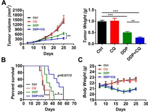
To investigate whether CQ could increase the efficacy of DDP in vivo, male BALB/c nude mice (5–6 weeks old) were used as an animal model. The mice were inoculated subcutaneously with 3 millions of human hypopharyngeal FaDu cells. The mice were divided into 4 groups (n = 7) 7 days after inoculation matched for tumor volume. Mice in groups were treated with vehicle control, CQ (60mg/kg/day), DDP (5mg/kg/6days), or a combination of DDP and CQ. Mice were sacrificed after treatment for 18 days and tumor tissues were collected. We monitored the tumor volume and final tumor weight for each group (Fig 1A). The mono-CQ therapy had no impact on the tumor volume or tumor weight compared with the control group. The mono-DDP group had a decreased tumor volume (657.0±66.2 mm3) and tumor weight (0.493 ±0.0496 g) compared with the control group (1405.0±117.2 mm3, 0.981±0.0577 g,p<0.001 for
both). The DDP+CQ treatment demonstrated a further decrease in the tumor volume (350.7 ±54.0 mm3) and tumor weight (0.263±0.0405 g) compared with the mono-DDP therapy (p<0.01 for both).
Kaplan-Meier curves were plotted to evaluate the survival of mice in each group (Fig 1B). The mono-CQ therapy had no effect on survival of the mice, and the mono-DDP therapy in-creased survival of the mice compared with that of control (p<0.01). The combination of CQ
and DDP significantly increased survival of the mice compared with that of the mono-DDP treatment (p= 0.0113 by log-rank test). DDP+CQ led to a robust 15.5-day increase in the me-dian survival compared with that of DDP alone.
with the control group. DDP caused a significant loss of body weight relative to the control group (p<0.001). DDP+CQ produced a notable body weight loss compared with that of the
control group (p<0.001), but the loss of body weight was not significant from that of the
mono-DDP therapy.
Autophagy was induced by DDP and suppressed by CQ in the
hypopharyngeal xenograft tumors
We used LC3 and p62 as markers of autophagy induction and inhibition. Groups of mice were sacrificed and tumor tissues were collected after 18 days of treatment, and the expression levels of LC3 and p62 were examined by Western blot and IHC (Fig 2,S2 Fig). (1) In the mono-CQ treated group, the levels of LC3 and p62 were substantially increased compared with that of control, suggesting a blockade of the autophagy flux by CQ administration. (2) In the mono-DDP treated group, the levels of LC3 were increased whereas accumulation of p62 was de-creased relative to control, which suggested that DDP, as a classical anti-tumor agent, caused autophagy induction in the tumors. (3) In the CQ+DDP treated group, a further increase of LC3 levels was observed compared with the mono-DDP treated group.
CQ addition increased apoptosis in DDP-treated xenograft mice
Bcl-2 family proteins are the main regulators of the mitochondrial pathway of apoptosis. Bax is pro-apoptotic and Bcl-2 is anti-apoptotic [29]. To assess the effect of CQ on tumor cell apopto-sis, we performed Western blot analysis of Bax and Bcl-2, and the Bax/Bcl-2 ratio was calculat-ed. A TUNEL assay was also performed to evaluate apoptosis (Fig 3). As expected, DDP
Fig 1. Chloroquine (CQ) enhances the efficacy of cisplatin (DDP) in xenograft tumors.Tumor-bearing mice (7 animals per group) were treated by intraperitoneal injection as follows: vehicle control, CQ (60 mg/kg/ day), DDP (5 mg/kg/6 days) and a combination of DDP and CQ for 18 days. (A) The tumor volume and tumor weight for each group. (B) Survival analysis of the treated mice in each group (n = 7). DDP+CQ led to a 15.5-day increase in the median survival compared with the mono-DDP treatment. (C) The body weight of mice during treatment.*p<0.05,**p<0.01,***p<0.001.
treatment caused marked apoptosis of tumor cells (p<0.05 relative to the control). CQ alone
did not have a pro-apoptosis effect. However, the addition of CQ significantly increased the ap-optosis of tumor cells induced by DDP compared with that of DDP alone (p<0.001).
Suppression of Beclin-1 expression by shRNA enhanced the effect of
DDP on xenograft tumor growth
To further confirm the importance of autophagy inhibition in sensitization to DDP treatment, we inhibited autophagy in FaDu cells with shRNA to Beclin-1 and assessed the effect on xeno-graft tumor growth. Beclin-1 expression was blocked by shRNA interference in xenoxeno-graft tu-mors (Fig 4A). Similar to the results of CQ treatment, suppression of Beclin-1 expression by shRNA did not impact tumor growth (Fig 4B). However, the combination of Beclin-1 suppres-sion and DDP treatment significantly decreased tumor growth compared with DDP plus
Fig 2. Inhibition of autophagy by CQ and induction of autophagy by DDPin vivo.Tumor tissues were harvested for autophagy analysis after treatment for 18 days. (A) Western blot analysis of autophagy markers LC3 and p62. (B) Representative micrographs of tumor sections: hematoxylin and eosin (H&E) stain (upper panel) and immunohistochemistry (IHC) for LC3 (middle panel) and p62 (lower panel). Scale bar = 20μm. (C)
Quantification of LC3 expression (left) and p62 expression (right) according to the Integrated Optical Density (IOD) (**p<0.01,***p<0.001).
scrambled control shRNA, as evidenced by the tumor volume and tumor weight data (p<0.01).
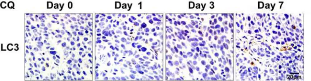
CQ delayed autophagy inhibition effects in xenograft tumors
To confirm the time needed for inhibition of autophagy, tumor bearing mice treated with CQ (60 mg/kg/day) were sacrificed after consecutive administrations for 1, 3 and 7 days separately. Tumor tissues were sectioned for LC3 Immunohistochemistry (Fig 5). LC3 accumulation on
Fig 3. Addition of CQ to DDP treatment increases apoptosis of the tumor cellsin vivo.(A) Representative photographs of Western blot analysis of Bax and Bcl-2 and Bax/Bcl-2 ratio levels by densitometric analysis of the Western blot bands (7 animals per group). (B) Representative TdT-mediated dUTP nick end labeling (TUNEL) staining of tumor tissues, scale bar = 20μm. (C) The average numbers of
TUNEL-positive cells per field in each group. Cells were counted in 10 randomly selected fields in 7 tumor samples from each group. The values represent the mean±SEM.*p<0.05,**p<0.01,***p<0.001.
doi:10.1371/journal.pone.0126147.g003
Fig 4. Suppression of Beclin-1 by shRNA decreases tumor growthin vivo.FaDu cells infected with lentiviral shRNA to Beclin-1or scrambled control were used for animal model building (n = 7 for each group). Established tumors were treated with DDP (5 mg/kg/6 days) or vehicle control. (A) Western blot of Beclin-1 suppression by shRNA. (B) Tumor volumes and tumor weights for each group.**p<0.01,***p<0.001.
day 1 and 3 was rare. A marked accumulation of LC3 was observed after CQ administration for 7 consecutive days, suggesting an autophagy inhibition effect by CQ till day 7.
Discussion
Previously, we reported that levels of Beclin-1 and LC3 were downregulated in the human HSCC tissues and low expression of beclin-1 and LC3 could correlate with poor prognosis for patients [28]. Both Beclin-1 and LC3 are closely related to the process of autophagy. In this study, we demonstrated that CQ, a mostly used autophagy inhibitor, could inhibit autophagy and increase apoptosis in DDP-treated hypopharyngeal tumor-bearing mice. CQ enhanced the efficacy of DDP, leading to decreased tumor growth and prolonged survival of mice. The inhi-bition of autophagy by lentiviral shRNA to Beclin-1 had an effect similar to that of CQ admin-istration. Autophagy is a complex cellular homeostatic process. The role of autophagy in cancer is still controversial, and it seems to have both antitumorigenesis and protumorigenesis effects. Though we are still in the early stages of understanding autophagy, targeting autophagy is becoming a new approach for treating cancers. The addition of CQ has been reported to en-hance the effectiveness of antitumor therapies in several tumors, including hepatocellular carci-noma [10], colorectal carcinoma [13], esophageal carcinoma [15], prostate carcinoma [8] and breast carcinoma [14]. However, CQ does not sensitize 4T1 tumors [17] or small cell lung can-cer [18].
Manyin vitroantitumor studies have shown evidence of autophagy inhibition by CQ and have mainly attributed the enhancement of antitumor efficacy by CQ to the suppression of autophagy [30]. However, few antitumor studies have provided evidence of autophagy inhibi-tion by CQin vivo. It is often unclear whether CQ reaches sufficient levels in the tumor tissues to effectively inhibit autophagy. Our data demonstrated that CQ administration (60 mg/kg/ day) caused significant autophagy inhibition in hypopharyngeal tumor-bearing mice after 18 days of treatment, as indicated by the increased accumulation of LC3 and p62 proteins (Fig 2). However, CQ failed to generate an autophagy inhibition effect at the start of the treatment. The LC3 level of days 1 and 3 did not differ from those of the control on IHC sections (Fig 5), which is in agreement with pharmacokinetic studies on CQ. The maximal plasma concentra-tion of CQ (daily administraconcentra-tion) falls within the range of 1.5 and 3μM. Based on thein vitro
CQ concentrations required for autophagy inhibition, that concentration is unlikely to effec-tively inhibit autophagyin vivoat the beginning of the treatment [17]. However, CQ has a long half-life and can accumulate to higher concentrations in various tissues over time, especially when it is administered for a long period of time. In our study, it required approximately 7 days for CQ (60 mg/kg/d) to accumulate sufficient concentrations to cause obvious LC3 accu-mulation in hypopharyngeal tumors. Therefore, it might be necessary to develop a more potent
Fig 5. Delayed effects of autophagy inhibition by CQ in xenograft tumors.Tumors treated with CQ (60 mg/kg/day) were collected after treatment for 1, 3 and 7 days and sectioned for LC3 immunohistochemistry. Scale bar = 20μm.
but less toxic autophagy inhibitor that can suppress autophagyin vivoat the beginning of intervention.
Although it effectively suppressed autophagy in hypopharyngeal tumors, CQ alone did not affect tumor growth compared with that of control. This result was in agreement with studies on non-small cell lung cancer [31]. In contrast, CQ administered as a mono-drug therapy was reported to cause obvious tumor suppression in pancreatic cancers bothin vitroandin vivo
[16]. This finding can be explained by the following. Pancreatic cancer cells have a relatively high level of basal autophagy and, even under normal conditions, rely substantially on autop-hagy for survival [16,32]. As in HSCC, the levels of Beclin-1 and LC3-II are often downregu-lated [28], suggesting a downregulation of basal autophagy activity. In other words,
hypopharyngeal cancer cells do not have to rely heavily on autophagy to survive under normal conditions. Therefore, when CQ was added alone, autophagy was inhibited, but hypopharyn-geal cancer cells could survive and there was no tumor suppression effect.
We also showed that autophagy was induced in tumors treated with DDP. The literature has demonstrated that chemotherapy agents can stress tumor cells and induce autophagy [33]. Tumor cells under stressful conditions may rely on autophagy for survival and to generate re-sistance to antitumor therapies [34,35]. When treated with DDP, hypopharyngeal cancer cells were severely stressed and had to resort to autophagy for survival. When CQ was added to the DDP treatment, autophagy was blocked and hypopharyngeal tumor cells were unable to utilize autophagy to promote survival. Tumors in the DDP+ CQ group had increased apoptosis com-pared with the DDP alone group (Fig 3). Tumor growth was decreased and the survival of mice was prolonged by the addition of CQ to DDP (Fig 1). Similar results were observed in the tumor-bearing mice with Beclin-1 suppression (Fig 4), suggesting that autophagy inhibition may enhance hypopharyngeal tumor sensitivity to DDP.
However, it is inappropriate to conclude that the increase in apoptosis and enhancement of efficacy with the addition of CQ only resulted from an inhibition of autophagy. In fact, CQ may even enhance the efficacy of chemotherapy independent of autophagy [36]. As an old drug, CQ still has alternative mechanisms for facilitating cancer therapies. For example, CQ could form a complex with DNA and cause defects in DNA synthesis and repair [37]. It could decrease the sequestration of anti-cancer drugs in endosomes by increasing the endosomal pH, thereby increasing the cytotoxic effects on tumor cells [38]. CQ could induce tumor cell differ-entiation and inhibit growth [39]. It could also cause tumor vessel normalization and restrain tumor invasion and metastasis while improving chemotherapy [40]. More studies should be performed to explore the detailed mechanisms of CQ-induced sensitization to chemo- and ra-diotherapies in various tumors.
It is important to note that the combination of CQ and DDP failed to completely control tumor growth. As shown inFig 1, the tumors finally grew to a large volume and the mice died. This finding may be because CQ was not able to block autophagy to a more severe and complete degree, which allowed the tumor cells to use the residual autophagy for life and avoid death, or because the combination of CQ with DDP therapy was too weak, indicat-ing that more antitumor drugs should be combined with CQ to further strengthen the antitumor effect.
Conclusions
improve human HSCC treatment. However, more studies should be performed to further elu-cidate the mechanism of CQ-induced sensitization to antitumor therapies.
Supporting Information
S1 Fig. Flow charts for scheduling of the drugs.CQ (60mg/kg/day) was administered intra-peritoneally (i.p.) for 18 consecutive days. DDP (5 mg/kg) was administered every 6 days. 3 in-jections were given in total.
(TIF)
S2 Fig. LC3 II levels by densitometric analysis of the Western blot bands.p<0.05, p<0.001.
(TIF)
S1 Table. The Guidelines Checklist for Animal Research: Reporting In Vivo Experiments (ARRIVE).The Balb/c nude mice were used for the in vivo experiments, all the related infor-mation was provided.
(PDF)
Acknowledgments
We thank professor Xiao-lan Cai (Department of Otolaryngology, Qilu Hospital of Shandong University, Jinan, Shandong, China) for helpful advice and comments on this work.
Author Contributions
Conceived and designed the experiments: XGZ XLP. Performed the experiments: XGZ XYY. Analyzed the data: XGZ DYL. Contributed reagents/materials/analysis tools: TJ DPL RJS. Wrote the paper: XGZ.
References
1. Hall SF, Groome PA, Irish J, O'Sullivan B (2008) The natural history of patients with squamous cell car-cinoma of the hypopharynx. Laryngoscope 118: 1362–1371. doi:10.1097/MLG.0b013e318173dc4a
PMID:18496152
2. Chang MF, Wang HM, Kang CJ, Huang SF, Lin CY, Fang KH, et al. (2010) Treatment results for hypo-pharyngeal cancer by different treatment strategies and its secondary primary—an experience in
Tai-wan. Radiat Oncol 5: 91. doi:10.1186/1748-717X-5-91PMID:20925962
3. Takes RP, Strojan P, Silver CE, Bradley PJ, Haigentz M Jr, Wolf GT, et al. (2012) Current trends in ini-tial management of hypopharyngeal cancer: the declining use of open surgery. Head Neck 34: 270–
281. doi:10.1002/hed.21613PMID:22228621
4. Adelstein DJ, Saxton JP, Rybicki LA, Esclamado RM, Wood BG, Strome M, et al. (2006) Multiagent concurrent chemoradiotherapy for locoregionally advanced squamous cell head and neck cancer: ma-ture results from a single institution. J Clin Oncol 24: 1064–1071. PMID:16505425
5. Kogashiwa Y, Yamauchi K, Nagafuji H, Matsuda T, Tsubosaka T, Tsutsumi T, et al. (2009) Concurrent chemoradiotherapy for organ function preservation in advanced patients with hypopharyngeal and la-ryngeal cancer. Oncol Rep 22: 1163–1167. PMID:19787235
6. Shen DW, Pouliot LM, Hall MD, Gottesman MM (2012) Cisplatin resistance: a cellular self-defense mechanism resulting from multiple epigenetic and genetic changes. Pharmacol Rev 64: 706–721. doi:
10.1124/pr.111.005637PMID:22659329
7. Thome R, Lopes SC, Costa FT, Verinaud L (2013) Chloroquine: modes of action of an undervalued drug. Immunol Lett 153: 50–57. doi:10.1016/j.imlet.2013.07.004PMID:23891850
8. Wu Z, Chang PC, Yang JC, Chu CY, Wang LY, Chen NT, et al. (2010) Autophagy Blockade Sensitizes Prostate Cancer Cells towards Src Family Kinase Inhibitors. Genes Cancer 1: 40–49. doi:10.1177/
9. Ghadimi MP, Lopez G, Torres KE, Belousov R, Young ED, Liu J, et al. (2012) Targeting the PI3K/ mTOR axis, alone and in combination with autophagy blockade, for the treatment of malignant peripher-al nerve sheath tumors. Mol Cancer Ther 11: 1758–1769. doi:10.1158/1535-7163.MCT-12-0015
PMID:22848094
10. Ding ZB, Hui B, Shi YH, Zhou J, Peng YF, Gu CY, et al. (2011) Autophagy activation in hepatocellular carcinoma contributes to the tolerance of oxaliplatin via reactive oxygen species modulation. Clin Can-cer Res 17: 6229–6238. doi:10.1158/1078-0432.CCR-11-0816PMID:21825039
11. Shi YH, Ding ZB, Zhou J, Hui B, Shi GM, Ke AW, et al. (2011) Targeting autophagy enhances sorafenib lethality for hepatocellular carcinoma via ER stress-related apoptosis. Autophagy 7: 1159–1172. doi:
10.4161/auto.7.10.16818PMID:21691147
12. Guo XL, Li D, Hu F, Song JR, Zhang SS, Deng WJ, et al. (2012) Targeting autophagy potentiates che-motherapy-induced apoptosis and proliferation inhibition in hepatocarcinoma cells. Cancer Lett 320: 171–179. doi:10.1016/j.canlet.2012.03.002PMID:22406827
13. Sasaki K, Tsuno NH, Sunami E, Tsurita G, Kawai K, Okaji Y, et al. (2010) Chloroquine potentiates the anti-cancer effect of 5-fluorouracil on colon cancer cells. BMC Cancer 10: 370. doi: 10.1186/1471-2407-10-370PMID:20630104
14. Cufi S, Vazquez-Martin A, Oliveras-Ferraros C, Corominas-Faja B, Cuyas E, Lopez-Bonet E, et al. (2013) The anti-malarial chloroquine overcomes primary resistance and restores sensitivity to trastuzu-mab in HER2-positive breast cancer. Sci Rep 3: 2469. doi:10.1038/srep02469PMID:23965851 15. Liu D, Yang Y, Liu Q, Wang J (2011) Inhibition of autophagy by 3-MA potentiates cisplatin-induced
apo-ptosis in esophageal squamous cell carcinoma cells. Med Oncol 28: 105–111. doi:
10.1007/s12032-009-9397-3PMID:20041317
16. Yang S, Wang X, Contino G, Liesa M, Sahin E, Ying H, et al. (2011) Pancreatic cancers require autop-hagy for tumor growth. Genes Dev 25: 717–729. doi:10.1101/gad.2016111PMID:21406549
17. Bristol ML, Emery SM, Maycotte P, Thorburn A, Chakradeo S, Gewirtz DA (2013) Autophagy inhibition for chemosensitization and radiosensitization in cancer: do the preclinical data support this therapeutic strategy. J Pharmacol Exp Ther 344: 544–552. doi:10.1124/jpet.112.199802PMID:23291713
18. Zinn RL, Gardner EE, Dobromilskaya I, Murphy S, Marchionni L, Hann CL, et al. (2013) Combination treatment with ABT-737 and chloroquine in preclinical models of small cell lung cancer. Mol Cancer 12: 16. doi:10.1186/1476-4598-12-16PMID:23452820
19. Choi AM, Ryter SW, Levine B (2013) Autophagy in human health and disease. N Engl J Med 368: 651–662. doi:10.1056/NEJMra1205406PMID:23406030
20. Kabeya Y, Mizushima N, Ueno T, Yamamoto A, Kirisako T, Noda T, et al. (2000) LC3, a mammalian ho-mologue of yeast Apg8p, is localized in autophagosome membranes after processing. EMBO J 19: 5720–5728. PMID:11060023
21. Pankiv S, Clausen TH, Lamark T, Brech A, Bruun JA, Outzen H, et al. (2007) p62/SQSTM1 binds di-rectly to Atg8/LC3 to facilitate degradation of ubiquitinated protein aggregates by autophagy. J Biol Chem 282: 24131–24145. PMID:17580304
22. Mathew R, Karp CM, Beaudoin B, Vuong N, Chen G, Chen HY, et al. (2009) Autophagy suppresses tu-morigenesis through elimination of p62. Cell 137: 1062–1075. doi:10.1016/j.cell.2009.03.048PMID:
19524509
23. Poole B, Ohkuma S (1981) Effect of weak bases on the intralysosomal pH in mouse peritoneal macro-phages. J Cell Biol 90: 665–669. PMID:6169733
24. Gozuacik D, Kimchi A (2004) Autophagy as a cell death and tumor suppressor mechanism. Oncogene 23: 2891–2906. PMID:15077152
25. Takamura A, Komatsu M, Hara T, Sakamoto A, Kishi C, Waguri S, et al. (2011) Autophagy-deficient mice develop multiple liver tumors. Genes Dev 25: 795–800. doi:10.1101/gad.2016211PMID:
21498569
26. Degenhardt K, Mathew R, Beaudoin B, Bray K, Anderson D, Chen G, et al. (2006) Autophagy promotes tumor cell survival and restricts necrosis, inflammation, and tumorigenesis. Cancer Cell 10: 51–64.
PMID:16843265
27. Peng YF, Shi YH, Shen YH, Ding ZB, Ke AW, Zhou J, et al. (2013) Promoting colonization in metastatic HCC cells by modulation of autophagy. PLoS One 8: e74407. doi:10.1371/journal.pone.0074407 PMID:24058558
28. Wang J, Pan XL, Ding LJ, Liu DY, Lei D, Jin T (2013) Aberrant expression of Beclin-1 and LC3 corre-lates with poor prognosis of human hypopharyngeal squamous cell carcinoma. PLoS One 8: e69038. doi:10.1371/journal.pone.0069038PMID:23935917
30. Han W, Sun J, Feng L, Wang K, Li D, Pan Q, et al. (2011) Autophagy inhibition enhances daunorubicin-induced apoptosis in K562 cells. PLoS One 6: e28491. doi:10.1371/journal.pone.0028491PMID: 22164300
31. Zou Y, Ling YH, Sironi J, Schwartz EL, Perez-Soler R, Piperdi B (2013) The autophagy inhibitor chloro-quine overcomes the innate resistance of wild-type EGFR non-small-cell lung cancer cells to erlotinib. J Thorac Oncol 8: 693–702. doi:10.1097/JTO.0b013e31828c7210PMID:23575415
32. Fujii S, Mitsunaga S, Yamazaki M, Hasebe T, Ishii G, Kojima M, et al. (2008) Autophagy is activated in pancreatic cancer cells and correlates with poor patient outcome. Cancer Sci 99: 1813–1819. doi:10.
1111/j.1349-7006.2008.00893.xPMID:18616529
33. Del BB, Toscano M, Moretti D, Maellaro E (2013) Cisplatin-induced apoptosis inhibits autophagy, which acts as a pro-survival mechanism in human melanoma cells. PLoS One 8: e57236. doi:10. 1371/journal.pone.0057236PMID:23437349
34. Ren JH, He WS, Nong L, Zhu QY, Hu K, Zhang RG, et al. (2010) Acquired cisplatin resistance in human lung adenocarcinoma cells is associated with enhanced autophagy. Cancer Biother Radio-pharm 25: 75–80. doi:10.1089/cbr.2009.0701PMID:20187799
35. Yu L, Gu C, Zhong D, Shi L, Kong Y, Zhou Z, et al. (2014) Induction of autophagy counteracts the anti-cancer effect of cisplatin in human esophageal anti-cancer cells with acquired drug resistance. Cancer Lett 355: 34–45. doi:10.1016/j.canlet.2014.09.020PMID:25236911
36. Maycotte P, Aryal S, Cummings CT, Thorburn J, Morgan MJ, Thorburn A (2012) Chloroquine sensitizes breast cancer cells to chemotherapy independent of autophagy. Autophagy 8: 200–212. doi:10.4161/
auto.8.2.18554PMID:22252008
37. Chatterjee T, Muhkopadhyay A, Khan KA, Giri AK (1998) Comparative mutagenic and genotoxic effects of three antimalarial drugs, chloroquine, primaquine and amodiaquine. Mutagenesis 13: 619–624.
PMID:9862194
38. Lee CM, Tannock IF (2006) Inhibition of endosomal sequestration of basic anticancer drugs: influence on cytotoxicity and tissue penetration. Br J Cancer 94: 863–869. PMID:16495919
39. Martirosyan A, Leonard S, Shi X, Griffith B, Gannett P, Strobl J (2006) Actions of a histone deacetylase inhibitor NSC3852 (5-nitroso-8-quinolinol) link reactive oxygen species to cell differentiation and apo-ptosis in MCF-7 human mammary tumor cells. J Pharmacol Exp Ther 317: 546–552. PMID:16497787
40. Maes H, Kuchnio A, Peric A, Moens S, Nys K, De Bock K, et al. (2014) Tumor vessel normalization by chloroquine independent of autophagy. Cancer Cell 26: 190–206. doi:10.1016/j.ccr.2014.06.025