Cathepsin Inhibition-Induced Lysosomal
Dysfunction Enhances Pancreatic Beta-Cell
Apoptosis in High Glucose
Minjeong Jung1, Jaemeun Lee1, Hye-Young Seo1, Ji Sun Lim1, Eun-Kyoung Kim1,2*
1Department of Brain Science, Daegu Gyeongbuk Institute of Science & Technology, Daegu, Korea,2
Neurometabolomics Research Center, Daegu Gyeongbuk Institute of Science & Technology, Daegu, Korea
Abstract
Autophagy is a lysosomal degradative pathway that plays an important role in maintaining cellular homeostasis. We previously showed that the inhibition of autophagy causes pancreaticβ-cell apoptosis, suggesting that autophagy is a protective mechanism for the survival of pancreaticβ-cells. The current study demonstrates that treatment with inhibitors and knockdown of the lysosomal cysteine proteases such as cathepsins B and L impair autophagy, enhancing the caspase-dependent apoptosis of INS-1 cells and islets upon exposure to high concentration of glucose. Interestingly, treatment with cathepsin B and L inhibitors prevented the proteolytic processing of cathepsins B, D and L, as evidenced by gradual accumulation of the respective pro-forms. Of note, inhibition of aspartic cathepsins had no effect on autophagy and cell viability, suggesting the selective role of cathepsins B and L in the regulation ofβ-cell autophagy and apoptosis. Lysosomal localization of accu-mulated pro-cathepsins in the presence of cathepsin B and L inhibitors was verified via immunocytochemistry and lysosomal fractionation. Lysotracker staining indicated that cathepsin B and L inhibitors led to the formation of severely enlarged lysosomes in a time-dependent manner. The abnormal accumulation of pro-cathepsins following treatment with inhibitors of cathepsins B and L suppressed normal lysosomal degradation and the processing of lysosomal enzymes, leading to lysosomal dysfunction. Collectively, our find-ings suggest that cathepsin defects following the inhibition of cathepsin B and L result in lysosomal dysfunction and consequent cell death in pancreaticβ-cells.
Introduction
The integrity of pancreaticβ-cell function and mass is critical for the pathogenesis of diabetes [1]. Although glucose is the main regulator of insulin biosynthesis and secretion, chronic hy-perglycemia is associated with impaired function of insulin secretion. The detrimental effect of excessive glucose concentration is referred to as 'glucotoxicity' [2,3], which can negatively affect β-cell mass by inducing apoptosis [4]. Glucotoxicity is associated with the induction of
OPEN ACCESS
Citation:Jung M, Lee J, Seo H-Y, Lim JS, Kim E-K (2015) Cathepsin Inhibition-Induced Lysosomal Dysfunction Enhances Pancreatic Beta-Cell Apoptosis in High Glucose. PLoS ONE 10(1): e0116972. doi:10.1371/journal.pone.0116972
Academic Editor:Srinivasa M. Srinivasula, IISER-TVM, INDIA
Received:August 2, 2014
Accepted:December 17, 2014
Published:January 27, 2015
Copyright:© 2015 Jung et al. This is an open access article distributed under the terms of the
Creative Commons Attribution License, which permits unrestricted use, distribution, and reproduction in any medium, provided the original author and source are credited.
Data Availability Statement:All relevant data are within the paper.
endoplasmic reticulum (ER) stress, mitochondrial dysfunction and oxidative damage to pro-teins [5,6].
Mounting evidence has indicated that autophagy plays an important role in cell survival and death in response to cellular stress. Under certain stress conditions, autophagy can protect cells against cytotoxicity [7,8]. For example, it provides a protective role by removing cellular components damaged by oxidative stress [9–11]. Autophagy is a dynamic process associated with the formation of autophagosomes, double-membrane vacuoles that engulf cellular com-ponents. The autophagosomes subsequently fuse with lysosomes to form autolysosomes, which degrade the dysfunctional cytoplasmic organelles and damaged proteins using lysosomal hydrolytic enzymes [12]. Therefore, autophagy maintains tissue homeostasis and ensures cell survival under stress conditions. [13–17]. Dysregulation of autophagy has been indicated in the pathogenesis of several diseases including neurodegenerative disease, heart disease, cancer and aging [7,18–20].
Microtubule-associated protein light-chain 3 (LC3), also called autophagy-related protein 8 (Atg8) in yeast, is processed to LC3-I, and then conjugated with phosphatidylethanolamine (PE) through the mediation of the Atg5/Atg12 complex to generate membrane-associated LC3-II [21–23]. LC3-II stays on the membrane until it is degraded by the lysosome, thus it is widely used as a marker for autophagic process [18].
The progression and resolution of autophagy critically depends on lysosomal function, as lysosomes play a role in the degradation of cellular compartments. Lysosomes contain many types of hydrolytic enzymes, such as peptidases, phosphatase, nucleases, glycosidases, protease and lipase, which can digest most macromolecules in the cell [24]. Cathepsins represent a major class of lysosomal proteases, especially important for the execution of autophagy [25–27]. The ca-thepsin family consists of aspartic, cysteine, and serine caca-thepsins. Aspartic caca-thepsins include cathepsin D and E, while cysteine cathepsins include cathepsin B, C, H, K, and L, and cathepsin A and G belong to serine cathepsins [25].
Cathepsins are synthesized as inactive (immature) pro-cathepsins and are proteolytically processed to form active (mature) cathepsins [28,29]. They contain a signal peptide which is cleaved within the ER, and are then transported into the endosome/lysosome compartment via mannose-6-phosphate receptors. Most lysosomal cathepsins are functionally optimized at low pH, as cathepsins are stable and active at acidic pH.
Recent studies have shown that autophagy is associated with diabetes through its effects on pancreaticβ-cells [30–32]. We previously reported that dysregulation of autophagy causes apo-ptotic cell death, suggesting that autophagy plays a protective role in the survival of pancreatic β-cells [33]. In this study, we investigate the mechanism by which inhibition of aspartic and cysteine cathepsins results in lysosomal dysfunction, enhancing pancreaticβ-cell apoptosis in conditions of high glucose.
Materials and Methods
Antibodies and chemical reagents
Antibodies against cleaved caspase-3, cleaved caspase-9, Bcl-2, phosphor-JNK (Thr183/Tyr185), JNK and GAPDH were obtained from Cell signaling. Antibodies against poly ADP ribose poly-merase (PARP) were purchased from BD Biosciences, and those against LC3 and lysosomal-associated membrane protein 2 (LAMP2) were from Sigma. Antibodies against cathepsin L and cathepsin D were purchased from Santa Cruz, while cathepsin B was from Millipore. Cathepsin B (CA074), K (Z-L-NHNHCONHNH-LF-Boc, II), and L (Z-FY(t-Bu)-DMK, III) inhibitors, along with E64d were purchased from Calbiochem. Pepstatin A and SP600125 (JNK inhibitor) were purchased from Sigma.
Cell culture
Rat insulinomaβ-cell line INS-1 (832/13) [34] (generously provided by Dr. Christopher New-gard, Department of Pharmacology and Cancer Biology, Duke University Medical Center, Durham, NC, U.S.A) and a stable INS-1 cell line (GFP-LC3/INS-1) expressing GFP-LC3 from INS-1 cells [35] were cultured in a 37°C incubator with 5% CO2in RPMI 1640 medium
(GIBCO) supplemented with 10% fetal bovine serum (Hyclone), 11 mM glucose (Sigma), 2 mM L-glutamine (GIBCO), 10mM 4-2-hydroxyethyl-1-piperazineethanesulfonic acid (HEPES) (GIBCO), 1 mM sodium pyruvate (GIBCO), 0.05 mM 2-mercaptoethanol (GIBCO), and 1% penicillin-streptomycin (Hyclone)..
Cell viability assay
To assess cell viability, INS-1 cells were seeded in 96-well plates at a density of 4 × 10⁵cells per mL in 11 mM glucose or 30 mM glucose medium treated with lysosomal protease inhibitors for the indicated times. After CellTiter-Blue (Promega) was added to each well and incubated for 3 hr, the absorbance of the samples was measured with a microplate reader at 560 nm exci-tation and 590 nm emission.
Annexin V staining
INS-1 cells were plated in 12-well plates at a density of 6.5 × 10⁵cells per mL in 11 mM glucose medium treated with E64d, cathepsin B and L inhibitors, and staurosporine. Cells were stained with incubation buffer containing Annexin V and Hoechst 33342 (Invitrogen) for 15 min at 37°C after removal of the medium. Apoptotic cells were assessed using an Alexa Fluor1
488 Annexin V/Dead Cell Apoptosis Kit (Invitrogen) according to the manufacturer's protocol. Images of the apoptotic cells were obtained with a fluorescence microscope (Zeiss).
Immunoblot analysis
INS-1 cells were plated in 6-well plates at a density of 6.5 × 10⁵cells per mL in 11 mM glucose or 30 mM glucose medium treated with E64d, and cathepsin B and L inhibitors. Samples were subjected to 10% SDS-polyacrylamide gel electrophoresis and subsequently transferred onto a polyvinylidene difluoride (PVDF) membrane. After transfer, the membrane was incubated with the specific primary antibodies at 4°C overnight. After three washes with Tris-Buffered Sa-line and Tween 20 (TBST) buffer, the membrane was incubated with horseradish peroxidase (HRP)-linked secondary antibody and visualized by Supersignal West Pico Chemiluminescent Substrate (Thermo), according to the recommended procedure.
Pancreatic islet culture
Rat islets were isolated as previously described [32] from adult male Sprague Dawley (SD) rats (200–250g). Animal experimentation was done in accordance with the guidelines on care and use as approved by the DGST institutional Animal Care and Use Committee. Islets were cultured in INS-1 medium containing 11 or 30 mM glucose, treated with cathepsin B and L inhibitors.
Immunofluorescence analysis
cells were washed with PBS, and blocked for 10 min at RT. Cells were incubated with anti-ca-thepsin B (Millipore), anti-caanti-ca-thepsin L (Santa Cruz) and anti-PDI (Abcam) for 3 hr at RT. Cells were incubated for an additional 3 hr at RT with secondary antibodies, and then stained with Hoechst 33342 (Invitrogen) for 10 min at RT. Coverslips were mounted with ProLong Gold antifade reagent (Invitrogen). The cells treated with E64d and cathepsin B and L inhibi-tors on coverslips were analyzed with an LSM700 Confocal microscope (Zeiss). The images were analyzed using the ZEN2009 software. The data illustrated were from one representative experiment of at least three independent repeats.
Cathepsin activity
INS-1 cells were plated in 6-well plates at a density of 6.5 × 10⁵cells per mL in 30 mM glucose medium in presence or absence of cathepsin inhibitors for 24, 48 and 72 hr. The activity of ca-thepsin B and L was assessed using the Caca-thepsin B and L activity kit (Calbiochem), as recom-mended by the manufacturer.
siRNA knock-down for cathepsin B and L
ON-TARGET plus SMARTpool (DHARMACON) composed of four different siRNAs against cathepsin B or L were used to obtain the higher knock-down efficiency and to reduce the off-target effects. siRNAs treatments were carried out on INS-1 cells seeded in 12-well plates con-taining 30 mM glucose medium using Lipofectamine 2000. The protein extracts were prepared at 48 and 72 hr post-transfection.
Nuclear and cytoplasmic fractionation
Cytoplasmic and nuclear fractions were prepared using a kit purchased from Thermo. INS-1 cells were harvested with trypsin-EDTA, centrifuged at 500 × g for 5 min, and washed by suspending the cell pellet with PBS. The cells were transferred to a 1.5 mL microcentrifuge tube and then centrifuged at 500 × g for 2–3 min. After buffers were added to the cell pellet and centrifuged, the supernatants (cytoplasmic extract) were transferred to a clean pre-chilled tube. The pellet, con-taining the nuclei, was suspended in ice-cold buffer. The pellet was centrifuged, and then the su-pernatants (nuclear extract) were transferred to a clean pre-chilled tube. Protein concentrations of the two fractions were assayed using the Bicinchoninic Acid (BCA) assay kit (Thermo).
Enriched lysosomal extraction
Extraction for lysosomal enrichment was carried out with a kit (PIERCE). INS-1 cells were har-vested and centrifuged at 850 × g for 2 min. After removal of the supernatants, the pellet was resuspended with Lysosome Enrichment Reagent A and placed on ice for 2 min. The cells were broken with a Dounce tissue grinder on ice, after which Lysosome Enrichment Reagent B was added. The cells were then centrifuged at 500 × g for 10 min at 4°C, and the supernatants were overlaid on the density gradients and further subjected to ultracentrifugation at 145,000 × g for 2 hr at 4°C. After ultracentrifugation, the lysosome band located at the top of the gradient was carefully removed. The samples were diluted with 2–3 volumes of PBS, and were centrifuged at 18,000 × g for 30 min at 4°C. The lysosome pellets were stored on ice until used. The lysosome pellets were then boiled with SDS-PAGE sample buffer and analyzed with western blotting.
Transfection of mRFP-GFP-LC3 constructs
using Lipofectamine 2000 (Invitrogen). Transfected INS-1 cells were treated daily with cathep-sin B and L inhibitors for 48 hr. The cells were fixed with 4% formaldehyde, and the coverslips were then mounted with ProLong Gold antifade reagent (Invitrogen). The images were ana-lyzed with an LSM700 Confocal microscope (Zeiss).
Statistical analysis
All results were represented as mean ± SEM. When comparisons between groups were re-quired, statistical significance was determined by an unpairedttest.Pvalues of<0.05 were considered statistically significant.
Results
Inhibition of cathepsin B and L increases
β
-cell apoptosis
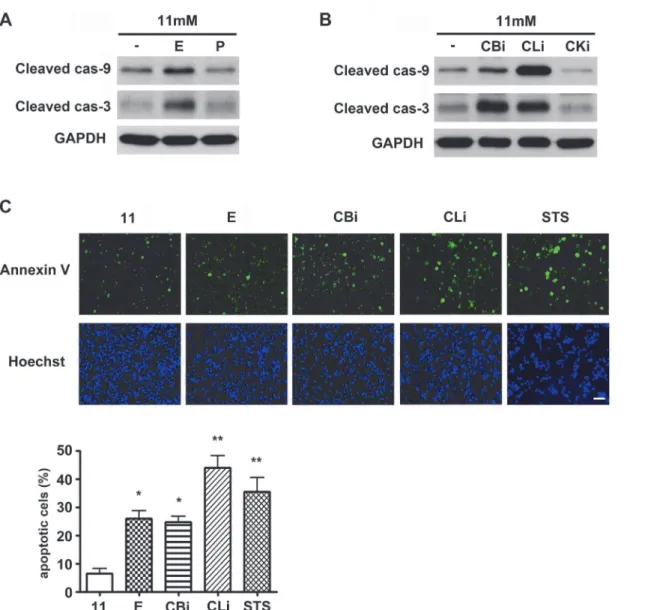
In this study, the effect of pharmacological inhibition of lysosomal proteases onβ-cell death was examined. INS-1 cells were cultured in the presence of lysosomal protease inhibitors in 11 mM glucose (normal culture concentration) medium for 48 hr, and then the activation of apoptosis was analyzed by western blotting with antibodies against the active, cleaved form of caspases (Fig. 1A). Among the inhibitors of lysosomal proteases, E64d has inhibitory effects on cysteine proteases such as cathepsin B, K, and L, whereas pepstatin A has inhibitory effects on aspartic proteases such as cathepsin D and E. E64d treatment was observed to increase caspase-9 and -3 activation, whereas pepstatin A had no effect on caspase activation (Fig. 1A). Among the lyso-somal cysteine proteases inhibitors, inhibitors of cathepsin B and L, but not cathepsin K, also in-creased caspase-9 and -3 activation (Fig. 1B). These results demonstrated that the inhibition of cathepsin B and L specifically triggers caspase-dependent apoptosis. The consequent increase in apoptotic cell death was next confirmed using Annexin-V staining, following treatment with ly-sosomal cysteine protease inhibitors. INS-1 cells treated with staurosporine (STS), a known pro-totypic inducer of apoptosis, were used as a positive control. Increases in cell death by E64d, cathepsin B and L inhibitors were represented by increasing numbers of Annexin V-positive cells (Fig. 1C). Compared to the 11 mM glucose control, apoptotic cell death was significantly in-creased by 4.0, 3.9, and 6.9 fold following treatment with E64d, cathepsin B and L inhibitors, re-spectively. These results suggest that cathepsin B and/or L play an important role inβ-cell survival, as inhibition of their activity increases caspase-dependent apoptosis.
Inhibition of cathepsin B and L enhances cell death under glucotoxicity
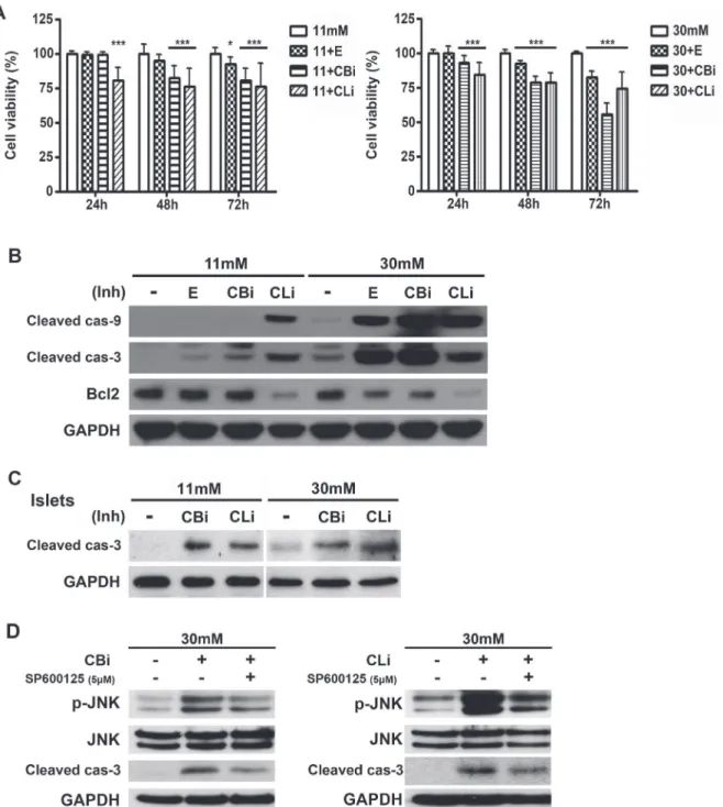
The detrimental effects of glucotoxicity negatively affectβ-cell mass by apoptosis [36]. In order to investigate whether the inhibition of lysosomal proteases worsensβ-cell apoptosis associated with glucotoxicity, hyperglycemic conditions were manipulated by culturing INS-1 cells in 30 mM glucose medium, which has been shown to decrease insulin mRNA levels [37] and induce ER stress in INS-1 cells by glucotoxicity [38].
Pancreatic islets from Sprague-Dawley (SD) rats cultured in 30 mM glucose medium also showed an increased caspase-3 activation when treated with cathepsin B and L inhibitors (Fig. 2C).
JNK activation is one of the upstream kinases in caspase-3 dependent apoptosis pathway [39,40]. Increases in phosphorylated JNK were observed in cathepsin B or L inhibitor-treated INS-1 cells in 30 mM glucose medium at 24 hr (Fig. 2D). The specific JNK inhibitor,
SP600125, reduces the cathepsin inhibitor-induced activation of caspase-3. The activated JNK might be involved in activation of caspase-3, resulting in the induction of apoptosis.
Taken together, these results indicate that cathepsin B and L inhibitors enhance caspase 3-dependent apoptosis, leading to a reduction in pancreaticβ-cell survival under glucotoxicity. Figure 1. Inhibition of cathepsins B and L triggers apoptosis in INS-1 cells.(A) INS-1 cells were treated with E64d (E, 20μg/mL) and pepstatin A (P, 20μg/mL) in 11 mM glucose medium for 48 hr, and caspase activation was monitored by western blotting using antibodies detecting cleaved, active forms of caspases. (B) INS-1 cells were treated with cathepsin B inhibitor (CBi, 20μM), cathepsin L inhibitor (CLi, 20μM), and cathepsin K inhibitor (CKi, 20μM) for 48 hr in 11 mM glucose medium, and caspase activation was monitored by western blotting. (C) INS-1 cells stained with Annexin-V and Hoechst were observed using a fluorescence microscope for quantification of apoptotic cells. Cells were treated with E, CBi, CLi and staurosporine (STS, 0.5μM) in 11 mM glucose medium. Lysosomal protease inhibitors were added daily, and STS was added 6 hr prior to staining (three independent counts). STS is a known prototypic inducer of apoptosis.*p<0.01,**p<0.05 compared with 11 mM glucose. The scale bar represents 20μm.
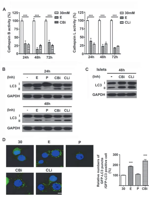
Inhibition of cathepsin B and L leads to LC3-II accumulation
We further focused on the effects of lysosomal cysteine cathepsin inhibition on the autophagy process in high glucose conditions. The inhibition of cathepsins was verified by measuring Figure 2. Inhibition of cathepsins B and L increases caspase-dependent apoptosis under glucotoxicity.(A) INS-1 cells were treated with E64d (E, 20μg/mL), cathepsin B inhibitor (CBi, 20μM), and cathepsin L inhibitor (CLi, 20μM) in INS-1 medium containing 11 mM or 30 mM glucose. Cell viability was assessed using the CellTiter-BlueⓇCell Viability assay and indicated as a percentage of the values measured at each time point (n = 15). Statistical significance was represented as*p<0.01, and***p<0.001 compared with 11 mM glucose and 30 mM glucose. (B) Protein levels of active caspase-9
(cleaved cas-9), caspase-3 (cleaved cas-3) and Bcl-2 were measured in INS-1 cells cultured in 11 or 30 mM glucose medium for 48 hr. Cells were treated daily with E, CBi, and CLi. (C) Immunoblot analysis of pancreatic islets of SD rats treated with CBi and CLi in 11 mM or 30 mM glucose medium for 48 hr. (D) JNK inhibitor (SP600125, 5μM) was treated with CBi and CLi in INS-1 cells cultured in 30 mM glucose medium for 24 hr.
their activities in the presence of the inhibitors (Fig. 3A). Cathepsin B and L activities were sig-nificantly reduced by 75% and 78% following treatment with inhibitors in 30 mM glucose, re-spectively (Fig. 3A). An increase in LC3-II level or LC3 puncta formation can occur due to either an increased rate of autophagy flux (“on-state”) or impaired autophagy flux accompa-nied by incomplete degradation of LC3 (“off-state”). When cells are treated with inhibitors of lysosomal proteases such as E64d and pepstatin A, the degradation of LC3-II is blocked, lead-ing to accumulation of LC3-II and the off-state of autophagy [41]. In line with the impairment of autophagy following the inhibition of lysosomal cathepsins, the treatment of INS-1 cells with lysosomal cysteine protease inhibitors, including E64d, and cathepsin B and L inhibitors, resulted in the incomplete degradation of LC3 and subsequent accumulation of LC3-II at 24 and 48 hr (Fig. 3B).
The inhibition of cathepsin B and L resulted in the accumulation of LC3-II not only in INS-1 cells, but also in the islets. Islets from SD rats showed similar results when treated with ca-thepsin B and L inhibitors in 30 mM glucose medium. Increased accumulation of LC3-II in the islets was detected at 48 hr (Fig. 3C).
The increase in LC3 puncta formation by treatment with E64d and cathepsin B and L inhib-itors is also indicative of impaired autophagy flux, as was the case of LC3-II protein level (Fig. 3D). To monitor LC3 puncta, ring-shaped or punctate green fluorescence signal was mea-sured in a stable INS-1 cell line expressing GFP–LC3 (GFP–LC3/INS-1) cultured in 30 mM glucose in the presence or absence of inhibitors. Compared with the 30 mM glucose control, treatment of GFP–LC3/INS-1 cells with E64d, cathepsin B and L inhibitors increased the num-ber of cells containing GFP–LC3 puncta up to 185%, 240% and 226% at 48 hr, respectively (Fig. 3D). This increase in GFP-LC3 puncta accumulation was due to the incomplete degrada-tion of LC3 by impaired autophagy, as seen inFig. 3B. In contrast, pepstatin A, an aspartic pro-tease inhibitor, had no effect on LC3 accumulation (Fig. 3B). In addition, no significant changes in punctate signals in GFP-LC3/INS-1 cells treated with pepstatin A were observed (Fig. 3D). These data strongly suggest the selective role of cathepsin B and L in the regulation of pancreaticβ-cell autophagy.
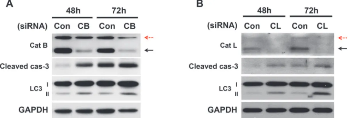
Knockdown of cathepsin B and L enhances caspase-3 activation and
increases LC3-II accumulation
The inhibitor studies demonstrated the critical role of cathepsins B and L in the apoptosis and autophagy of pancreaticβ-cells. To confirm the role of cathepsins B and L via a genetic method, cathepsin B or L was depleted through the use of siRNA (Fig. 4). Cathepsin B and L were knocked-down in INS-1 for 48 and 72 hr. The knockdown with siRNA in 30 mM glucose me-dium resulted in an increase in apoptotic cell death through caspase-3 activation. This result is consistent with the pharmacological inhibition of cathepsins, as shown inFig. 2. In addition, the depletion of cathepsins B and L also increased LC-3 accumulation, which is consistent with the results obtained by the treatment with inhibitors inFig. 3. In contrast, when cathepsin K was knocked down with siRNAs there were no changes in activation of caspase-3 and LC-3 ac-cumulation as expected (data not shown).
Inhibition of cathepsin B and L results in accumulation of pro-cathepsins
in the lysosomes
Figure 3. Inhibition of cathepsins B and L leads to LC3 accumulation.(A) Activity of cathepsins B and L in 30 mM glucose with E64d (E, 20μg/mL), cathepsin B inhibitor (CBi, 20μM) or cathepsin L inhibitor (CLi, 20μM) was measured at 24 hr, 48 hr and 72 hr (n = 9). Statistical significance is represented as***p<0.001 compared with 30 mM glucose. (B) INS-1 cells cultured in 30 mM glucose for 24 or 48 hr were treated daily with E, P, CBi, and CLi. LC3-II
accumulation was assessed by immunoblot analysis. (C) Islets from SD rat cultured in 30 mM glucose and treated with CBi, and CLi. Level of LC3 was measured by immunoblot assay. (D) GFP-LC3/INS-1 stable cells were treated daily with E, P, CBi, and CLiin in 30 mM glucose for 48 hr. The graph indicates the number of GFP-LC3 puncta counted among GFP positive cells (n = 5).***p<0.001 compared with 30 mM glucose. The scale bar represents 5μm.
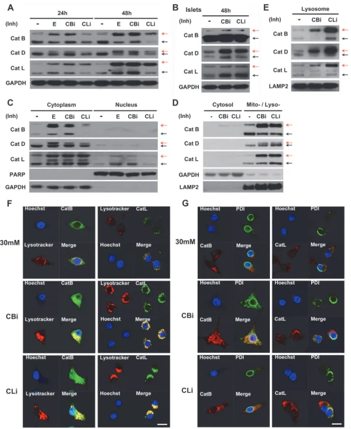
As shown inFig. 5A, the proteolytic processing of both cysteine proteases (cathepsins B and L) and aspartic proteases (cathepsin D) were inhibited by the cathepsin B and L inhibitors. The inhibitors of cathepsin B and L led to the accumulation of pro-cathepsin B, L and D, as denoted by a red dashed arrow. Pro-cathepsin B, L and D exhibited greater accumulation at 48 hr than 24 hr in INS-1 cells.
Similarly, the inhibition of cathepsins B and L showed incomplete processing of the cathep-sins in the pancreatic islets of SD rats (Fig. 5B).
To determine the subcellular localization of the accumulated pro-cathepsins, the cyto-plasmic (non-nuclear) and nuclear fractions were first analyzed. Both pro-cathepsins and ca-thepsins (B, D and L) were found to be predominantly localized in the cytoplasmic fraction in the presence of cathepsin B and L inhibitors (Fig. 5C). Next, the cytoplasmic fraction was fur-ther separated to assess the levels of pro-cathepsin accumulation in the cytosolic and mito-chondrial/lysosomal fractions (Fig. 5D). Much higher accumulation of the pro-cathepsins was observed in the mitochondrial/lysosomal fractions compared with the cytosolic fraction. The lysosomal localization of cathepsins was further verified using a lysosomal enrichment kit. The enriched lysosomal fraction contained a high accumulation of pro-cathepsins when treated with inhibitors (Fig. 5E). In addition, fluorescence imaging by staining with Lysotracker, a lyso-some marker, showed co-localization of the lysolyso-somes and cathepsins in 30 mM glucose cul-ture conditions, which was further increased upon treatment with cathepsin B or L inhibitors (Fig. 5F).
Cathepsins become mature by cleavage of the N-terminal signal peptide within the ER, and are then activated through proteolytic processing in the endosome and lysosome [47–53]. To observe whether the pro-cathepsins accumulated in the ER after treatment with cathepsin B and L inhibitors, the co-localization of cathepsin B/L and protein disulfide isomerase (PDI), a known ER marker, was examined using immunofluoresence (Fig. 5G). The results indicated that PDI and cathepsin B/L do not co-localize in the ER following the inhibition of cathepsins B and L, which is in sharp contrast to the substantial co-localization of cathepsin with the lysosomes.
Taken together, these findings indicate that the inhibition of cathepsins B and L blocks the proteolytic processing of cathepsins B, L and D, resulting in accumulation of pro-cathepsins B, L and D in the lysosomes.
Figure 4. Cathepsin B- and L-knockdown with siRNA enhances apoptosis and impairs autophagy.INS-1 cells were transfected with scrambled siRNA (con) or with either the cathepsin B (A) or cathepsin L (B) siRNA and cultured in 30 mM glucose for 48 and 72 hr. Immunoblot analysis was performed at the indicated time points post-transfection. Red dashed arrows indicate immature cathepsins, while block solid arrows indicate the mature active form of cathepsins.
Abnormal accumulation of pro-cathepsins in the lysosomes results in
severe lysosomal dysfunction
Interestingly, cathepsin inhibition by treatment with cathepsin B and L inhibitors resulted in enlargement of the lysosomes in a time-dependent manner compared with the control (Fig. 6A). It was next tested whether the enlarged lysosomes had the normal ability to form autolysosomes with autophagosomes, using mRFP and GFP tandem fluorescent-tagged LC3. mRFP-GFP-LC3 shows both GFP and mRFP fluorescence in the autophagosome before fusion with the lysosome. However, the GFP signal is quenched in acidic lysosomal conditions [54,55] whereas the mRFP-LC3 can be readily detected in autolysosomes with more stable fluorescence [56]. Therefore, the mRFP-GFP-LC3 tandem construct is a useful tool to trace the maturation process of autophagosomes into autolysosomes, by labeling the autophagosomes and autolyso-somes in yellow and red, respectively [57]. Our results showed that only the yellow puncta were increased after treatment without an increase in the red puncta, indicating that the ca-thepsin B and L inhibitors blocked the maturation of autophagosomes into autolysosomes (Fig. 6B).
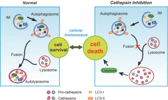
Our results suggest that the impaired processing of cathepsins following the inhibition of ca-thepsins B and L leads to the accumulation of immature forms of caca-thepsins. Failure of cathep-sin procescathep-sing will prevent lysosomal degradation, resulting in enlargement of the lysosomes. Consequent impaired maturation of the autophagosome with lysosomes which may be respon-sible for the enhanced caspase-dependent apoptosis of pancreaticβ-cells when exposed to high glucose (Fig. 7).
Discussion
In our previous study, we showed that the chronic exposure of INS-1 cells and rat islets to high concentration of glucose induces autophagy as a protective mechanism [35]. In this study, pharmacological and genetic studies targeting lysosomal cathepsins demonstrated that the cys-teine cathepsins, but not aspartic cathepsins, play a protective role in INS-1 cells and islets under glucotoxicity (Figs.1and2). This suggests that the cysteine cathepsins may play an im-portant role in the balance between cell death and survival in pancreaticβ-cells under hyperglycemic conditions.
A normal lysosomal process is crucial for the clearance of long-lived proteins and impaired organelles via autophagic degradation. The process of protein degradation and organelle turn-over is required for cell survival. Dysfunction of this process can result in abnormal cell growth or cell death, leading to various pathological conditions [58]. Recently, many studies have shown that the impairment of autophagy in pancreatitis is caused by defective lysosomal degra-dation [59–62]. These findings indicate that cathepsin processing is altered in pancreatitis, no-tably indicated by a decrease in the amount of mature cathepsins and an accumulation of immature pro-cathepsin [60–62]. Likewise, the defective processing of cathepsins causes lyso-somal dysfunction in pancreatitis [61]. This lysosomal dysfunction leads to an impairment of autophagy, inflammation, and cell death, all of which are the known features of pancreatitis. Other studies reported that deficiency of lysosomal enzymes induces the impairment of autoly-sosome formation, leading to the accumulation of unprocessed substrates in the lyautoly-sosomes. (E) Lysosome-enrichment extraction was prepared and separated by 10% SDS-PAGE. Fraction purity and loading were controlled by immunoblotting for LAMP2 (a lysosomal marker). Red dashed arrows indicate immature cathepsins, while block solid arrows indicate mature cathepsins. (F) Co-localization of cathepsin B (CatB) and L (CatL) with lysotracker was observed in INS-1 cells treated daily with CBi and CLi in 30 mM glucose for 48 hr, as detected by immunofluorescence analysis. (G) Co-localization of CatB / CatL and protein disulfide isomerase (PDI) observed in INS-1 cells treated daily with CBi and CLi in 30 mM glucose for 48 hr. Nuclei were stained with Hoechst 33342 dye. The scale bar represents 5μm.
This has also been implicated in neurodegeneration, such as Alzheimer's, Parkinson's and Huntington's diseases [63,64]. Failure of protein degradation due to lysosomal dysfunction me-diates neuronal cell death [65]. Therefore, autophagy is relevant to cell survival by manipulat-ing lysosomal function through cathepsins and their processmanipulat-ing of lysosomal enzymes.
Lysosomes include various types of enzymes such as peptidases, phosphatases, and prote-ases. Among these enzymes, cathepsins are a major class of lysosomal protease, cleaved from pro-cathepsins and activated in the lysosomes. The cathepsins function optimally at low pH. In this study, the role of lysosomal cathepsins in pancreaticβ-cell death was assessed upon inhibi-tion of autophagy using lysosomal protease inhibitors (Fig. 3). Cysteine lysosomal proteases in-hibitors triggered caspase-dependent cell death (Figs.1and2), and the specific inhibition of cathepsin B and L using siRNAs enhanced apoptosis in high glucose conditions compared to normal glucose (Fig. 4).
Our data indicated that the pharmacological inhibition of cathepsins B and L blocks cathep-sin procescathep-sing (Fig. 5). Another study found abnormal function of the lysosomal enzymes to be present in models of pancreatitis [66]. Impaired processing of cathepsin was observed in exper-imental pancreatitis. As mentioned earlier, many studies have indicated that the accumulation of pro-cathepsins was caused by a blockade of cathepsin processing [60–62]. Here, interesting-ly, the inhibition of cathepsins B and L failed to proceed to mature forms not only of them-selves, but also of other cysteine and aspartic proteases. Previous studies reported cathepsins B Figure 6. Inhibition of cathepsins B and L causes severe lysosomal dysfunction.(A) Lysotracker staining was performed in INS-1 cells cultured in 30 mM glucose for 24 or 48 hr, treated daily with E64d (E, 20μg/mL), cathepsin B inhibitor (CBi, 20μM), and cathepsin L inhibitor (CLi, 20μM). Nuclei were stained with Hoechst 33342 dye. (B) INS-1 cells were transfected mRFP-GFP-LC3 construct and then treated with cathepsin B and L inhibitors in 30 mM glucose for 48 hr. 30 mM glucose medium alone increased red and yellow puncta (autolysosome, left panel), whereas treatment with cathepsin inhibitors increased only yellow puncta (autophagosome, middle and right panel). The scale bar represents 5μm.
and L to be processed by cathepsin D [44,45]. Accordingly, we suggest that cathepsin matura-tion is involved in the cleavage and maturamatura-tion of other cysteine and aspartic proteases.
In addition, accumulation of LC3-II and enlarged lysosomes were indicative of impaired ly-sosomal degradation caused by cysteine cathepsin inhibitors. Our results indicated that the in-hibition of cathepsins B and L leads to the impairment of autophagy not only by decreased degradation activity, but also by inhibiting the processing of cathepsins and decreasing the lev-els of lysosomal membrane protein (Fig. 6).
Localization of the pro-cathepsins and cathepsins following the inhibition of cathepsins B and L was predominantly restricted to the lysosomes (Fig. 5E and 5F). Accumulation of unde-graded and unprocessed cathepsins induces the malfunction of lysosomes, causing lysosomal storage disorders [67,68]. Enlarged and swollen morphology of the lysosome was seen when ca-thepsin B and L were inhibited. A similar effect was shown with mucolipidosis (ML-II), a defi-ciency of GlcNAc-phosphotransferase which mediates the mannose-6-phosphate recognition signal on lysosomal enzymes [69]. Proliferated lysosomes were observed to be filled with undi-gested substrates, suggesting impairment of autophagy [69,70]. These results indicate that defective lysosomal hydrolases and acidification lead to the disruption of lysosomal maturation and induction of lysosomal storage. In our study, the inhibition of lysosomal cysteine cathep-sins in pancreaticβ-cells led to enlarged lysosomes, implying defective lysosomal function. Defective lysosomal function is also related to impaired autophagic vesicle turnover. The mRFP-GFP-LC3 tandem construct is a useful tool to monitor the various stages of autophagic vesicle turnover, including fusion of autophagosomes with lysosomes and formation of an Figure 7. Lysosomal dysfunction by inhibition of cathepsins B and L causes cell death.In the macroautophagy pathway, impaired organelles are enclosed by a phagophore or isolation-membrane (IM), expansion of which gives rise to the autophagosome, a double-membrane vacuole that engulfs cellular components. Subsequently, the autophagosome fuses with lysosomes, in which lysosomal cathepsins, i.e. cathepsin B, D, L, etc. play a key role to allow normal function of the lysosome. Fusion of autophagosomes with lysosomes forms autolysosomes, playing a role in the degradation of cytoplasmic organelles. Inhibition of cathepsins B and L resulted in the accumulation of abnormal unprocessed cathepsins (pro-cathepsins) in the lysosomes. Abnormal accumulation of pro-cathepsins in the lysosomes leads to impaired autophagic process, especially fusion with autophagosomes, resulting in enlarged lysosomes. This lysosomal dysfunction indicates the phenomenon manifesting in lysosomal storage, finally inducing cell death by the activation of caspases.
autolysosome. Expression of both RFP and GFP fluorescence signals in the autophagosome yields a yellow punctate signal, while the autolysosome exhibits only an RFP signal due to efficient quenching of the GFP signal in the acidic autolysosome. A substantial increase in the yellow signals, but not RFP, was observed in the cathepsin B and L inhibitor-treated cells, demonstrating impaired autophagic progression (Fig. 6B). Collectively, these data suggest that the defects in the maturation of autolysosomes mediated by cathepsin inhibitors may be due to a blockade of the fusion between autophagosomes and lysosomes.
Proper function of the lysosomal compartment is crucial to preserve cellular homeostasis. In early endosomes, pro-cathepsins are cleaved into active cathepsins. This maturation of ca-thepsins by autocatalysis or by other proteases is followed by translocation to the lysosomes [47]. It is important to note that the accumulation of pro-cathepsins upon treatment with in-hibitors was limited to the lysosomes, not but to the ER. Many studies reported ER stress to cause pancreaticβ-cell death [14,17]. However, no changes in the ER stress markers such as ROS, NO, PERK, and XBP-1 were observed after treatment ofβ-cells with lysosomal protease inhibitors (data not shown). Failure to process the lysosomal cathepsins and dysfunction of the lysosome are closely related to apoptotic cell death [71,72]. It is likely that a blockade by the in-hibition of lysosomal proteases causes "lysosomal stress" that induces apoptosis, by a mecha-nism which is unknown as of yet (Fig. 7). Interestingly, however, our preliminary results indicated that cathepsin B and L inhibitors induced JNK activation at 24 hr (Fig. 2D). Although further studies are needed to determine the role of JNK activation, it will be intriguing to inves-tigate whether the activation of JNK might be involved in the cellular pathway in response to lysosomal stress in the future.
In summary, our findings suggest that the impairment of autophagy by inhibition of cathep-sins B and L induces cell death through lysosomal dysfunction, which prevents the maturation of these proteases in the lysosomes. As the first deleterious effect, the inhibition of cathepsins can have a functional impact, such as accumulation of pro-cathepsins in pancreaticβ-cells and concurrent inhibition of the processing of other cathepsins in the lysosomes. Second, malfunc-tion of cathepsins causes the accumulamalfunc-tion of undegraded substrates and disrupmalfunc-tion of lyso-somal function, such as blocking fusion with autophagosomes. Finally, lysolyso-somal dysfunction is associated with cell death, which has a detrimental effect in the pathogenesis of diseases. Therefore, mechanistic studies to provide an understanding of lysosomal function (system) will help to understand the pathogenesis in many diseases.
Author Contributions
Conceived and designed the experiments: MJ JL HS JSL EK. Performed the experiments: MJ JL HS JSL. Analyzed the data: MJ JL HS JSL. Wrote the paper: MJ JL EK.
References
1. Maedler K, Donath MY (2004) Beta-cells in type 2 diabetes: a loss of function and mass. Horm Res 62 Suppl 3: 67–73. PMID:15539803
2. Laybutt DR, Kaneto H, Hasenkamp W, Grey S, Jonas JC, et al. (2002) Increased expression of antioxi-dant and antiapoptotic genes in islets that may contribute to beta-cell survival during chronic hypergly-cemia. Diabetes 51: 413–423. PMID:11812749
3. Laybutt DR, Sharma A, Sgroi DC, Gaudet J, Bonner-Weir S, et al. (2002) Genetic regulation of metabol-ic pathways in beta-cells disrupted by hyperglycemia. J Biol Chem 277: 10912–10921. PMID:
11782487
4. Kaiser N, Leibowitz G, Nesher R (2003) Glucotoxicity and beta-cell failure in type 2 diabetes mellitus. J Pediatr Endocrinol Metab 16: 5–22. PMID:12585335
6. Robertson RP (2004) Chronic oxidative stress as a central mechanism for glucose toxicity in pancreatic islet beta cells in diabetes. J Biol Chem 279: 42351–42354. PMID:15258147
7. Rodriguez-Enriquez S, He L, Lemasters JJ (2004) Role of mitochondrial permeability transition pores in mitochondrial autophagy. Int J Biochem Cell Biol 36: 2463–2472. PMID:15325585
8. Shintani T, Klionsky DJ (2004) Autophagy in health and disease: a double-edged sword. Science 306: 990–995. PMID:15528435
9. Gonzalez CD, Lee MS, Marchetti P, Pietropaolo M, Towns R, et al. (2011) The emerging role of autophagy in the pathophysiology of diabetes mellitus. Autophagy 7: 2–11. PMID:20935516 10. Lee J, Giordano S, Zhang J (2012) Autophagy, mitochondria and oxidative stress: cross-talk and redox
signalling. Biochem J 441: 523–540. doi:10.1042/BJ20111451PMID:22187934
11. Victor VM, Rocha M, Herance R, Hernandez-Mijares A (2011) Oxidative stress and mitochondrial dys-function in type 2 diabetes. Curr Pharm Des 17: 3947–3958. PMID:22188447
12. Suzuki K, Ohsumi Y (2010) Current knowledge of the pre-autophagosomal structure (PAS). FEBS Lett 584: 1280–1286. doi:10.1016/j.febslet.2010.02.001PMID:20138172
13. Codogno P, Meijer AJ (2006) Atg5: more than an autophagy factor. Nat Cell Biol 8: 1045–1047. PMID:
17013414
14. Kaneto H, Xu G, Fujii N, Kim S, Bonner-Weir S, et al. (2002) Involvement of c-Jun N-terminal kinase in oxidative stress-mediated suppression of insulin gene expression. J Biol Chem 277: 30010–30018.
PMID:12011047
15. Klionsky DJ, Abdalla FC, Abeliovich H, Abraham RT, Acevedo-Arozena A, et al. (2012) Guidelines for the use and interpretation of assays for monitoring autophagy. Autophagy 8: 445–544. PMID:
22966490
16. Levine B, Klionsky DJ (2004) Development by self-digestion: molecular mechanisms and biological functions of autophagy. Dev Cell 6: 463–477. PMID:15068787
17. Solinas G, Naugler W, Galimi F, Lee MS, Karin M (2006) Saturated fatty acids inhibit induction of insulin gene transcription by JNK-mediated phosphorylation of insulin-receptor substrates. Proc Natl Acad Sci U S A 103: 16454–16459. PMID:17050683
18. Klionsky DJ, Abeliovich H, Agostinis P, Agrawal DK, Aliev G, et al. (2008) Guidelines for the use and in-terpretation of assays for monitoring autophagy in higher eukaryotes. Autophagy 4: 151–175. PMID:
18188003
19. Levine B, Kroemer G (2008) Autophagy in the pathogenesis of disease. Cell 132: 27–42. doi:10.1016/
j.cell.2007.12.018PMID:18191218
20. Mizushima N, Levine B, Cuervo AM, Klionsky DJ (2008) Autophagy fights disease through cellular self-digestion. Nature 451: 1069–1075. doi:10.1038/nature06639PMID:18305538
21. Hanada T, Noda NN, Satomi Y, Ichimura Y, Fujioka Y, et al. (2007) The Atg12-Atg5 conjugate has a novel E3-like activity for protein lipidation in autophagy. J Biol Chem 282: 37298–37302. PMID:
17986448
22. Mizushima N, Yoshimori T (2007) How to interpret LC3 immunoblotting. Autophagy 3: 542–545. PMID:
17611390
23. Tanida I, Ueno T, Kominami E (2004) LC3 conjugation system in mammalian autophagy. Int J Biochem Cell Biol 36: 2503–2518. PMID:15325588
24. Saftig P (2006) Physiology of the lysosome. NBK11604 [bookaccession].
25. Kaminskyy V, Zhivotovsky B (2012) Proteases in autophagy. Biochim Biophys Acta 1824: 44–50. doi:
10.1016/j.bbapap.2011.05.013PMID:21640203
26. Punnonen EL, Autio S, Marjomaki VS, Reunanen H (1992) Autophagy, cathepsin L transport, and acid-ification in cultured rat fibroblasts. J Histochem Cytochem 40: 1579–1587. PMID:1326577
27. Uchiyama Y (2001) Autophagic cell death and its execution by lysosomal cathepsins. Arch Histol Cytol 64: 233–246. PMID:11575420
28. Erickson AH (1989) Biosynthesis of lysosomal endopeptidases. J Cell Biochem 40: 31–41. PMID:
2663888
29. Ishidoh K, Kominami E (2002) Processing and activation of lysosomal proteinases. Biol Chem 383: 1827–1831. PMID:12553719
30. Jung HS, Chung KW, Won Kim J, Kim J, Komatsu M, et al. (2008) Loss of autophagy diminishes pan-creatic beta cell mass and function with resultant hyperglycemia. Cell Metab 8: 318–324. doi:10.1016/
j.cmet.2008.08.013PMID:18840362
31. Jung HS, Lee MS (2009) Macroautophagy in homeostasis of pancreatic beta-cell. Autophagy 5: 241–
32. Quan W, Jung HS, Lee MS (2013) Role of autophagy in the progression from obesity to diabetes and in the control of energy balance. Arch Pharm Res 36: 223–229. doi:10.1007/s12272-013-0024-7PMID:
23371805
33. Quan W, Lim YM, Lee MS (2012) Role of autophagy in diabetes and endoplasmic reticulum stress of pancreatic beta-cells. Exp Mol Med 44: 81–88. doi:10.3858/emm.2012.44.2.030PMID:22257883 34. Hohmeier HE, Mulder H, Chen G, Henkel-Rieger R, Prentki M, et al. (2000) Isolation of INS-1-derived
cell lines with robust ATP-sensitive K+ channel-dependent and -independent glucose-stimulated insu-lin secretion. Diabetes 49: 424–430. PMID:10868964
35. Han D, Yang B, Olson LK, Greenstein A, Baek SH, et al. (2010) Activation of autophagy through modu-lation of 5'-AMP-activated protein kinase protects pancreatic beta-cells from high glucose. The Bio-chemical journal 425: 541–551. doi:10.1042/BJ20090429PMID:19903169
36. Robertson R, Zhou H, Zhang T, Harmon JS (2007) Chronic oxidative stress as a mechanism for glu-cose toxicity of the beta cell in type 2 diabetes. Cell Biochem Biophys 48: 139–146. PMID:17709883 37. Karin M, Gallagher E (2005) From JNK to pay dirt: jun kinases, their biochemistry, physiology and
clini-cal importance. IUBMB Life 57: 283–295. PMID:16036612
38. Bennett BL, Satoh Y, Lewis AJ (2003) JNK: a new therapeutic target for diabetes. Curr Opin Pharmacol 3: 420–425. PMID:12901952
39. Schwabe RF, Uchinami H, Qian T, Bennett BL, Lemasters JJ, et al. (2004) Differential requirement for c-Jun NH2-terminal kinase in TNFα-and Fas-mediated apoptosis in hepatocytes. The FASEB journal 18: 720–722. PMID:14766793
40. Que J, Cao Q, Sui T, Du S, Kong D, et al. (2013) Effect of FK506 in reducing scar formation by inducing fibroblast apoptosis after sciatic nerve injury in rats. Cell death & disease 4: e526. doi:10.1038/ki. 2014.374PMID:25566502
41. Tanida I, Minematsu-Ikeguchi N, Ueno T, Kominami E (2005) Lysosomal turnover, but not a cellular level, of endogenous LC3 is a marker for autophagy. Autophagy 1: 84–91. PMID:16874052
42. Chan SJ, San Segundo B, McCormick MB, Steiner DF (1986) Nucleotide and predicted amino acid se-quences of cloned human and mouse preprocathepsin B cDNAs. Proc Natl Acad Sci U S A 83: 7721–
7725. PMID:3463996
43. Ishidoh K, Imajoh S, Emori Y, Ohno S, Kawasaki H, et al. (1987) Molecular cloning and sequencing of cDNA for rat cathepsin H. Homology in pro-peptide regions of cysteine proteinases. FEBS Lett 226: 33–37. PMID:3691815
44. Katunuma N (2010) Posttranslational processing and modification of cathepsins and cystatins. Journal of signal transduction 2010: 375345. doi:10.1155/2010/375345PMID:21637353
45. Katunuma N, Kominami E (1983) Structures and functions of lysosomal thiol proteinases and their en-dogenous inhibitor. Curr Top Cell Regul 22: 71–101. PMID:6347528
46. Kido H, Izumi K, Otsuka H, Fukusen N, Kato Y, et al. (1986) A chymotrypsin-type serine protease in rat basophilic leukemia cells: evidence for its immunologic identity with atypical mast cell protease. J Immunol 136: 1061–1065. PMID:2416825
47. Reiser J, Adair B, Reinheckel T (2010) Specialized roles for cysteine cathepsins in health and disease. The Journal of clinical investigation 120: 3421–3431. doi:10.1172/JCI42918PMID:20921628 48. Repnik U, Stoka V, Turk V, Turk B (2012) Lysosomes and lysosomal cathepsins in cell death. Biochim
Biophys Acta 1824: 22–33. doi:10.1016/j.bbapap.2011.08.016PMID:21914490
49. Turk B, Stoka V, Rozman-Pungercar J, Cirman T, Droga-Mazovec G, et al. (2002) Apoptotic pathways: involvement of lysosomal proteases. Biol Chem 383: 1035–1044. PMID:12437086
50. Turk B, Turk D, Turk V (2000) Lysosomal cysteine proteases: more than scavengers. Biochim Biophys Acta 1477: 98–111. PMID:10708852
51. Turk B, Turk V (2009) Lysosomes as "suicide bags" in cell death: myth or reality? J Biol Chem 284: 21783–21787. doi:10.1074/jbc.R109.023820PMID:19473965
52. Turk V, Turk B, Turk D (2001) Lysosomal cysteine proteases: facts and opportunities. EMBO J 20: 4629–4633. PMID:11532926
53. Zaidi N, Maurer A, Nieke S, Kalbacher H (2008) Cathepsin D: a cellular roadmap. Biochem Biophys Res Commun 376: 5–9. doi:10.1016/j.bbrc.2008.08.099PMID:18762174
54. Bampton ET, Goemans CG, Niranjan D, Mizushima N, Tolkovsky AM (2005) The dynamics of autophagy visualized in live cells: from autophagosome formation to fusion with endo/lysosomes. Autophagy 1: 23–36. PMID:16874023
55. Kabeya Y, Mizushima N, Ueno T, Yamamoto A, Kirisako T, et al. (2000) LC3, a mammalian homologue of yeast Apg8p, is localized in autophagosome membranes after processing. EMBO J 19: 5720–5728.
56. Katayama H, Yamamoto A, Mizushima N, Yoshimori T, Miyawaki A (2008) GFP-like proteins stably ac-cumulate in lysosomes. Cell Struct Funct 33: 1–12. PMID:18256512
57. Kimura S, Noda T, Yoshimori T (2007) Dissection of the autophagosome maturation process by a novel reporter protein, tandem fluorescent-tagged LC3. Autophagy 3: 452–460. PMID:17534139 58. Klionsky DJ, Emr SD (2000) Autophagy as a regulated pathway of cellular degradation. Science 290:
1717–1721. PMID:11099404
59. Gukovskaya AS, Gukovsky I (2012) Autophagy and pancreatitis. Am J Physiol Gastrointest Liver Phy-siol 303: G993–G1003. doi:10.1152/ajpgi.00122.2012PMID:22961802
60. Gukovsky I, Gukovskaya AS (2010) Impaired autophagy underlies key pathological responses of acute pancreatitis. Autophagy 6: 428–429. PMID:20215882
61. Gukovsky I, Pandol SJ, Gukovskaya AS (2011) Organellar dysfunction in the pathogenesis of pancrea-titis. Antioxid Redox Signal 15: 2699–2710. doi:10.1089/ars.2011.4068PMID:21834686
62. Mareninova OA, Hermann K, French SW, O'Konski MS, Pandol SJ, et al. (2009) Impaired autophagic flux mediates acinar cell vacuole formation and trypsinogen activation in rodent models of acute pan-creatitis. J Clin Invest 119: 3340–3355. doi:10.1172/JCI38674PMID:19805911
63. Boland B, Nixon RA (2006) Neuronal macroautophagy: from development to degeneration. Mol As-pects Med 27: 503–519. PMID:16999991
64. Rubinsztein DC (2006) The roles of intracellular protein-degradation pathways in neurodegeneration. Nature 443: 780–786. PMID:17051204
65. Settembre C, Fraldi A, Rubinsztein DC, Ballabio A (2008) Lysosomal storage diseases as disorders of autophagy. Autophagy 4: 113–114. PMID:18000397
66. Gukovsky I, Pandol SJ, Mareninova OA, Shalbueva N, Jia W, et al. (2012) Impaired autophagy and organellar dysfunction in pancreatitis. Journal of gastroenterology and hepatology 27 Suppl 2: 27–32.
doi:10.1111/j.1440-1746.2011.07004.xPMID:22320913
67. Elrick MJ, Lieberman AP (2013) Autophagic dysfunction in a lysosomal storage disorder due to im-paired proteolysis. Autophagy 9: 234–235. doi:10.4161/auto.22501PMID:23086309
68. Winchester B, Vellodi A, Young E (2000) The molecular basis of lysosomal storage diseases and their treatment. Biochemical Society transactions 28: 150–154. PMID:10816117
69. Otomo T, Higaki K, Nanba E, Ozono K, Sakai N (2011) Lysosomal storage causes cellular dysfunction in mucolipidosis II skin fibroblasts. J Biol Chem 286: 35283–35290. doi:10.1074/jbc.M111.267930
PMID:21846724
70. Kollmann K, Pohl S, Marschner K, Encarnacao M, Sakwa I, et al. (2010) Mannose phosphorylation in health and disease. Eur J Cell Biol 89: 117–123. doi:10.1016/j.ejcb.2009.10.008PMID:19945768 71. Muller S, Dennemarker J, Reinheckel T (2012) Specific functions of lysosomal proteases in endocytic
and autophagic pathways. Biochim Biophys Acta 1824: 34–43. doi:10.1016/j.bbapap.2011.07.003
PMID:21767668
72. Parkinson-Lawrence EJ, Shandala T, Prodoehl M, Plew R, Borlace GN, et al. (2010) Lysosomal stor-age disease: revealing lysosomal function and physiology. Physiology (Bethesda) 25: 102–115. doi: