The Role of Aquaporins in pH-Dependent
Germination of
Rhizopus delemar
Spores
Tidhar Turgeman1,2, Arava Shatil-Cohen2, Menachem Moshelion2, Paula Teper-Bamnolker1, Christopher D. Skory3, Amnon Lichter1, Dani Eshel1*
1Department of Postharvest Sciences of Fresh Produce, Agricultural Research Organization (ARO), The Volcani Center, Bet-Dagan, Israel,2Department of Plant Sciences and Genetics in Agriculture, The Robert H. Smith Faculty of Agriculture, Food and Environment, The Hebrew University of Jerusalem, Rehovot, Israel,3Renewable Product Technology Research Unit, NTL Center for Agricultural Utilization Research, Peoria, Illinois, United States of America
*dani@agri.gov.il
Abstract
Rhizopus delemarand associated species attack a wide range of fruit and vegetables after harvest. Host nutrients and acidic pH are required for optimal germination ofR. dele-mar, and we studied how this process is triggered. Glucose induced spore swelling in an acidic environment, expressed by an up to 3-fold increase in spore diameter, whereas spore diameter was smaller in a neutral environment. When suspended in an acidic envi-ronment, the spores started to float, indicating a change in their density. Treatment of the spores with HgCl2, an aquaporin blocker, prevented floating and inhibited spore swelling and germ-tube emergence, indicating the importance of water uptake at the early stages of germination. Two putative candidate aquaporin-encoding genes—RdAQP1and
RdAQP2—were identified in theR.delemargenome. Both presented the conserved NPA motif and six-transmembrane domain topology. ExpressingRdAQP1andRdAQP2in Ara-bidopsisprotoplasts increased the cells' osmotic water permeability coefficient (Pf) com-pared to controls, indicating their role as water channels. A decrease inR.delemar aquaporin activity with increasing external pH suggested pH regulation of these proteins. Substitution of two histidine (His) residues, positioned on two loops facing the outer side of the cell, with alanine eliminated the pH sensing resulting in similarPfvalues under acidic and basic conditions. Since hydration is critical for spore switching from the resting to activate state, we suggest that pH regulation of the aquaporins can regulate the initial phase ofR.delemarspore germination, followed by germ-tube elongation and host-tissue infection.
Introduction
Fungal diseases are one of the major causes of fruit and vegetable losses, withRhizopusspp. having a major impact [1,2].Rhizopusinfection occurs before and after harvest, and one of its characteristic features is the rapid establishment of infection on diverse hosts with a preference
OPEN ACCESS
Citation:Turgeman T, Shatil-Cohen A, Moshelion M, Teper-Bamnolker P, Skory CD, Lichter A, et al. (2016) The Role of Aquaporins in pH-Dependent
Germination ofRhizopus delemarSpores. PLoS ONE 11(3): e0150543. doi:10.1371/journal. pone.0150543
Editor:Graça Soveral, Faculty of Pharmacy, University of Lisbon, PORTUGAL
Received:October 12, 2015
Accepted:February 15, 2016
Published:March 9, 2016
Copyright:This is an open access article, free of all copyright, and may be freely reproduced, distributed, transmitted, modified, built upon, or otherwise used by anyone for any lawful purpose. The work is made available under theCreative Commons CC0public domain dedication.
Data Availability Statement:All relevant data are within the paper.
Funding:This work was supported by Chief Scientist, Ministry of Agriculture, Israel Grant 430-0421-12 to D.E.,http://agriscience.co.il/, and Israel Science Foundation Grant 1131/12 to M.M.,http:// www.isf.org.il/.
for fleshy fruit and vegetables. The large amount of fungal spores which are produced during the infection increase the probability of infecting wounds in subsequent disease cycles and the resting state ensures the preservation of fungal viability under the action of unfavorable factors (reviewed by [3]). The first developmental stage in the life cycle of all filamentous fungi and in host–pathogen interactions of pathogenic fungi initiates with germination of the resting spore
in response to suitable environmental conditions [4,5]. The factors triggering spore germina-tion are diverse; they are based on the adaptagermina-tions and requirements of the fungal species and they differ among species. Wound-invading fungi such asRhizopus delemarandPenicillium expansumrequire nutrients that often originate from host wounds to induce spore germination [6,7]. Fungal spores have been optimally induced for germination in the presence of a mixture of amino acids, as in the case ofRhizopus oligosporus[8] andAspergillus flavus[9]. Alterna-tively, single amino acids can act as stimulators of germination, as found, for example, for Tri-chophyton mentagrophytesmicroconidia [10–12]. Mixtures of nitrogen and carbon sources
have been found to enhance germination in several pathogenic fungi: glucose combined with ammonium chloride (NH4Cl) induced a high percentage ofA.flavusconidial germination [9].
Optimal germination ofRhizopus arrhizusspores has been observed in the presence of carbon and nitrogen sources together with phosphate, sulfate, potassium and magnesium ions [13]. Hydration, attachment to hydrophobic surfaces, simple sugars and minerals can be additional stimulators for spore germination [14,15].
Apart from nutrient availability, spore germination is critically influenced by environmen-tal conditions, e.g., temperature, relative humidity and water activity [16–20]. Fungal ability
to sense and respond to the environment is critical for their survival. For species with a tight association to host organisms (pathogens, symbionts or commensals), adaptation and response to the host's microenvironment are particularly important. One key environmental factor to which fungi must respond is ambient pH. Changes in the surrounding pH can poten-tially affect cellular mechanisms at both the transcriptional and functional levels (e.g. by alter-ing micronutrient availability, protein function or membrane potential) and can thus
determine the fate of microorganisms [21,22]. Spores of various fungi are known to germinate in a specific pH range [8,23–27]. For several pathogenic fungi, such asPenicillium,Aspergillus
andRhizopusspp., the optimal pH values for germination are acidic, ranging between 4.0 and 6.0 [25,27].
Materials and Methods
Fungal strains and growth conditions
TheRhizopus delemarstrain used in this study was 99–880 from the collection of Dr. Skory.
Fungal cultures were routinely grown at 25°C on potato dextrose agar (PDA) plates. PDA was prepared by dissolving 39 g/l of PDA powder (Difco Laboratories, Detroit, MI) and 25 mg/l of chloramphenicol (Sigma, Rehovot, Israel) in purified water, sterilizing it with an autoclave and pouring the medium into 90-mm Petri dishes. Spores were harvested from 1- to 2-week-old PDA plates by gently rubbing the mycelia with a Drigalski spatula and sterile water. The sus-pension was filtered through eight layers of gauze cloth and spore concentration was deter-mined by counting with a hemacytometer and light microscope. The spore concentration was adjusted to the desired value by adding sterile water.
Identification of germination inducers in purified sweet potato extract
Preparation, extraction and purification of sweet potato (Ipomoea batatasL. cv. Georgia Jet) active fraction (SPAF) were performed according to Turgeman et al. [7]. Fungal spores were treated with SPAF in purified water as the test treatment or purified water as a negative control. To examine the effect of pH onR.delemarspore germination, 20 mg/ml SPAF solution was adjusted for pH with 0.1 M HCl or 1 M NaOH and the level of osmolarity was 40+/-2 mOsm for pH 2.5–8.5 (using a Vapro 5600 vapor pressure osmometer, Wescor, Logan, UT). Spore
solution (0.5 ml) at a final concentration of 5 x 10⁵spore/ml was transferred into 50-ml tubes containing 0.5 ml SPAF and 4 ml purified water, or 4.5 ml purified water. The treated spores were then incubated in an orbital shaker at 30°C and 100 rpm. Spore germination was exam-ined after 3 h and 24 h by mixing the solution and placing a 10-μl drop (in duplicate) onto a glass slide (Diagnostic microscope slides, Marienfeld-Superior, Lauda, Germany) and observ-ing it under a light microscope (Eclipse 50i, Nikon, Japan). For photography, 1 ml of the sam-ple at each studied time point was spun down (4°C, 2500 xg) and the spores were fixed with 3.7% (v/v) formaldehyde to prevent further growth. Pictures were taken at the same magnifica-tion (X100) with a digital camera (DS-Fi1 Nikon, Japan) mounted on the microscope.
HgCl
2treatment
To determine whether aquaporin (AQP) proteins are involved in water uptake during the early stage of germination, spores were incubated for 5 min with 40μM of the AQP blocker HgCl2
(Sigma-Aldrich), followed by two washes in double-distilled water and suspension in 20 mg/ml SPAF to induce germination. Spores were incubated with 5μM of the reducing agent 2-β -mer-captoethanol (2ME) (Sigma-Aldrich) for 5 min to reverse the inhibition effect.
RNA extraction and cDNA synthesis
Quantitative real-time PCR
Quantitative real-time PCR (qPCR) was performed on cDNA reverse-transcribed from the RNA extracted fromR.delemarspores. TheRhizopus 18Sribosomal RNA served as the refer-ence gene for RNA amounts, and was amplified using specific primers:50-GACGCAAGGCTGAA ACTTAAAGG-30(F) and50-CCCCGTGTTGAGTCAAATTAAGC-30(R) [34]. For the
determi-nation ofR.delemarAQP [GenBank accession numbers EIE90948.1 (RdAQP1) and EIE91236.1 (RdAQP2)] expression, the following primers were used:50-TCAACTGTTGGGTGCATTTGC-30
(F) and50-TGACCGCCATCGAACTGAAC-30(R) and50-AACCTCTTCCCTTGGTTCAGG-30
(F) and50-GGATTCAGATGACCGCCAGA-30(R), respectively. qPCR amplification conditions
were as previously described [35]. The expression data were analyzed by the relative standard curve method using theΔΔCT method [36]. All experiments were carried out with a non-tem-plate control and repeated three times, each with four technical repeats.
Phylogenetic analysis
231 fungal major intrinsic proteins (MIPs) from 88 fungal species representing four phyla (Basidiomicota, Ascomycota, Glomerumycota and Zygomycota) (reviewed in [37]) were phy-logenetically analyzed and a phylogenetic tree was constructed. Deduced amino acid sequences were aligned using ClustalW, followed by analysis using neighbor-joining in MEGA 4.0.
Expression plasmids. The coding regions ofR.delemar RdAQP1andRdAQP2[ATG to stop codon according to GenBank accession numbers EIE90948.1 (918 bp) and EIE91236.1 (951 bp), respectively] were chemically synthesized (Genewiz, Plainfield, NJ). In addition, two His residues of the geneRdAQP1in positions 85 and 275 (H85 and H275, respectively) were replaced with alanine (A) and synthesized as well. All fragments were cloned separately into plasmid pDONR221 using the standard BP clonase2 reaction of the Gateway cloning sys-tem (Invitrogen, Carlsbad, CA). The insert of the entry clone was verified by sequencing. All three fragments were subcloned into expression vector pK7WG2D1 (35S promoter and 35S terminator; 50μg/ml kanamycin as a selection marker) for constitutive expression in Arabi-dopsis protoplasts. In addition,RdAQP1andRdAQP2, fused to N-terminal GFP, were sub-cloned into pK7FWG2 (35S promoter and 35S terminator) for constitutive expression inR.
delamarprotoplasts, using standard LR clonase2 according to a Gateway Vector Conversion protocol (Invitrogen). The nucleotide sequences of all constructs were confirmed by
sequencing.
Arabidopsis
protoplast isolation and transformation
Arabidopsis thaliana(Col-0) seeds were sterilized, cold-treated (4°C) and germinated in pots. Plants were grown in a 20°C to 22°C growth chamber under short-day conditions (8 h light and 16 h dark) for 1 month. For protoplast isolation, the lower leaf epidermis was peeled off at the leaf center. The peeled leaves were cut into small squares and incubated in enzyme solution [3.3% w/w of an enzyme mix containing the following enzymes: 0.55 g cellulase (Worthington, Lakewood, NJ), 0.1 g pectolyase (Karlan, Phoenix, AZ), 0.33 g polyvinylpyrrolidone K 30 (Sigma-Aldrich), 0.33 g BSA (Sigma-Aldrich)] in solution containing 10 mM KCl, 1 mM CaCl2, 540 mM D-sorbitol and 8 mM 2-(N-morpholine)-ethanesulfonic acid (MES), pH 5.7.
epifluorescence inverted microscope (Olympus-IX8 Cell-R, Tokyo, Japan) with the following features: objective lens, plan apochromat, 60X, oil immersion, and a numerical aperture of 1.42. The CCD camera was a 12-bit Orca-AG (Hamamatsu, Hamamatsu city, Japan). The filter sets were GFP-3035B-000 and TXRED-4040B, with zero pixel shift (Semrock, Rochester, NY). All images were processed using Olympus imaging software CELL-R for Windows. For detailed description (video article) of the protoplast isolation andPfmeasurement, please see
Shatil-Cohen et al. [39].
R
.
delemar
protoplast isolation and transformation
Fungal transformation of protoplasts was performed as previously described [40]. Briefly,R.
delemarspores originated from 7-day-old cultures (5 × 10⁷spores/ml) were germinated in 20 mg/ml SPAF solution for 4 h and filtered through Whatman No. 1 filter paper. The collected spores were then transferred to a filtered (0.45-μm filter) enzyme mixture prepared in 15 ml of osmotic medium (147.9 g MgSO4in 10 mM NaPO4buffer adjusted to pH 5.8 in 500 ml)
con-taining 0.1 g lysing enzyme fromTrichoderma harzianum(Sigma-Aldrich), 0.05 g cellulase fromAspergillus niger(Fluka Japan, Tokyo, Japan), 0.1 g novozyme (InterSpex Products, Inc., Foster City, CA), and 0.1 g Yatalase (Takara, Shiga, Japan). The spores were shaken overnight at 27°C and 50 rpm and transferred to osmotic buffer that enabled protoplast collection. For PEG-mediated transformation, 1–9 x 10⁶protoplasts in 100μl STC (1.2 M sorbitol, 10 mM
Tris–HCl pH 7.5) were mixed with 10μg of the constructs and incubated for 20 min at room
temperature. Three 400-ml aliquots of 60% PEG 4000 (Sigma) containing 10 mM CaCl2and
10 mM Tris–HCl (pH 7.5) were gently mixed with the protoplasts and incubated at room
tem-perature for 20 min. The protoplasts were then pelleted at 800 xgfor 5 min, washed with 2 ml of STC, collected by centrifugation at 800 xgfor 5 min and resuspended in 225 ml of 20 mg/ml SPAF solution. The cell suspension was plated on PDA with 0.3% (w/v) yeast extract contain-ing 0.6 M sucrose. After 7 days, spores were harvested and screened for GFP fluorescence uscontain-ing an epifluorescence inverted microscope.
Osmotic water permeability coefficient (Pf) measurements
To identify the GFP-labeled protoplasts, we screened the protoplast population using the above filter sets. The Pf was measured in single protoplasts from the initial (recorded) rate of their volume increase in response to hypo-osmotic challenge (a 0.25 MPa change from 600 mOsm isotonic bath solution to 500 mOsm hypotonic solution). Isotonic (600 mOsm) and hypotonic (500 mOsm) solutions containing 10 mM KCl,1 mM CaCl2, and 8 M
2-(N-morpho-line)-ethanesulphonic acid (MES), pH 5.7 and osmolarity was adjusted with the appropriate amounts of D-sorbitol: 540 mM for the isotonic and 440 mM for the hypotonic solution. pH was adjusted using 0.1M HCl or NaOH with no detected change in osmolarity. Osmolarity ver-ification of the solution was done within 3% of the target value using a vapor pressure osmome-ter (Wescor).
Pfwas determined using a numerical approach, in an offline curve-fitting procedure of the
PfFit program, as described previously [39,41–44] and detailed in Moshelion et. al. [42].
Briefly, the instantaneous, initial value of the osmotic permeability of the membrane, Pfi, was
determined from theinitialrate of volume increase, i.e., from the slope dV/dt of the linear phase (first few seconds) of the volumevs. time plot, using specific equations. This determina-tion was based on the premise that the rate of bath soludetermina-tion exchange was instantaneous, such that the external concentration, Cout, attained immediately its final value, and that the internal
In-silico topology and homology model
In-silicoprediction of RdAQP1 and RdAQP2 topology was done using the TMHMM method (http://www.cbs.dtu.dk/services/TMHMM). A hypothetical three-dimensional structure of the RdAQP1, was based on the crystal structure of theSaccharomyces cerevisiaeAQY1. The Swiss-Model server was used to create the model [45].
Results
Glucose induces
R
.
delemar
spore swelling under acidic conditions
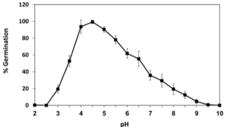
In a previous work, we isolated a fraction within the sweet potato extract (SPAF) that was found to induceR.delemarspore germination [7]. The pH of the SPAF was acidic (pH 4.7) and when it was modified, the optimal pH range forR.delemarspore germination was 4.0 to 5.0, with maximal germination at pH 4.5 (Fig 1). Germination was reduced to less than 20% at pH values above 8.0 and below 3.0.
In our previous study, we identified sucrose, glucose, fructose, organic acids and amino acids as the major constituents of SPAF [7]. Incubation ofR.delemarspores with the separated major HPLC fractions of SPAF did not result in germination (Table 1). The pH of the fractions containing the sugar peaks (sucrose, glucose and fructose) was neutral (~7.0), whereas the frac-tions containing the organic acid and amino acid peaks had a pH of 4.7. As acidic pH was deemed essential for germination, the sugar fraction was acidified to pH 4.7 and incubated withR.delemarspores. The acidified glucose fraction induced spore swelling, without germ-tube emergence (Table 1), whereas acidification of the sucrose or fructose fractions did not have any effect on spore swelling or germination. Only incubation ofR.delemarspores with the glucose fraction combined with amino acid and organic acid fractions enabled germ-tube emergence (Table 1). In both cases, i.e. SPAF or combined peak treatments, acidic pH caused spore swelling and a higher percentage of germ-tube emergence.
Inhibition of
R
.
delemar
spore swelling
For the spore-germination experiments, the solutions were acidified with 0.1 M HCl, implicat-ing that pH, rather than the organic or amino acids, induced swellimplicat-ing ofR.delemarspores. The fact that the spores swell in acidified solution led to the hypothesis of regulated water uptake by the spores in an acidic conditions and a potential involvement of aquaporin water channels on
Fig 1. Effect of pH onRhizopus delemarspore germination.Percentage ofR.delemarspores germinated under different pH conditions. Spores were incubated in SPAF solution (20 mg/ml, 42°C) and scored at 6 h. Values are means±SE (n = 500).
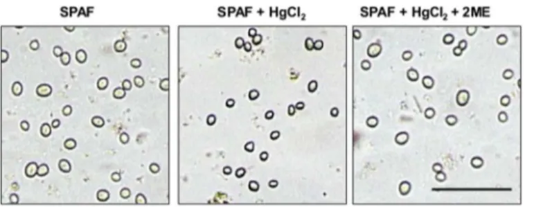
top of water influx by osmotic gradient [46]. To determine whether AQPs are involved in water uptake during the early stage of germination, we treated the spores with mercury chlo-ride (HgCl2), which is expected to decrease AQP water permeability by binding to cysteine
(Cys) residues in the AQP channel [41,43,47]. Adding HgCl2to the spore suspension in the
presence of SPAF led to inhibition of swelling and germination (Fig 2andTable 2). Treatment of the spores with 5μM of the reducing agent 2ME reversed the effect of HgCl2[48,49] and
the spores developed in a pattern similar to controls (Fig 2andTable 2).
AQPs in the
R
.
delemar
genome
A search of theR.delemargenome database [Mucorales Sequencing Project, Broad Institute of Harvard and MIT (http://www.broadinstitute.org)] revealed only two candidate AQP genes, Table 1. Effect of major peaks resulting from HPLC fractionation of sweet potato active fraction (SPAF) onRhizopus delemarspore swelling and germination.The pH of each HPLC fraction was modified to 4.7 or 7. The peaks contained: 1–a mixture of organic and amino acids, 2–sucrose, 3- glucose, 4–fructose, or the combination of 1 and 3. SPAF and water served as controls. Values are means±SE (n = 500).
HPLC Fraction pH Spore diameter (μm) Germ-tube emergence (%)
No. Content
Water 4.7 8.5±0.3*Bb 0
7.0 8.1±0.7 Bb 0
SPAF 4.7 22.2±0.6 Aa 96.9±2.5 Aa
7.0 15.7±2.1 Ab 44.2±3.3 Bb
1 Amino & organic acids 4.7 8.6±0.6 Bb 0
7.0 8.1±0.2 Bb 0
2 Sucrose 4.7 7.9±0.7 Bb 0
7.0 8.3±0.8 Bb 0
3 Glucose 4.7 19.5±1.9 Aa 0
7.0 14.3±2.4 Ab 0
4 Fructose 4.7 8.9±0.3 Bb 0
7.0 8.4±0.6 Bb 0
1+3 Amino & organic acids & Glucose 4.7 21.1±0.7 Aa 97.3±1.8 Aa
7.0 14.6±1.5 Ab 35.7±5.7 Bb
*Within each pH level (4.7 or 7.0) different upper case letters are significantly different from each other (P0.05). Within each pair of pH level in the same fraction content (Water, SPAF, etc.), lowercase letters are significantly different from each other (P0.05).
doi:10.1371/journal.pone.0150543.t001
Fig 2. Effect of HgCl2, an inhibitor of AQPs function on swelling of spore ofRhizopus delemar.
Treatment ofR.delemarspores with 40μM HgCl2for 5 min inhibited spore swelling and germination. Additional treatment (5 min) with 5μM of the reducing agent 2-β-mercaptoethanol (2ME) fully reversed the inhibition effect. Pictures were taken 15 min and 3 h, in the upper and lower rows, respectively, after incubation in water or sweet potato active fraction (SPAF). Bar = 100μm.
RdAQP1andRdAQP2, which are common among fungi but are considered to be members of a very small gene family compared to organisms from other kingdoms. Phylogenetic analysis of
R.delemarAQPs using the MEGA 4.0 program (http://www.megasoftware.net/mega4) showed that both RdAQP1 and RdAQP2 are located in Cluster III, of MIPs, that putatively act as water and small neutral molecule transport channels (Fig 3).
Table 2. Effect of HgCl2on swelling ofRhizopus delemarspores incubated in sweet potato active
frac-tion (SPAF) and germ-tube emergence. Spores were treated with 40μM HgCl2to inhibit aquaporin activity. Additional treatment with the reducing agent 2-β-mercaptoethanol (2ME, 5μM) was used to reverse the HgCl2inhibition effect. The assays were performed at pH 4.7. Values are means±SE (n = 500)
Treatment pH Diameter (μm) Germ-tube emergence (%)
SPAF 4.7 23.1±1.6*A 97.2±1.2 A
SPAF + HgCl2 4.7 7.8±0.6 B 0 B
SPAF + HgCl2+ 2ME 4.7 20.5±0.4 A 98.2±1.4 A
*Within each measured parameter different upper case letters are significantly different from each other (P0.05).
doi:10.1371/journal.pone.0150543.t002
Fig 3. Phylogenetic tree of 231 fungal major intrinsic proteins (MIPs).The MIPs clustered into four distinct groups: Cluster I—putative water channels MIPs (represented by ADC55259|Saccharomyces
cerevisiae[Black circle] and JF491353|Terfezia claveryi[black square]), cluster II—putative
aquaglyceroporins MIPs that preferentially transporting small neutral molecules (represented by Lacbi2| 671860|Laccaria bicolor[open triangle]), cluster III—MIPs that putatively act as water and small neutral molecule transport channels (represented by GAA23030|S.cerevisiae[black rhombus]), and cluster IV— putative fungal X intrinsic proteins (XIPs) (represented by TmeAQP2|Tuber melanosporum[black triangle]). BothRdAQP1andRdAQP2(open circles) are located in Cluster III, pointing on their potential capability to act as a water channels. Bar represents 0.2 changes.
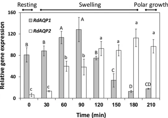
Differential expression of
RdAQP1
and
RdAQP2
The differential expression of the two AQP-encoding genes was monitored in spores from the resting stage, through the swelling stage and to the polar growth stage (Fig 4). Differential expression patterns were revealed for the two genes:RdAQP1transcripts were abundant at the resting stage and their expression increased about 1.5-fold during swelling, up to 90 min after induction of germination, followed by a 6.5-fold decrease before and during polar growth (Fig 4). In contrast,RdAQP2transcript levels were relatively low at the resting stage and its expres-sion increased steadily from the induction of germination to a peak after 180 min, at the onset of the polar growth stage (Fig 4).
Predicted topology and localization of RdAQP1 and RdAQP2 proteins
In-silicoprediction of RdAQP1 and RdAQP2 topology using the TMHMM method (http:// www.cbs.dtu.dk/services/TMHMM) confirmed the typical AQP structure of six transmem-brane domains (Fig 5a) (reviewed by [51]). For both RdAQP1 and RdAQP2, the predicted pro-tein sequence contained the highly conserved NPA domain which forms the water-specific channel [52]. Fusion of RdAQP1 and RdAQP2 to GFP and transformation intoR.delemarspores suggested that both of the proteins are confined to the intracellular part with higher intensity in the limits of the cell (Fig 5b).
Fig 4. Differential expression ofRhizopus delemarputative aquaporin genes (RdAQP1and RdAQP2) during germination.R.delemarspores were incubated in SPAF and harvested at three main stages: resting (time 0), swelling (30–180 min) and polar growth (210 min). qPCR was performed on cDNA reverse-transcribed from the RNA extracted from the spores.Rhizopus 18SrRNA served as the reference gene. Values of the steady-state levels of gene transcripts were determined as the ratio between two conditions using the 2Δ-ΔCt method [50]. Values are means±SE. Different upper or lowercase letters
above the bars denote significant differences between measurement times inRdAQP1orRdAQP2
respectively (P<0.05).
Functional analysis of RdAQP1 and RdAQP2 in relation to pH conditions
AQPs are integral membrane proteins belonging to a larger gene family that functions as water-channel activity or in transport of non-charged molecules such as glycerol, urea, ammo-nia and CO2[54]. To determine whether the two AQP genes found in theR.delemargenome
Fig 5. Predicted topology and localization of RdAQP1 and RdAQP2 proteins.(a) Hypothetical prediction of RdAQP1 and RdAQP2 topology by the TMHMM method, drawn by theProtter-visualize proteoforms
program [53], showing the proteins' six transmembrane domains. The highly conserved NPA domains are circled. The Cys and His amino acids are filled in gray and black color, respectively. (b) Expression of RdAQP1::GFP and RdAQP2::GFP inR.delemarspores. Bar = 10μm.
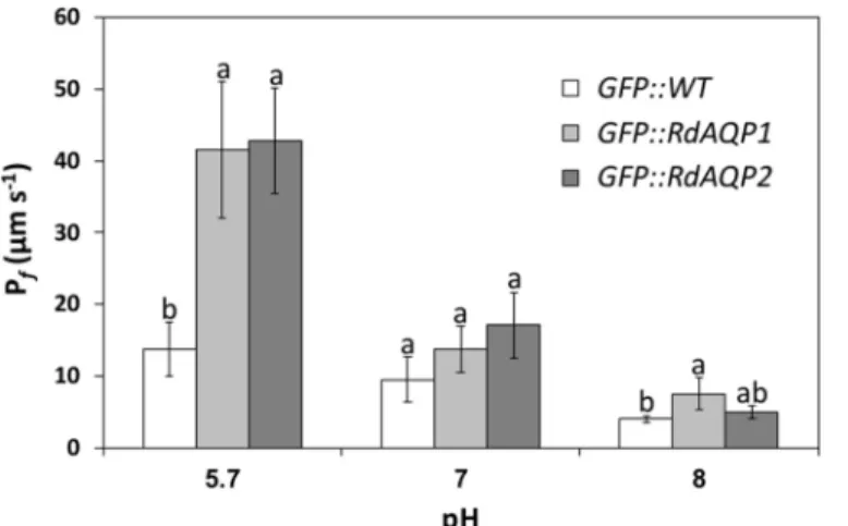
function as water channels, each gene was cloned under a constitutive 35S promoter and tran-siently expressed in the heterologous expression system ofArabidopsisprotoplasts. Then, the impact of RdAQP1 and RdAQP2 on the osmotic water permeability coefficient (Pf) of the
transformed cells was measured. At pH 5.7, thePfvalue of the protoplasts expressingR.
dele-marAQPs was significantly higher than for control cells (3.4- and 3-fold for RdAQP1 and RdAQP2, respectively). These results confirmed the role ofR.delemarAQPs as water channels (Fig 6).
As spore swelling was affected by pH, the osmotic water permeability assay was conducted at different pHs (Fig 6). While at pH 5.7, thePfvalue of cells expressing both RdAQP1- and
RdAQP2 was about 3-fold higher than in controls, at pH 7.0, thePfwas similar to that in
con-trol cells (Fig 6). A further reduction inPfvalues was observed at pH 8.0. These results
demon-strated the critical role of external pH on the water-channel activity ofR.delemarAQPs.
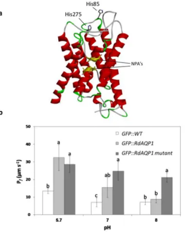
The role of His residues in pH sensing by RdAQP1
Previous studies have proposed a gating mechanism in which the translated AQP protein is regulated by inner or outer cell pH [46,55]. Those authors suggested that protonation of a con-served His residue will increase or decrease water conductivity of the AQP channel, depending on that residue's position [56]. A hypothetical three-dimensional structure of the RdAQP1pro-tein revealed two His residues at positions 85 and 275, predicted to be located externally and hence be subject to pH-dependent protonation (Fig 7a). A double His mutant (substitution of H85 and H275 with A) was constructed and transformed intoArabidopsisprotoplasts. The RdAQP1 mutant lost its pH sensitivity as thePfvalues of the transformed protoplasts showed a
minor and non-significant decrease when pH was raised from 5.7 to 8.0 (Fig 7b). At pH 8.0, thePfvalue of the RdAQP1 mutant was significantly higher than those of bothRdAQP1
-trans-formed protoplasts and control protoplasts (Fig 7b). As expected at pH 5.7, theRdAQP1 -trans-formed protoplasts differed significantly from the control, while at pH 7.0 and 8.0, no
differences were observed between treatments. This experiment suggested that the His residues facing the outside of the cell are involved in RdAQP1 pH sensing.
Fig 6. The effect of pH onRhizopus delemaraquaporin (AQP) water permeability.The protoplast osmotic water permeability coefficient (Pf) determined inArabidopsis thalianaprotoplasts transiently expressing GFP::AQP (GFP, green fluorescent protein) and in control protoplasts transiently expressing GFP. Values are means±SE (n = 15). Different lowercase letters above the bars denote significant differences for each pH level in each transformed gene (P<0.05).
Discussion
Spore formation is a very important stage in the fungal life cycle that enables these organisms better preservation under unfavorable conditions [5]. The spore stays in a resting state until it encounters environmental conditions that can support its development. Thus, the spore must be capable of sensing and responding to environmental signals. Spores of different fungal spe-cies have diverse requirements for the triggering of germination [5,8,9,14,15,57–59].
Never-theless, spore dehydration during maturation seems to be a fundamental process that is common to all spore-producing fungi. The question of regulation of water uptake during ger-mination is of prime significance for the fungal life cycle [31].
In this study, spore germination ofR.delemardecreased markedly below pH 3.5 and above pH 6.5 (Fig 1), while the optimal pH for germination was between 4 and 5. The time point selected for assessment of the pH effect was 6 h, when the peak germination efficacy was above 90%. As expected, at later time points, the efficacy at the marginal pH values was higher (not shown) suggesting that pH has a modulatory rather than strictly restrictive activity. The effect of pH on spore germination has been documented in previous studies [25,27]. The intense decrease in germination at low pH compared to the moderate decrease at higher pH implies different mechanisms by which pH affects spore germination. Efficiency of enzyme activities, nutrient availability and proton gradient across the plasma membrane are likely involved in Fig 7. The role of the outer cell His residues in pH sensing.(a) Predicted three-dimensional structure of RdAQP1 showing two His residues (His85 and His275) positioned on the loops of the protein and facing the outside of the cell. (b)Pfvalues of GFP::WT (wild type), GFP::RdAQP1 and GFP::RdAQP1 mutant,. Different lowercase letters above the bars denote significant differences for each pH level between fungal types (P<0.05).
modulation of germination [28,29]. According to the results of this study, the presence of glu-cose, at low pH, triggers spore swelling. These results are in accordance with previous studies showing that glucose induces spore swelling inRhizopus,FusariumandPenicilliumspp. [8,13, 59–61]. Thanh et al. [59] suggested that glucose and amino acids play important roles in
activa-tion and germinaactiva-tion of sporangiospores ofRhizopus oligosporusand this depends on the uptake potential of specific amino acids and/or glucose [59]. Such mechanism may explain why only glucose inducedR.delemarspore swelling in an acidic environment, while glucose under neutral conditions had reduced effect on the spores (Table 1). Interestingly, acidified sucrose or fructose fractions did not have any effect on spore swelling or germination. Glucose can be replace by fructose or sucrose as a carbon source, but still maximum germination and biomass production required a nitrogen source, phosphate ions and potassium or sodium ions [62,63]. The specific effect of different sugars on germination has been documented in fungi: while the uptake of glucose is higher in many fungal species, fructose is a better inducer of ger-mination inB.cinerea([64] and references therein). Knockout of specific fructose transporter in Botrytis demonstrated delayed fructose-induced germination but both mutants and wild type conidia showed higher affinity to glucose. These results suggest that fructose-induced ger-mination did not depend on transport but possibly on intracellular sensing mechanisms.
R.delemarspores treated with acidified water (Table 1) did not respond by swelling or ger-mination, and did not aggregate. In addition, a treatment with the AQP blocker HgCl2
inhib-ited spore swelling and germination (Fig 2andTable 2) pointed to water uptake and hydration of the spore as a crucial step in the very early stages of fungal spore germination. These results are in agreement with a study in whichFusarium graminearumandFusarium poaespore hydration was inhibited as a means of preventing spore germination [65].
To better understand the relation between water uptake and pH conditions and the mecha-nism that enables water uptake by the spore at an early stage of germination, we analyzed the
R.delemargenome to identify genes that could potentially act as water channels. The presence of only two candidate AQPs in theR.delemargenome corresponded with other fungi which present a relatively small number of these genes compared to mammals and plants [66]. Phylo-genetic analysis clustered these proteins with a group of fungal major intrinsic proteins (MIPs) suggested to act as water channels (Fig 3). Taking into account the important activity of AQPs in the transitioning of small and uncharged molecules such as water, acids, carbon dioxide, glycerol and ammonia, the small number of these genes inR.delemarand other fungal genomes suggests a non-selective role for these proteins [67–70]. The protein structure of
AQPs is conserved and can be found in organisms from all kingdoms [69,71–74]. According
to the transmembrane domains and the expected role in water uptake, fusion of the two genes to GFP and transformation intoR.delemarspores localized the two encoded proteins to the cell membrane (Fig 5b). The proteins' similarity ofR.delemarAQPs to the plasma membrane-intrinsic protein (PIP;Fig 3), AQP subfamily in other organisms [75,76], is another indication for localization on the outer membrane.
Expression analysis ofRdAQP1andRdAQP2revealed opposite patterns for the two genes as germination progressed (Fig 5). WhileRdAQP1transcripts were abundant in the spore rest-ing stage and durrest-ing the early stages of germination,RdAQP2transcripts were low in the rest-ing stage and increased as germination progressed (Fig 5). These results suggest thatRdAQP1
glycerol and/or NH3uptake [80,81]. The role of AQPs in yeast and filamentous fungi has been
reviewed [66,82] with respect to three main aspects: sporulation, freeze tolerance and mycor-rhizal associations [77,80,82–85]. Since AQPs might act as a transfer channel for small and
non-charged molecules rather than water channels, it was important to perform a biological assay that would elucidate their role (Fig 6).RdAQP1andRdAQP2were expressed in Arabi-dopsisprotoplasts which were then exposed to hypotonic solution to calculate plasma mem-branePfpotential [39]. Although thePfvalues of the plasma membrane, whenRdAQP1and
RdAQP2expressed, is lower than those found in other fungal AQPs [80], they were found to be significantly higher than controls and probably contributed to water uptake ofR.delemar
spores during the early stages of spore germination. In contrast to an earlier work that did not find any correlation between environmental osmotic pressure and water-uptake capacity [13], the current work demonstrated rapid water uptake by the transformed protoplasts as the osmotic pressure of the solutions changed from isotonic (600 mOsm) to hypotonic (500 mOsm). To the best of our knowledge, this is the first evidence for regulated water uptake in fungi during the early stages of spore germination.
In this work, we found thatR.delemar, like several other pathogenic fungi, requires a spe-cific pH range for optimal germination (Fig 1) [25,27]. In addition, water uptake occurred at the early stages of germination and inhibition of water uptake using HgCl2caused inhibition of
spore swelling and germination (Fig 2). Both of RdAQP1 and RdAQP2 present Cys residues located near to the conserved NPA motifs (C240 and C256, respectively). Due to their proxim-ity to these motifs and according to their trans-membranal predicted localization they are assumed to be targets for HgCl2binding.
To understand whether water uptake byRdAQP1andRdAQP2is regulated by ambient pH, water-uptake experiments were carried out under several pH conditions (Fig 6). Reduction in
RdAQP1andRdAQP2 Pflevels as the ambient pH increased suggests pH regulation ofR.
dele-marwater channels and might provide a practical explanation for the specific range of ambient pH required for optimal spore germination. pH regulation of AQPs has been vastly docu-mented in the literature [46,55,56,86–88].
Different regulation mechanisms that directly affect the spatial structure of AQPs have been proposed (reviewed by [89]): (i) phosphorylation of S residues within the N or C termini; (ii) oligomeric state of the protein (heteromerization); (iii) gating by high solute concentration and pressure pulses, and (iv) regulation by pH. With respect to pH regulation, several works have shown that His residues are protonated in acidic environments and contribute to the "gating mechanism" of the water channel [46,87]. Protonation on a conserved His residue of AQPs could either lead to activation or inhibition of the water channels activity. Most of plant's aqua-porins of the PIP subgroup are inhibited by cytosol acidosis [46,55] while external acidic pH or low Ca2+ increased the water permeability in bovine's AQP0 [56]. Depending on the posi-tion of the His residue (on the loop, inside or outside the cell), the protonaposi-tion will switch the water channel's permeability on or off [87]. It seems that the flexibility of pH regulation through the His residue position enables an efficient mechanism of adaptation to environmen-tal conditions. To better understand the mechanism by which pH regulates RdAQP1 function, two His residues positioned on the outer cell loops (Fig 7a) were replaced with A. In agreement with earlier studies, theRdAQP1mutant lost its pH sensitivity and itsPfbecame unaffected by
pH (Fig 7b), suggesting that one (or both) of the His play a role in pH sensing [46].
Acknowledgments
This work was supported by grant 430-421-12 of the Chief Scientist, Ministry of Agriculture, Israel and by grant 1131/12 of the Israel Science Foundation (ISF). This manuscript is contri-bution no. 716/15 from ARO, the Volcani Center, P.O Box 6, Bet-Dagan 20125, Israel.
Author Contributions
Conceived and designed the experiments: DE TT PT-B. Performed the experiments: TT ASC. Analyzed the data: DE MM TT. Contributed reagents/materials/analysis tools: CDS PT-B. Wrote the paper: TT AL DE.
References
1. Ghosh B, Ray RR. Current commercial perspective ofRhizopus oryzae: a review. J Appl Sci. 2011; 11:2470–86.
2. Zheng R-Y, Chen G-Q, Huang H, Liu X-Y. A monograph of Rhizopus. Sydowia. 2007; 59(2):273–372.
3. Feofilova E, Ivashechkin A, Alekhin A, Sergeeva YE. Fungal spores: Dormancy, germination, chemical composition, and role in biotechnology. App Biochem Microbiol. 2012; 48(1):1–11.
4. d'Enfert C. Fungal spore germination: Insights from the molecular genetics ofAspergillus nidulansand
Neurospora crassa. Fungal Genet Biol. 1997; 21(2):163–72.
5. Griffin D. Spore dormancy and germination. Fungal physiology. New York, NY: John Wiley & Sons,; 1994. p. 375–98.
6. Janisiewicz WJ, Korsten L. Biological control of postharvest diseases of fruits. Ann Rev Phytopathol. 2002; 40(1):411–41. doi:10.1146/annurev.phyto.40.120401.130158
7. Turgeman T, Kakongi N, Schneider A, Vinokur Y, Teper-Bamnolker P, Carmeli S, et al. Induction of
Rhizopus oryzaegermination under starvation using host metabolites increases spore susceptibility to heat stress. Phytopathology. 2014; 104:240–7. doi:10.1094/PHYTO-08-13-0245-RPMID:24093921
8. Medwid RD, Grant DW. Germination ofRhizopus oligosporussporangiospores. App Environ Microbiol. 1984; 48(6):1067–71.
9. Pass T, Griffin G. Exogenous carbon and nitrogen requirements for conidial germination byAspergillus flavus. Can J Microbiol. 1972; 18(9):1453–61. PMID:4627197
10. Hashimoto T, Wu C, Blumenthal H. Characterization of L-leucine-induced germination ofTrichophyton mentagrophytesmicroconidia. J Bacteriol. 1972; 112(2):967–76. PMID:4117583
11. Weber DJ, Ogawa JM. Specificity of proline in germination of spores ofRhizopus arrhizus. Phytopathol-ogy. 1965; 55(3):262–&.
12. Weber D. The role of proline in the germination of Rhizopus stolonifer spores. Phytopathol. 1962; 52:756.
13. Ekundayo J. Further studies on germination of sporangiospores of Rhizopus arrhizus. J General Micro-biol. 1966; 42(2):283–91.
14. Chaky J, Anderson K, Moss M, Vaillancourt L. Surface hydrophobicity and surface rigidity induce spore germination inColletotrichum graminicola. Phytopathology. 2001; 91(6):558–64. doi:10.1094/PHYTO. 2001.91.6.558PMID:18943944
15. Holmes GJ, Stange RR. Influence of wound type and storage duration on susceptibility of sweetpota-toes to Rhizopus soft rot Plant Dis. 2002; 86(4):345–8.
16. Estrada A, Dodd J, Jeffries P. Effect of humidity and temperature on conidial germination and appresso-rium development of two Philippine isolates of the mango anthracnose pathogenColletotrichum gloeosporioides. Plant Pathol. 2000; 49(5):608–18.
17. Kope HH, Alfaro RI, Lavallée R. Effects of temperature and water activity on Lecanicillium spp. conidia germination and growth, and mycosis of Pissodes strobi. BioControl. 2008; 53(3):489–500.
18. Mcquilken M, Budge SP, Whipps JM. Effects of culture media and environmental factors on conidial germination, pycnidial production and hyphal extension ofConiothyrium minitans. Mycol Res. 1997; 101(01):11–7.
20. Sautour M, Rouget A, Dantigny P, Divies C, Bensoussan M. Application of Doehlert design to deter-mine the combined effects of temperature, water activity and pH on conidial germination of Penicillium chrysogenum. J App Microbiol. 2001; 91(5):900–6.
21. Bignell E. The molecular basis of ph sensing, signaling, and homeostasis in fungi. Adv App Microbiol. 2012; 79(79):1–18.
22. Manteau S, Abouna S, Lambert B, Legendre L. Differential regulation by ambient pH of putative viru-lence factor secretion by the phytopathogenic fungusBotrytis cinerea. FEMS Microbiol Ecol. 2003; 43 (3):359–66. doi:10.1111/j.1574-6941.2003.tb01076.xPMID:19719667
23. Gock MA, Hocking AD, Pitt JI, Poulos PG. Influence of temperature, water activity and pH on growth of some xerophilic fungi. Int J Food Microbiol. 2003; 81(1):11–9. PMID:12423914
24. Jackson A, Whipps J, Lynch J. Effects of temperature, pH and water potential on growth of four fungi with disease biocontrol potential. World J Microbiol Biotechnol. 1991; 7(4):494–501. doi:10.1007/ BF00303376PMID:24425136
25. Lopez-Malo A, Alzamora S, Argaiz A. Effect of vanillin concentration, pH and incubation temperature on Aspergillus flavus, Aspergillus niger, Aspergillus ochraceus and Aspergillus parasiticus growth. Food Microbiol. 1997; 14(2):117–24.
26. Magan N, Lacey J. Effect of temperature and pH on water relations of field and storage fungi. T Brit Mycol Soc. 1984; 82(1):71–81.
27. Pelser PdT, Eckert J. Constituents of orange juice that stimulate the germination of conidia of Penicil-lium digitatum. Phytopathol. 1977; 67(6):747–54.
28. Li B, Lai T, Qin G, Tian S. Ambient pH stress inhibits spore germination ofPenicillium expansumby impairing protein synthesis and folding: a proteomic-based study. J Proteome Res. 2009; 9(1):298– 307.
29. Schmidt P, Walker J, Selway L, Stead D, Yin Z, Enjalbert B, et al. Proteomic analysis of the pH response in the fungal pathogenCandida glabrata. Proteomics. 2008; 8(3):534–44. doi:10.1002/pmic. 200700845PMID:18186024
30. Barer R, Joseph S. Concentration and mass measurements in microbiology. J App Bacteriol. 1958; 21 (1):146–59.
31. Nehls U, Dietz S. Fungal aquaporins: cellular functions and ecophysiological perspectives. App Micro-biol Biotech. 2014; 98(21):8835–51.
32. Delp CJ. Effect of temperature and humidity on the grape powdery mildew fungus. Phytopathol. 1954; 44(11):615–26.
33. Yarwood C. Water content of fungus spores. Am J Bot. 1950:636–9.
34. Ibrahim AS, Gebremariam T, Lin L, Luo G, Husseiny MI, Skory CD, et al. The high affinity iron permease is a key virulence factor required forRhizopus oryzaepathogenesis. Mol Microbiol. 2010; 77(3):587– 604. doi:10.1111/j.1365-2958.2010.07234.xPMID:20545847
35. Mayzlish-Gati E, LekKala SP, Resnick N, Wininger S, Bhattacharya C, Lemcoff JH, et al. Strigolactones are positive regulators of light-harvesting genes in tomato. J Exp Bot. 2010; 61(11):3129–36. doi:10. 1093/jxb/erq138PMID:20501744
36. Livak KJ, Schmittgen TD. Analysis of relative gene expression data using real-time quantitative PCR and the 2−ΔΔCTmethod. Methods. 2001; 25(4):402–8. PMID:11846609
37. Xu H, Cooke JE, Zwiazek JJ. Phylogenetic analysis of fungal aquaporins provides insight into their pos-sible role in water transport of mycorrhizal associations. Botany. 2013; 91(8):495–504.
38. Locatelli F, Vannini C, Magnani E, Coraggio I, Bracale M. Efficiency of transient transformation in tobacco protoplasts is independent of plasmid amount. Plant Cell Rep. 2003; 21(9):865–71. PMID: 12789504
39. Shatil-Cohen A, Sibony H, Draye X, Chaumont F, Moran N, Moshelion M. Measuring the osmotic water permeability coefficient (Pf) of spherical cells: isolated plant protoplasts as an example. J Vis Exper 2014; 92:e51652–e.
40. Barad S, Horowitz SB, Kobiler I, Sherman A, Prusky D. Accumulation of the mycotoxin patulin in the presence of gluconic acid contributes to pathogenicity of Penicillium expansum. Mol Plant-Microbe Interact. 2014; 27(1):66–77. doi:10.1094/MPMI-05-13-0138-RPMID:24024763
41. Moshelion M, Becker D, Biela A, Uehlein N, Hedrich R, Otto B, et al. Plasma Membrane aquaporins in the motor cells of samanea saman diurnal and circadian regulation. Plant Cell. 2002; 14(3):727–39. PMID:11910017
43. Shatil-Cohen A, Attia Z, Moshelion M. Bundle-sheath cell regulation of xylem-mesophyll water transport via aquaporins under drought stress: a target of xylem-borne ABA? The Plant J. 2011; 67(1):72–80. doi:10.1111/j.1365-313X.2011.04576.xPMID:21401747
44. Volkov V, Hachez C, Moshelion M, Draye X, Chaumont F, Fricke W. Water permeability differs between growing and non-growing barley leaf tissues. J Exp Bot. 2007; 58(3):377–90. PMID:17122408
45. Schwede T, Kopp J, Guex N, Peitsch MC. SWISS-MODEL: an automated protein homology-modeling server. Nucleic Acids Res. 2003; 31(13):3381–5. PMID:12824332
46. Tournaire-Roux C, Sutka M, Javot H, Gout E, Gerbeau P, Luu D-T, et al. Cytosolic pH regulates root water transport during anoxic stress through gating of aquaporins. Nature. 2003; 425(6956):393–7. PMID:14508488
47. Pou A, Medrano H, Flexas J, Tyerman SD. A putative role for TIP and PIP aquaporins in dynamics of leaf hydraulic and stomatal conductances in grapevine under water stress and re-watering. Plant Cell Environ. 2013; 36(4):828–43. doi:10.1111/pce.12019PMID:23046275
48. Ohrui T, Nobira H, Sakata Y, Taji T, Yamamoto C, Nishida K, et al. Foliar trichome-and aquaporin-aided water uptake in a drought-resistant epiphyteTillandsia ionanthaPlanchon. Planta. 2007; 227 (1):47–56. PMID:17674031
49. Ishida Y, Nagae T, Azuma M. A water-specific aquaporin is expressed in the olfactory organs of the blowfly, Phormia regina. J Chem Ecol. 2012; 38(8):1057–61. doi:10.1007/s10886-012-0157-zPMID: 22767214
50. Arocho A, Chen B, Ladanyi M, Pan Q. Validation of the 2-ΔΔCt calculation as an alternate method of data analysis for quantitative PCR of BCR-ABL P210 transcripts. Diagnostic Mol Pathol. 2006; 15 (1):56–61.
51. Maurel C. Aquaporins and water permeability of plant membranes. Ann Rev Plant Biol. 1997; 48 (1):399–429.
52. Walz T, Hirai T, Murata K, Heymann JB, Mitsuoka K, Fujiyoshi Y, et al. The three-dimensional structure of aquaporin-1. Nature. 1997; 387(6633):624–6. PMID:9177353
53. Omasits U, Ahrens CH, Müller S, Wollscheid B. Protter: interactive protein feature visualization and integration with experimental proteomic data. Bioinformatics. 2013; 30:884–6. doi:10.1093/ bioinformatics/btt607PMID:24162465
54. Kaldenhoff R, Bertl A, Otto B, Moshelion M, Uehlein N. Characterization of plant aquaporins. Methods Enzymol. 2007; 428:505–31. PMID:17875436
55. Fischer M, Kaldenhoff R. On the pH regulation of plant aquaporins. J Biol Chem. 2008; 283(49):33889– 92. doi:10.1074/jbc.M803865200PMID:18818207
56. Németh-Cahalan KL, Kalman K, Hall JE. Molecular basis of pH and Ca2+regulation of aquaporin water permeability. J General Physiol. 2004; 123(5):573–80.
57. Carlile M, Watkinson S. Spores, dormancy and dispersal. The Fungi, Academic Press, New York. 1994:153–201.
58. Cook R, Baker K. Why biological control. In: Cook R, Baker K, editors. The nature and practice of bio-logical control of plant pathogens. St. Paul, MN: American Phytopathobio-logical Society.; 1983. p. 1–29.
59. Thanh N, Rombouts F, Nout M. Effect of individual amino acids and glucose on activation and germina-tion ofRhizopus oligosporussporangiospores in tempe starter. J App Microbiol. 2005; 99(5):1204–14.
60. Gottlieb D, Tripathi R. The physiology of swelling phase of spore germination inPencillium atrovene-tum. Mycologia. 1968:571–90.
61. Marchant R, White MF. Spore swelling and germination in Fusarium culmorum. J General Microbiol. 1966; 42(2):237–44.
62. Amiri A, Chai W, Schnabel G. Effect of nutrient status, pH, temperature and water potential on germina-tion and growth ofRhizopus stoloniferandGilbertella persicaria. J Plant Pathol. 2011; 93(3):603–12.
63. Ekundayo J, Carlile M. The germination of sporangiospores ofRhizopus arrhizus; spore swelling and germ-tube emergence. Microbiology. 1964; 35(2):261–9.
64. Doehlemann G, Molitor F, Hahn M. Molecular and functional characterization of a fructose specific transporter from the gray mold fungusBotrytis cinerea. Fungal Genetics Biol. 2005; 42(7):601–10.
65. Wang X, Liu X, Chen J, Han H, Yuan Z. Evaluation and mechanism of antifungal effects of carbon nanomaterials in controlling plant fungal pathogen. Carbon. 2014; 68:798–806.
66. Pettersson N, Filipsson C, Becit E, Brive L, Hohmann S. Aquaporins in yeasts and filamentous fungi. Biology of the Cell. 2005; 97(7):487–500. PMID:15966864
68. Katsuhara M, Hanba YT. Barley plasma membrane intrinsic proteins (PIP aquaporins) as water and CO2 transporters. Pflügers Arch-Eur J Physiol. 2008; 456(4):687–91.
69. Kruse E, Uehlein N, Kaldenhoff R. The aquaporins. Genome Biol. 2006; 7(2):206. PMID:16522221
70. Wu B, Beitz E. Aquaporins with selectivity for unconventional permeants. Cellular Mol Life Sci. 2007; 64(18):2413–21.
71. de Groot BL, Grubmüller H. Water permeation across biological membranes: mechanism and dynam-ics of aquaporin-1 and GlpF. Science. 2001; 294(5550):2353–7. PMID:11743202
72. Engel A, Fujiyoshi Y, Agre P. The importance of aquaporin water channel protein structures. EMBO J 2000; 19(5):800–6. PMID:10698922
73. Hill A, Shachar-Hill B, Shachar-Hill Y. What are aquaporins for? J Membrane Biol. 2004; 197(1):1–32.
74. Murata K, Mitsuoka K, Hirai T, Walz T, Agre P, Heymann JB, et al. Structural determinants of water per-meation through aquaporin-1. Nature. 2000; 407(6804):599–605. PMID:11034202
75. Chaumont F, Barrieu F, Wojcik E, Chrispeels MJ, Jung R. Aquaporins constitute a large and highly divergent protein family in maize. Plant Physiol. 2001; 125(3):1206–15. PMID:11244102
76. Sakurai J, Ishikawa F, Yamaguchi T, Uemura M, Maeshima M. Identification of 33 rice aquaporin genes and analysis of their expression and function. Plant Cell Physiol. 2005; 46(9):1568–77. PMID: 16033806
77. Sidoux-Walter F, Pettersson N, Hohmann S. The Saccharomyces cerevisiae aquaporin Aqy1 is involved in sporulation. P Nstl Acad Sci-Biol. 2004; 101(50):17422–7.
78. Aroca R, Porcel R, Ruiz-Lozano JM. How does arbuscular mycorrhizal symbiosis regulate root hydrau-lic properties and plasma membrane aquaporins in Phaseolus vulgaris under drought, cold or salinity stresses? New Phytol. 2007; 173(4):808–16. PMID:17286829
79. Porcel R, Aroca R, Azcón R, Ruiz-Lozano JM. PIP aquaporin gene expression in arbuscular mycor-rhizal Glycine max andLactuca sativaplants in relation to drought stress tolerance. Plant Mol Biol. 2006; 60(3):389–404. PMID:16514562
80. Dietz S, von Buelow J, Beitz E, Nehls U. The aquaporin gene family of the ectomycorrhizal fungus Lac-caria bicolor: lessons for symbiotic functions. New Phytologist. 2011; 190(4):927–40. doi:10.1111/j. 1469-8137.2011.03651.xPMID:21352231
81. Maurel C, Plassard C. Aquaporins: for more than water at the plant–fungus interface? New Phytol. 2011; 190(4):815–7. doi:10.1111/j.1469-8137.2011.03731.xPMID:21561457
82. Tanghe A, Van Dijck P, Thevelein JM. Why do microorganisms have aquaporins? Trends Microbiol. 2006; 14(2):78–85. PMID:16406529
83. Aroca R, Bago A, Sutka M, Paz JA, Cano C, Amodeo G, et al. Expression analysis of the first arbuscu-lar mycorrhizal fungi aquaporin described reveals concerted gene expression between salt-stressed and nonstressed mycelium. Mol Plant-Microbe Interact. 2009; 22(9):1169–78. doi: 10.1094/MPMI-22-9-1169PMID:19656051
84. Tanghe A, Van Dijck P, Dumortier F, Teunissen A, Hohmann S, Thevelein JM. Aquaporin expression correlates with freeze tolerance in baker's yeast, and overexpression improves freeze tolerance in industrial strains. App Environ Microbiol. 2002; 68(12):5981–9.
85. Will JL, Kim HS, Clarke J, Painter JC, Fay JC, Gasch AP. Incipient balancing selection through adaptive loss of aquaporins in natural Saccharomyces cerevisiae populations. PLoS Genet. 2010; 6(4): e1000893. doi:10.1371/journal.pgen.1000893PMID:20369021
86. Gerbeau P, Amodeo G, Henzler T, Santoni V, Ripoche P, Maurel C. The water permeability of Arabi-dopsis plasma membrane is regulated by divalent cations and pH. Plant J. 2002; 30(1):71–81. PMID: 11967094
87. Németh-Cahalan KL, Hall JE. pH and calcium regulate the water permeability of aquaporin 0. J Biol Chem. 2000; 275(10):6777–82. PMID:10702234
88. Törnroth-Horsefield S, Wang Y, Hedfalk K, Johanson U, Karlsson M, Tajkhorshid E, et al. Structural mechanism of plant aquaporin gating. Nature. 2005; 439(7077):688–94. PMID:16340961
![Fig 3. Phylogenetic tree of 231 fungal major intrinsic proteins (MIPs). The MIPs clustered into four distinct groups: Cluster I — putative water channels MIPs (represented by ADC55259|Saccharomyces cerevisiae [Black circle] and JF491353|Terfezia claveryi [](https://thumb-eu.123doks.com/thumbv2/123dok_br/18403379.358956/8.918.302.684.500.887/phylogenetic-intrinsic-proteins-clustered-represented-saccharomyces-cerevisiae-terfezia.webp)
![Fig 5. Predicted topology and localization of RdAQP1 and RdAQP2 proteins. (a) Hypothetical prediction of RdAQP1 and RdAQP2 topology by the TMHMM method, drawn by the Protter-visualize proteoforms program [53], showing the proteins' six transmembrane domain](https://thumb-eu.123doks.com/thumbv2/123dok_br/18403379.358956/10.918.308.831.121.848/predicted-topology-localization-hypothetical-prediction-visualize-proteoforms-transmembrane.webp)