Listeria monocytogenes
Jesper S. Nielsen1, Marianne Halberg Larsen2, Eva Maria Sternkopf Lillebæk1, Teresa M. Bergholz3, Mie H. G. Christiansen1, Kathryn J. Boor3, Martin Wiedmann3, Birgitte H. Kallipolitis1*
1Department of Biochemistry and Molecular Biology, University of Southern Denmark, Odense, Denmark,2Department of Veterinary Disease Biology, Faculty of Life Sciences, University of Copenhagen, Frederiksberg, Denmark,3Department of Food Science, Cornell University, Ithaca, New York, United States of America
Abstract
In recent years, more than 60 small RNAs (sRNAs) have been identified in the gram-positive human pathogenListeria monocytogenes, but their putative roles and mechanisms of action remain largely unknown. The sRNA LhrA was recently shown to be a post-transcriptional regulator of a single gene,lmo0850, which encodes a small protein of unknown function. LhrA controls the translation and degradation of thelmo0850mRNA by an antisense mechanism, and it depends on the RNA chaperone Hfq for efficient binding to its target. In the present study, we sought to gain more insight into the functional role of LhrA inL. monocytogenes. To this end, we determined the effects of LhrA on global-wide gene expression. We observed that nearly 300 genes inL. monocytogenesare either positively or negatively affected by LhrA. Among these genes, we identified lmo0302 and chiAas direct targets of LhrA, thus establishing LhrA as a multiple target regulator.
Lmo0302encodes a hypothetical protein with no known function, whereaschiAencodes one of two chitinases present inL. monocytogenes.We show here that LhrA acts as a post-transcriptional regulator oflmo0302and chiAby interfering with ribosome recruitment, and we provide evidence that both LhrA and Hfq act to down-regulate the expression oflmo0302
andchiA. Furthermore,in vitrobinding experiments show that Hfq stimulates the base pairing of LhrA tochiAmRNA. Finally, we demonstrate that LhrA has a negative effect on the chitinolytic activity ofL. monocytogenes. In marked contrast to this, we found that Hfq has a stimulating effect on the chitinolytic activity, suggesting that Hfq plays multiple roles in the complex regulatory pathways controlling the chitinases ofL. monocytogenes.
Citation:Nielsen JS, Larsen MH, Lillebæk EMS, Bergholz TM, Christiansen MHG, et al. (2011) A Small RNA Controls Expression of the Chitinase ChiA inListeria monocytogenes. PLoS ONE 6(4): e19019. doi:10.1371/journal.pone.0019019
Editor:Tanya Parish, Queen Mary University of London, United Kingdom
ReceivedJanuary 22, 2011;AcceptedMarch 14, 2011;PublishedApril 18, 2011
Copyright:ß2011 Nielsen et al. This is an open-access article distributed under the terms of the Creative Commons Attribution License, which permits unrestricted use, distribution, and reproduction in any medium, provided the original author and source are credited.
Funding:This research was supported by grants from the Danish Agency for Science Technology and Innovation (to BHK) and National Institutes of Health-National Institute of Allergy and Infectious Diseases R01 AI052151 (to KJB). The funders had no role in study design, data collection and analysis, decision to publish, or preparation of the manuscript.
Competing Interests:The authors have declared that no competing interests exist.
* E-mail: [email protected]
Introduction
Small RNAs (sRNAs) in bacteria play regulatory roles in a wide range of physiological processes, such as virulence [1,2], iron homeostasis [3], cell envelope stress [4] and sugar metabolism [5]. Many sRNAs act as post-transcriptional regulators by base pairing to specific target mRNAs, thereby affecting their translation and/ or stability [6,7]. A single sRNA may target multiple mRNAs and typically binds to short complementary regions overlapping the ribosome-binding site and/or start codon. In these cases, sRNA-mRNA pairing prevents the ribosome from binding to the sRNA-mRNA, resulting in repression of translation and rapid degradation of the mRNA. Although base-paring sRNAs are primarily known as negative regulators, some sRNAs have been found to exert a positive effect on gene expression. In these cases, sRNAs may activate translation by liberating the ribosome-binding site from an inhibitory stem-loop structure situated at the 59 mRNA region [6,8]. Alternatively, sRNA binding to the 59mRNA region may lead to processing and/or stabilization of the mRNA [9,10].
The gram-positive human pathogenListeria monocytogenessurvives and multiplies in many different environments, including soil and water, food processing environments and the eukaryotic host cell cytosol [11]. Controlled expression of genes supporting the growth
unknown function, to repress translation and stimulate degrada-tion oflmo0850mRNA in an Hfq-dependent manner [23]. These findings demonstrated that LhrA acts as an Hfq-dependent anti-sense RNA, however, the functional role of LhrA inL. monocytogenes remained unclear.
In this work, we studied the effects of LhrA on global gene expression inL. monocytogenesEGD-e by a comparative microarray analysis of wild type andDlhrAmutant strains. We found that lack oflhrAresults in the altered expression of approximately 300 genes and we further demonstrate a direct effect of LhrA on two genes: lmo0302, encoding a hypothetical protein with no known function, and the chitinase-encoding gene chiA. The chitinases ChiA and ChiB ofL. monocytogenescatalyze the hydrolysis of the carbohydrate polymer chitin, a highly abundant carbon and nitrogen source found in the environment [24,25]. Furthermore, chiA and chiB contribute to the pathogenesis ofL. monocytogenesin mice, possibly through the recognition of glycoproteins or other carbohydrate moieties present in the infected host [26]. Here, we show that LhrA acts to down regulate the expression oflmo0302andchiAat the post-transcriptional level in an Hfq-dependent manner, demonstrating that LhrA is a multiple target regulator in L. monocytogenes.
Results
Dissecting thelhrApromoter region
sRNAs are often highly regulated at the transcriptional level, and identification of the environmental signals and transcription factors controlling their expression may provide important clues to their biological function. Upon growth in rich media, LhrA accumulates in an Hfq-dependent manner at the entry into stationary phase, suggesting that LhrA plays a role in the transition from actively growing to resting cells ([15,23]). To gain further insight into the transcriptional regulation oflhrA, we performed a lhrApromoter deletion analysis. To this end, truncated versions of thelhrApromoter were fused to a promoter-lesslacZgene in the transcriptional fusion vector pTCV-lac. The lhrA promoter fragments range from position2157,283, 261,236 or 229, to position +71, relative to the transcriptional start site of lhrA (Figure 1A). The lhrA-lacZ fusion plasmids were introduced into theL. monocytogenesEGD-e wild type strain and the level of specific b-galactosidase activity was determined during growth in rich medium (Figure 1B). Very high and comparable levels of b -galactosidase activity were recorded throughout growth for all constructs containing deletions of thelhrApromoter region down to position 261. The construct plhr36-lacZ, containing the core promoter sequence oflhrA, displayed a 5 fold lower level of activity throughout growth (Figure 1B). Further deletion of the promoter region was expected to abolish the promoter activity, and accordingly, cells containing the construct plhr29-lacZ, which lacks the235 box, displayed background levels of activity (Figure 1B). As a control experiment, the transcription start site and RNA level of the variouslhrA-lacZtranscripts were tested by primer extension analysis using alacZ-specific primer. For all lhrA-lacZconstructs, only a single transcription start site was observed, corresponding to the expected start site forlhrA, and furthermore, the level of lhrA-lacZ transcript appeared constant throughout growth (data not shown).
Collectively, these results show that LhrA is expressed throughout growth from a highly active promoter, and that the region located between position 261 and position 236 has a stimulating effect on transcription. Within this region, we noticed a sequence motif similar to that recognized by the response regulator ResD (see Figure 1A), which is known to affect the transcription of
multiple genes in L. monocytogenes [27]. We therefore tested the effect of ResD on transcription of plhrA36-lacZand plhrA61-lacZin a DresD mutant background. No difference in b-galactosidase activity was observed between the wild type andresDmutant strain (data not shown) suggesting that expression of lhrA is not stimulated by ResD. We also noticed the presence of an AT-rich region between position 261 and position 236 (Figure 1A). In Escherichia coli and Bacillus subtilis, AT-rich regions called UP elements, which are located upstream of the235 region, are known to facilitate binding of the RNA polymerase to a promoter, resulting in an enhanced transcription activity [28–31]. The AT-rich element located between position261 and 236 in thelhrA promoter region may play a similar role resulting in a highly efficient transcription of lhrA. No other putative regulatory elements were observed within this region.
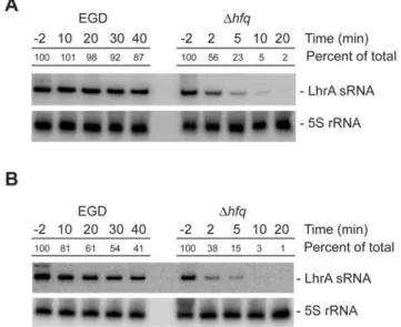
Since lhrA appears to be transcribed at a relatively high and constant level throughout growth, we hypothesized that the growth phase dependent accumulation of LhrA observed in Northern blots [15,23] is likely to be the result of a post-transcriptional control mechanism. To test this hypothesis, we compared the stability of LhrA in early stationary phase (OD600= 2.2) and early exponential phase cells (OD600= 0.4) (Figure 2A and 2B). In both cases, LhrA appears to be extremely stable in the wild type background; however, in exponential cells, the turnover of LhrA appears to be faster (half-life approximately 30 minutes) than in early stationary cells (half-life.60 minutes). Regardless of the growth phase, the stability of LhrA is clearly facilitated by the Hfq protein. Thus, it appears that the level of LhrA is controlled mainly at the post-transcriptional level.
LhrA affects gene expression on a global level
To learn more about the physiological role of LhrA, we performed a genome-wide transcriptome comparison of wild type andDlhrAmutant cells grown in rich growth medium until early stationary phase. Under these conditions, the transcript level of 284 genes differed significantly in theDlhrAstrain in comparison to the wild type strain ($1.5 fold; adjusted P,0.05). Of these, 191 genes were expressed at a lower level in the mutant strain (Table S1) whereas 93 genes were expressed at a higher level compared to the wild type strain (Table S2). In general, with the exception of genes encoding ribosomal proteins, no functional groups were overrepresented, suggesting that LhrA does not target any one specific group of genes under these growth conditions. The normal response to reaching stationary phase is characterized by a down-regulation of ribosome numbers in order to conserve energy [32], so a decreased level of ribosomal gene expression in the DlhrAstrain may indicate that the timing of the stationary response is compromised in the absence of LhrA, although no differences in growth of the two strains could be observed, as described previously [23]. When comparing our transcriptomic data with the results from other genome-wide expression experiments inL. monocytogenes, we noticed an overlap with genes identified as belonging to thesBregulon [12,33]. As indicated in Tables S1 and S2, approximately one half of the genes identified as being differentially expressed in theDlhrAmutant (131 out of 285 genes), have also been found to be affected in aDsigBstrain. Strikingly, there is a high degree of inverse correlation between the effects of LhrA and sB
on gene expression, suggesting a putative link between the two regulons, although it should be noted, that data on the sB regulon in L. monocytogenes strain 10403S was used for the comparison presented in Table S1 and S2.
S1 and Tables S1 and S2). In general, the results obtained by qRT-PCR and Northern blotting were consistent with the microarray data, however, some of the effects observed by microarrays could not be verified by other methods. These include the genesglpDandpflA, which based on the microarray analysis were expected to be positively affected by LhrA (Figure S1 and Table S1). Furthermore, we have previously shown that LhrA acts to destabilize the lmo0850 mRNA [23], but to our surprise,lmo0850was not identified as being affected by LhrA in our microarray analysis. The reasons for these discrepancies are currently unknown.
Identification of genes directly targeted by LhrA. Hfq-dependent sRNAs typically act by binding to the 59-region of target mRNAs, leading to repression of translation initiation and degradation of the mRNA. To identify genes directly controlled by LhrA, we therefore searched for potential base-pairing between the 59-regions of mRNAs, showing at least a 2-fold increased abundance in theDlhrAmutant strain (Table S2), and the single stranded region in LhrA, shown to be important for base-pairing to the 59-region of the lmo0850 mRNA [23]. The three best candidates are presented in Table 1. In all three cases, LhrA is proposed to pair with the Shine Dalgarno (SD) sequence
Figure 1. Promoter deletion analysis oflhrA.(A) DNA sequence showing thelhrApromoter region. The235 and210 regions are boxed, and+1
corresponds to the transcriptional start site of LhrA. The truncatedlhrApromoter fragments fused tolacZrange from229,236,261,283 or2157, to+71, as indicated in the sequence. A putative binding site for ResD, identified by searching theBacillus subtilisDBTBS Release 5 website (http://
dbtbs.hgc.jp/) [50], is boxed. (B) Specificb-galactosidase activity of wild type cells containing transcriptionallhrA-lacZfusions. Samples were drawn at the indicated time points through out growth. At 4 hours, the cells were in the mid-exponential growth phase; the 6 hour sample corresponds to late exponential growth phase cells, and the 8 and 24 hour samples correspond to early and late stationary phase cells, respectively. Means and standard error of the means from three independent experiments performed in duplicate are shown.
and/or translational start site (AUG), which is likely to cause translational repression. To test whether LhrA affects the expression of these candidates, in-frame translational fusions to lacZ were constructed in the vector pCK-lac. The resulting lacZ fusion vectors were transferred into wild type, Dhfq and DlhrA mutant cells, and b-galactosidase activity was measured during growth in rich medium. Thelmo0880-lacZfusion was found to be expressed throughout growth at similar levels in all three strains tested (data not shown), suggesting that this gene may not be a direct target of LhrA and therefore was not considered further in the present study. In contrast, the lmo0302-lacZ fusion was expressed at a higher level in theDlhrAand Dhfqmutant strains,
suggesting that LhrA acts to down regulate the expression of lmo0302in an Hfq-dependent manner (see Figure 3A).
According to the genome sequence of L. monocytogenesEGD-e, lmo0302is the first gene in an operon consisting of two genes, lmo302and lmo0303. Thelmo0302gene is predicted to encode a hypothetical protein of 94 amino acids, whereaslmo0303encodes a putatively secreted, lysine rich protein of 184 amino acids. The results of the microarray analysis suggested that bothlmo0302and lmo0303are negatively affected by LhrA (2.5 fold and 2.2 fold, respectively, see Table S2). To map the 59-end of the putative lmo0302-lmo0303 transcript, we performed a primer extension analysis, using total RNA purified from early stationary phase cells (Figure 3B). For theDlhrAand Dhfqmutant strains, we observed two bands corresponding to putative 59-ends mapping to position 238 and –34 relative to the translation start site for lmo0302. Putative210 and 235 sequences are located 8 bp upstream of position238 (Figure 3B) suggesting that transcription oflmo0302 starts at this site. Furthermore, the primer extension analysis clearly showed that the level of transcription is higher in theDlhrA andDhfqmutant strains, relative to the wild type.
To study the expression of thelmo0303gene, which is expected to be co-transcribed withlmo0302, we performed a Northern blot experiment using a radio-labeled probe directed againstlmo0303 RNA. We observed a single transcript of around 1000 nucleotides, which could be expected to encompass both genes (Figure 3C). Furthermore, we note that the level of the lmo0302-lmo0303 transcript was clearly higher in theDlhrA mutant relative to the wild type strain in both exponential phase and stationary phase cells. Identical results were obtained when using a probe directed againstlmo0302mRNA (data not shown). From these results we conclude that LhrA acts to down-regulatelmo0302andlmo0303at the RNA level. To investigate whether this regulatory effect occurs at the level of transcription initiation, thelmo0302promoter region was fused tolacZ in the transcriptional fusion vector pTCV-lac. The resulting plasmid was introduced into the wild type,DlhrAand Dhfqmutant strains, and theb-galactosidase activity was measured throughout growth in rich medium. The three strains displayed no difference inb-galactosidase activity (Figure 3A), suggesting that the regulation oflmo0302andlmo0303by LhrA and Hfq indeed occurs at the post-transcriptional level.
According to the predicted interaction between LhrA and lmo0302 mRNA, LhrA binds to a region overlapping the SD region as well as the start codon (Table 1). To investigate if LhrA inhibits the formation of a translation initiation complex on the lmo0302mRNA, we performed a toeprint experiment (Figure 3D).
Figure 2. The stability of LhrA depends on the growth phase and the presence of Hfq.Northern blot analysis of LhrA and 5S rRNA (control). Total RNA samples were prepared from wild type andDhfq mutant cells grown to early stationary phase (A) or mid-exponential phase (B) and treated with rifampicin. Cells were harvested 2 minutes before (22) and at indicated time points (in minutes) after rifampicin treatment. The experiment was repeated twice with similar results. Northern blots were quantified using ImageQuant by measuring the amount of radioactivity in each band. The numbers given are relative to the amount observed in the wild type strain at time 22 minutes. Relative expression levels were corrected with respect to the levels of 5S rRNA.
doi:10.1371/journal.pone.0019019.g002
Table 1.Putative LhrA target genes.
Gene Function
Fold of regulation
(DlhrA/wt) Potential base pairinga
lmo0302 Hypothetical protein of unknown function
2.5 lmo0302mRNA 59-CAUAG-GAGAUGAAUAGAUGAUGAAAAAAA-39 :|: ||||||||| || :|::|| ||
LhrA 39-CGGUUACUCUACUUA-CUUUUGUUUAUUAC-59
lmo0880 Putative cell wall associated protein precursor (LPXTG motif)
2.0 lmo0880mRNA 59-UUUUAA-GGGG-GAAUG-AAACAAAAUGAA-39 ::|| |:|| ||||| ||||||| :
LhrA 39-CCGGUUACUCUACUUACUUUUGUUUAUUAC-59
chiA(lmo1883) Chitinase ChiA 3.3 chiAmRNA 59-AGUAGAUGAAUGGAAAACAAGUAAUGGUUGG-39 |||||||| ||||||||||||||:||
LhrA 39-UACUCUACUUA-CUUUUGUUUAUUACUAAAA-59
aStart codons (AUG) are indicated in bold. In the predicted duplex formed bychiAmRNA and LhrA, the underlined nucleotides in LhrA were substituted from UGUU to ACAA in the mutant version LhrA-Mut3*.
An in vitro transcribed lmo0302mRNA fragment was incubated with 30S ribosomes in the absence or presence of uncharged tRNAfMet followed by primer extension. In the presence of tRNAfMet, specific binding of 30S ribosomes to the lmo0302 mRNA generates a toeprint signal downstream from the start codon (Figure 3D, lane 3). When LhrA was added, the toeprint signal was clearly diminished (Figure 3D, lane 4), demonstrating that LhrA efficiently prevents the formation of a translation initiation complex at thelmo0302mRNA.
The third candidate predicted to base pair with LhrA, thechiA mRNA, was identified previously in a computational search for putative LhrA target genes [23]. In our previous study, we constructed a translational fusion ofchiA(lmo1883) tolacZin pCK-lac, but no detectableb-galactosidase activity was recorded, and the chiA gene was not investigated any further [23]. However, according to the microarray analysis, the expression ofchiAis 3.3 fold higher in a DlhrA mutant strain relative to the wild type, suggesting that LhrA has a negative effect on the level ofchiA
Figure 3. LhrA inhibits the expression oflmo0302-lmo0303.(A) Specificb-galactosidase activity of wild type,DhfqandDlhrAcells containing a translational (pCK-lac) or transcriptional (pTCV-lac)lmo0302-lacZfusion. Cells were harvested in the mid-exponential growth phase (OD600= 0.4). Means and standard error of the means from three independent experiments performed in duplicate are shown. (B) Primer extension analysis showing the transcription start site oflmo0302and the effect of Hfq and LhrA onlmo0302expression. Lane 1: wild type; lane 2:Dhfq; lane 3:DlhrA. Samples were drawn from early stationary phase cells (OD600= 1.0 plus 3 hours). The experiment was repeated twice with similar results. (C) Northern blot analysis showing the steady state levels oflmo0302-lmo0303mRNA in wild type andDlhrAmutant cells, using a probe directed againstlmo0302. The results from two independent experiments (1 and 2) are shown. Samples were drawn in the mid-exponential growth phase and in the early stationary growth phase. The experiment was repeated twice with similar results. Northern blots were quantified using ImageQuant by measuring the amount of radioactivity in each band. Numbers given are relative to the amount found in the EGD wild type strain (exponential phase, experiment 1). Relative expression levels were corrected with respect to the levels of 5S rRNA. (D) Toeprint experiment oflmo0302RNA in the absence or presence of LhrA.In vitrotranscribedlmo0302RNA was mixed with 30S ribosomes in the absence or presence of 10 fold excess of LhrA. Lanes 1 and 2 correspond to control reactions containinglmo0302RNA only, in the absence (lane 1) or presence (lane 2) of 30S ribosomes. Lane 3: In the presence oflmo0302RNA, 30S ribosomes and tRNAfMet, a specific toeprint is generated approximately 14 nucleotides downstream from the lmo0302start codon (AUG). Lane 4: The addition of LhrA diminishes the formation of thelmo0302toeprint signal. The experiment was repeated twice with similar results.
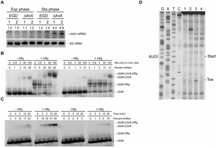
mRNA. To investigate this in more detail, we performed a Northern blot analysis to evaluate the expression ofchiAmRNA in wild type andDlhrAcells during growth in rich medium. As shown in Figure 4A, the expression ofchiAis very low in the wild type strain, irrespective of the growth phase tested. Likewise, the expression of chiA in the DlhrA mutant strain is minimal in exponentially growing cells, however, at the entry into stationary phase, the level ofchiAmRNA increases (Figure 4A). Thus, LhrA indeed has a negative affect the level ofchiAmRNA.
LhrA shows extensive complementarity to the translation initiation region the 59-end ofchiAmRNA (see Table 1). To test whether LhrA binds to this region in the chiA mRNA, we performed gel mobility shift assays (Figure 4B). In these experiments, a 59-end-labelledin vitro-transcribedchiARNA was mixed with different concentrations of wild type LhrA or mutant
LhrA-Mut3* carrying four nucleotide substitutions within the region predicted to interact withchiARNA (see Table 1 and [23] for details). The binding experiments were performed in the absence or presence of Hfq. In the absence of Hfq, less than half of thechiARNA had shifted at the highest concentration of wild type LhrA, whereas in the presence of Hfq, almost all of thechiARNA had shifted (Figure 4B, left panel). When adding LhrA-Mut3*, RNA duplex formation was clearly diminished, both in the absence and presence of Hfq (Figure 4B, right panel). These results show that LhrA interacts with the 59-end of thechiAmRNA, and that Hfq promotes the formation of an LhrA-chiARNA duplex. To further investigate this issue, we conducted a time course experiment in which a 59-end-labelled chiA RNA fragment was mixed with 5-fold excess LhrA or LhrA-Mut3*, in the absence or presence of Hfq (Figure 4C). The results clearly show that Hfq
Figure 4. LhrA specifically targets thechiAmRNA.(A) Northern blot analysis ofchiAexpression in wild type andDlhrAmutant cells grown in BHI medium. The results from two independent experiments (1 and 2) are shown. Samples were drawn in the mid-exponential growth phase and in the early stationary growth phase. Northern blots were quantified using ImageQuant by measuring the amount of radioactivity in each band. Numbers given are relative to the amount found in the EGD wild type strain (exponential phase, experiment 1). Relative expression levels were corrected with respect to the levels of 5S rRNA. (B)In vitrobinding assay of LhrA andchiARNA in the absence (2) or presence (+) of Hfq. In all
samples, end-labelledin vitrotranscribedchiARNA was used, andin vitrotranscribed LhrA (left part) or LhrA-Mut3* (right part) was added to the indicated final concentrations. The experiment was repeated twice with similar results. Quantification of the gelshifts was carried out using ImageQuant by measuring the amount of RNA that had shifted in each lane. The numbers presented in the figure are relative to the total amount of unshifted RNA found in the first lane, from the left. (C) Time course experiment showing the association ofchiARNA with LhrA (left part) or LhrA-Mut3* (right part). The experiments were carried out with end-labelledin vitrotranscribedchiARNA mixed within vitrotranscribed LhrA or LhrA-Mut3*, in the absence (2) or presence (+) of Hfq. The samples were incubated at 37uC for 0, 2, 5, 10 or 20 minutes and then loaded onto a gel. The experiment was repeated twice with similar results. Quantification of the bands was carried out as described for 4B. (D) Toeprint experiment ofchiA RNA in the absence or presence of LhrA. Lanes 1 and 2 correspond to control reactions containingchiARNA only, in the absence (lane 1) or presence (lane 2) of 30S ribosomes. Lane 3: In the presence ofchiARNA, 30S ribosomes and tRNAfMet, a specific toeprint is generated approximately 18 nucleotides downstream from thechiAstart codon (AUG). Lane 4: The addition of LhrA diminishes the formation of thechiAtoeprint signal. The experiment was repeated twice with similar results.
stimulates the rate of association between LhrA and chiARNA, and that substitutions in LhrA predicted to disrupt this interaction diminishes RNA duplex formation.
Since LhrA binds to a region overlapping the start codon on the chiAmRNA (see Table 1), we performed a toeprint experiment to investigate the effect of LhrA on the formation of a translation initiation complex. In the presence of both 30S and tRNAfMeta distinct toeprint signal was observed downstream from the start codon (Figure 4D, lane 3). The formation of the toeprint was strongly inhibited by the addition of LhrA RNA (Figure 4D, lane 4), showing that binding of LhrA tochiAmRNA efficiently blocks the formation of a translation initiation complex.
LhrA and Hfq affect the chitinolytic activity ofL. monocytogenes
L. monocytogenescontains two genes encoding chitinases,chiAand chiB, and a single gene (lmo2467) encoding a putative chitin binding protein [24]. The chitinase activity of L. monocytogenes EGD-e has been shown to depend on bothchiAandchiB, and both chitinases contribute to growth in the livers and spleens of mice, with chiA playing the most significant role in L. monocytogenes virulence [26]. Expression of the chitinase genes is induced in the presence of chitin [34]. In order to study the effect of LhrA onchiA andchiBexpression under conditions known to stimulate chitinase activity, we performed a Northern blot on RNA isolated from late exponential and early stationary phase cells grown in LB medium containing chitin (Figure 5A, lanes 7–12). In agreement with the results presented previously [34] the chiA and chiB mRNAs are readily detected in the wild type strain in the presence of chitin. We note that chiA expression is higher in the DlhrA and Dhfq mutant strains in comparison to the wild type strain, in particular in the early stationary phase cells (Figure 5A, lanes 7–9, lower band), whereas the effect of Hfq and LhrA on chiB expression appears to be minimal (upper band). These results demonstrate that LhrA and Hfq act to repress the expression ofchiA, also in the presence of the substrate of the chitinases.
In order to study the contribution of LhrA and Hfq to the chitinolytic activity ofL. monocytogenes, bacterial suspensions of the wild type,DlhrAandDhfqmutant strains were spotted on LB agar plates supplemented with chitin (Figure 5B). We observed that the chitinolytic activity of the DlhrA mutant strain is slightly higher compared to that of the wild type strain. The average zone size of the wild type strain (1.5 mm, SD = 0.4) was found to be significantly smaller (P,0.01) compared to the average zone size of theDlhrAmutant strain (1.9 mm, SD = 0.4). This result shows that the regulatory effect of LhrA onchiAindeed serves to decrease the chitinolytic activity ofL. monocytogenes. Surprisingly, we found that the clearing zone for theDhfqmutant was markedly reduced when compared to the wild type strain (Figure 5B). Since Hfq acts to down-regulate the chiA levels in the presence of chitin (Figure 5A) we expected Hfq to display a negative effect on the chitinolytic activity to approximately the same extent as LhrA. In contrast to our expectations, the average zone diameter of theDhfq mutant (0.9 mm, SD = 0.3) was significantly smaller (P,0.01) compared to that of the wild type strain. Thus, it appears that Hfq has a stimulating effect on the chitinolytic activity of L. monocytogenes, which is not reflected at the RNA level.
Discussion
In the present work we studied the role of the Hfq-binding sRNA LhrA in L. monocytogenes. We demonstrate that LhrA is expressed throughout growth in rich medium from a highly active promoter, and that LhrA is more stable in stationary phase cells in
comparison to exponentially growing cells. Furthermore, we show that LhrA affects the expression of approximately 300 genes in early stationary phase cells ($1.5 fold; adjusted P,0.05), and we provide evidence that two additional genes,lmo0302andchiA, are a direct target for LhrA, extending the regulatory capacity of LhrA to multiple target genes.
To identify genes affected by LhrA, we compared the total gene expression of aDlhrAmutant and wild type strain by microarray analysis. These types of analyses are often complicated by secondary effects resulting from regulation of primary targets. Furthermore, deletion oflhrA, which is one of the most abundant RNAs in stationary phase cells [12], may have indirect effects on other Hfq-dependent processes inL. monocytogenes. We also note that several genes encoding regulatory proteins were among those differentially expressed in theDlhrA strain (Table S1 and Table S2), and that some of the observed differences could potentially be due to differences in the regulation of entry into stationary phase between the strains tested. Thus, the LhrA-mediated regulation of 284 genes inL. monocytogenesis most likely the result of both direct and indirect effects. In order to find genes directly targeted by LhrA, we searched the 59-region of putative mRNAs encoded by the genes down-regulated at least 2 fold by LhrA for potential RNA-duplex formation. By this strategy, we expected to find mRNAs that interact with LhrA, leading to repression of translation initiation and degradation of the mRNA. Further analyses of the top three candidates confirmed that LhrA down-regulates the expression of the lmo0302-lmo0303 operon and of chiA. Usingin vivoandin vitroanalyses, we showed that LhrA acts to prevent the formation of a translation initiation complex at the 59 -end of the lmo0302-lmo0303 and chiA mRNAs, resulting in a decrease in the mRNA levels. We observed that the expression of these genes is affected by the RNA chaperone Hfq as well, and that Hfq stimulates the base-pairing between LhrA and chiA mRNA.
Some Hfq-binding sRNAs act by binding to the coding region of mRNAs, as exemplified by MicC in Salmonella [35], or by binding to the 59-region more upstream from the translational start site, resulting in activation of translation, as shown for DsrA inE. coli[36,37]. We therefore searched within the coding region as well as the far 59-upstream regions of genes affected at least 2-fold by LhrA for potential base-pairing with LhrA, but no obvious targets were identified. Curiously, we note that approximately half of the genes identified in the microarray analysis were found in other studies to belong to thesB
regulon, suggesting a regulatory link between LhrA and the alternative stress sigma factorsB. In L. monocytogenes,sBplays an important role in stress tolerance and virulence, and several sRNA-encoding genes are known to depend on sB
for their expression, including sbrA [14] and sbrE (rli47) [12,13], but the level of LhrA is not affected by sB
(our unpublished data). Further studies will be needed in order to clarify the extensive regulatory networks involvingsBand sRNAs inL. monocytogenes.
pneumophila, a chitinase was shown to promote bacterial persistence in the lung [41]. In L. monocytogenes, the chitin-binding protein Lmo2467 and the chitinases ChiA and ChiB contribute to pathogenesis in mice, but appeared not to influence bacterial invasion or replication within selected mammalian cell lines [26]. The expression of chiA andchiB is induced by the presence of chitin and depends on at least two regulatory proteins: The central virulence regulator PrfA and the alternative stress sigma factorsB[34]. Furthermore, glucose has a negative effect onchiAandchiBexpression, suggesting that the chitinases are subject to catabolite repression [34]. We show that LhrA acts as a post-transcriptional regulator of chiA thus adding another layer of complexity to the gene regulatory networks controlling the expression of chitinases in L. moncoy-togenes. We have shown that LhrA acts to down-regulate the
expression of chiA by an antisense-mechanism. However, the environmental signal and molecular mechanism leading to alleviation of LhrA repression, remains to be determined. We speculate that under specific growth conditions, the LhrA level may decrease via repression of transcription oflhrA, and/or by removal of cellular LhrA by degradation. Interestingly, the utilization of chito-sugars by E. coli was recently shown to involve a small Hfq-dependent sRNA named MicM [42], and a similar sRNA, ChiX, was characterized in Salmonella [43]. Under normal growth conditions, MicM down-regulates its target geneybfM, encoding a chito-sugar porin. In the presence of chitobiose, an RNA trap is produced from the chitobiose operon. The RNA trap binds to MicM, leading to MicM degradation and alleviation of repression of the YbfM chito-sugar porin [42,43]. It is tempting to speculate that a similar
Figure 5. The effect of LhrA and Hfq onchiAandchiB.(A) Northern blot showing the levels ofchiAandchiBmRNA in wild type (lanes 1, 4, 7, 10),DlhrAmutant (lanes 2, 5, 8, 11) andDhfqmutant cells (lanes 3, 6, 9, 12) grown in LB medium (lanes 1–6) or LB medium supplemented with chitin (lanes 7–12). Cells were harvested in the late exponential growth phase (lanes 1–3 and 7–9) or early stationary growth phase (lanes 4–6 and 10–12). The experiment was repeated twice with similar results. As loading control, we probed for 16S rRNA. Northern blots were quantified using ImageQuant by measuring the amount of radioactivity in each band. Numbers given are relative to the amount found in the EGD wild type strain grown to exponential phase in the presence of chitin (lane 7). Relative expression levels were corrected with respect to the levels of 16S rRNA. (B) Comparison of the chitinolytic activity of wild type,DlhrAandDhfqmutant cells on agar plates containing chitin. Similar results were observed in ten independent experiments.
mechanism could apply to the LhrA-chiAregulatory case, but if so, what is the potential signal leading to alleviation of LhrA repression? In the presence of chitin, the expression ofchiA is clearly down-regulated by LhrA, so the putative signal does not appear to be linked directly to the substrate (chitin) or its degradation products. Although both ChiA and ChiB contribute to the chitinolytic activity of L. monocytogenes, the full range of their potential substrates remains to be determined, including the identification of host-related targets. Since onlychiA, and not chiB, is targeted by LhrA, we speculate that LhrA-regulation could be linked to a ChiA-specific substrate in the external environment, or possibly during infection, which would require differential regulation of the two genes. This hypothesis is supported by recent findings showing that a Salmonella Typhimurium chitinase shows activity towards a N-acetyllacto-samine-conjugate which is a model substrate to LAcNAc terminating glycoproteins and glycolipids on vertebrate cells [44]. Future work will focus on defining the mechanism leading to de-repression of LhrA-regulated genes.
The role of LhrA as an antisense regulator of the expression oflmo0850 [26],lmo0302-lmo0303 and chiA is closely linked to the RNA chaperone Hfq. The stability and function of LhrA depends on Hfq and the levels of all three target mRNAs are diminished in the presence of Hfq. The formation of an RNA duplex between LhrA and target mRNA is clearly stimulated by the Hfq protein, as shown forlmo0850[23] andchiAmRNA (this study). We were therefore surprised to find that theDhfqmutant was less chitinolytic in comparison to wild type strain. Importantly, this stimulatory effect of Hfq on the chitinolytic activity of L. monocytogeneswas not reflected at mRNA level of chiAorchiB. This result points to a more complex role of Hfq in L. monocytogenesand suggests that Hfq exerts a stimulating effect on the protein level, activity and/or secretion of the chitinases by a mechanism that may involve the action of other Hfq-dependent sRNAs inL. monocytogenes.
Materials and Methods
Bacterials strains and growth media
Listeria monocytogenesEGD-e seroptype 1/2a was used as the wild type strain; construction of the isogenic mutant derivatives Dhfq and DlhrA were described in previous work [16,23]. The DresD mutant strain was described previously [27]. All L. monocytogenes strains were grown in either Brain Heart Infusion media (BHI, Oxoid), at 37uC, or Luria broth (LB, Oxoid), at 30uC. The effect of chitin was examined by supplementing LB with acid-hydrolyzed chitin (2.5 g/L) (catalog nr. C9213; Sigma-Aldrich). Acid-hydrolyzed/colloidal chitin was prepared as described previously [34]. When appropriate, cultures were supplemented with Kanamycin (50mg/ml). For cloning purposes, E. coli TOP10 (Invitrogen) grown in LB medium was used.
Construction of lacZ fusions and b-galactosidase assays For analysis of the lhrA promoter activity, DNA fragments corresponding to various lengths of regions upstream oflhrAwere constructed by PCR using different LhrA forward primers in combination with the reverse primer sRNA1-13, listed in Table S3. The resulting PCR fragments were digested with EcoRI and BamHI and ligated into the low-copy number promoter-lesslacZ transcriptional fusion vector pTCV-lac [45]. For the construction of in-frame translationallacZfusions, DNA fragments containing 59-regions oflmo0880orlmo0302were amplified by PCR using the primers listed in Table S3. The resulting PCR fragments were digested with EcoRI and BamHI and ligated into pCK-lac, a
derivative of pTCV-lac containing alacZ gene without a Shine-Dalgarno sequence or start codon allowing for translational analysis of the gene in question. For construction of a transcriptional fusion between lmo0302 and lacZ, the lmo0302 PCR fragment digested with EcoRI and BamHI was ligated into pTCV-lac. b-galactosidase assay was carried out as described previously [16].
RNA techniques
RNA used for microarray or TaqMan RT-PCR was purified using the RNeasy mini or midi kit from Qiagen as described by the manufacturer. Cells grown to early-stationary phase were first treated with RNA protect as instructed by the manufacturer (Qiagen) and subsequently disrupted by sonication on ice (3630 seconds, each round followed by a 30 second pause). For primer extension analysis and Northern blotting experiments, total RNA was extracted from L. monocytogenes using TRI reagent (MR CGENE). Cells were disrupted using the FastPrep instrument and RNA was purified as described previously [23]. The integrity of the RNA was confirmed by agarose gel electrophoresis and the concentration and purity was determined on a NanoDrop 2000.
Northern blotting and primer extension analysis on total RNA purified from cells grown in BHI medium was performed as previously described [23]. The primers used as probes for Northern blotting on LhrA, 5S RNA,lmo0302,lmo0303andchiA, and primers used for primer extension analysis of lhrA-lacZ, lmo0302andchiA, are listed in Table S3. Northern blotting analysis of chiAand chiB on total RNA purified from cells grown in LB medium, with or without chitin, was performed at described previously [34].
For gel shift experiments, the template forin vitrotranscription of chiARNA was prepared by PCR using the primers listed in Table S3. The 59-end corresponds to the putative transcription start site from asBdependent promoter, located 50 base pairs upstream of the start codon [33]. Templates forin vitrotranscription of LhrA and LhrA-Mut3* were prepared as described previously [23]. In each case, the 59-end primer contains a T7-RNA Polymerase binding site for subsequentin vitrotranscription.In vitrotranscribed RNA was prepared using the MegaScript kit from Ambion as described by the manufacturer. Following transcription, the RNA was separated on a denaturing polyacrylamide gel and the largest transcript (identified by UV shadowing) was excised from the gel and subsequently purified by electro-elution followed by phenol-chloroform extraction. RNA to be used for 59-end labeling was dephosphorylated using the KinaseMax kit from Ambion as described by the manufacturer. The purity and concentration ofin vitrotranscribed RNA was determined using a NanoDrop 2000. Gelshifts were conducted as previously described [23]. Briefly, 40 fmol 59-end labeled chiARNA was incubated in a total of 10ml
without or with 0.8, 4, 20 or 100 nM unlabelled LhrA or LhrA-Mut3* in the absence or presence of 2.5mM Hfq and 10mg of non-specific tRNA. The samples were incubated 20 min at 37uC followed by 10 min on ice and subsequently separated on a 5% non-denaturing polyacrylamide gel at 4uC with the current running. For time-course experiments, 40 fmol 59-end labeled chiARNA was mixed with 20 nM LhrA in the presence or absence of 2.5mM Hfq and incubated at 37uC for 0, 2, 5, 10 or 20
minutes. The samples were then loaded onto a 5% non-denaturing polyacrylamide gel at 4uC with the current running.
For toeprinting experiments,in vitrotranscribedlmo0302RNA andchiARNA was prepared using the primers listed in Table S3. Toeprinting experiments were performed as described in [23] using 0.35mM lmo0302 RNA or 0.05mM chiA RNA; 10 fold
shift experiments), relative tolmo0302orchiARNA, and 0.4 pmol of 59-end labelledlmo0302orchiAprimer (see Table S3).
Quantitative RT-PCR was essentially performed as described previously [46]. Briefly, TaqMan primers and probes (Table S3) were designed using Primer Express 2.0 software (Applied Biosystems). qRT-PCR was performed using TaqMan one-step RT-PCR master mix reagent, Multiscribe RT, and an ABI Prism 7000 sequence detection system (Applied Biosystems). Each qRT-PCR experiment was run in triplicate. The housekeeping genes rpoB and gapwere used for normalization of absolute transcript levels. Data analysis was conducted with ABI Prism 7000 SDS software. Significant differences in RNA levels were determined by ANOVA as described previously [46].
cDNA labeling and microarray hybridization
RNA was extracted from wild type andDlhrAcells grown in BHI medium at 37uC to early stationary phase (OD600= 1.0+3 -hours) as described above. The experiment was conducted with four biological replicates which were compared in pairs on four microarray slides. cDNA from each strain was labeled twice with Cy3 and twice with Cy5 to minimize any bias. cDNA labeling was performed as previously described [46]. Briefly, cDNA synthesis and labeling of total RNA were performed using the SuperScript Plus indirect cDNA labeling system for DNA microarrays (Invitrogen). 10mg total RNA was mixed with 5mg random hexamers and incubated for 10 minutes at 70uC, with a subsequent chill on ice for at least 5 minutes. Superscript III RT, amino-modified deoxynucleoside triphophates, dithiothreitol, RNaseOUT, and buffer was then added and the reaction mix incubated at 42uC for 17 hours. RNA was hydrolyzed by the addition of 10ml 1 M NaOH and 10ml 0.5 M EDTA, followed by incubation at 65uC for 15 minutes. The mixture was neutralized with 10ml 1 M HCl and cDNA purified using the
Qiagen PCR purification kit. Labeling reactions with Alexa Fluor 555 or Alexa Fluor 647 fluorescent dyes were performed for 2 h at room temperature. Differentially labeled cDNAs from the two strains to be cohybridized were combined, dried in a Savant SVC100 Speed-Vac (Farmingdale) and stored at 280uC until hybridization.
Microarrays were constructed as previously described [47]. Briefly, 70-mer probes targeting 2,857L. monocytogenesORFs were spotted onto Corning UltraGAPS slides (Corning Inc) at the Microarray Core Facility at Cornell University.
Microarray hybridization was performed as described previous-ly [46]. Spotted microarray slides were first incubated for 1 h in a 1% bovine serum albumin-5X SSC-0.1% sodium dodecyl sulfate solution pre-warmed to 42uC. Subsequently, slides were washed twice in 0.1X SSC and twice in filtered water and then dried. The combined cDNA targets were reconstituted in 55ml hybridization
buffer and denatured at 95uC for 5 minutes. Targets were applied to microarray slides and overlaid with mSeries LifterSlips (Erie Scientific) followed by overnight hybridization at 42uC. Slides were then washed 5 minutes in 42uC pre-warmed 2X SSC plus 0.1% SDS, 5 minutes in 2X SCC, and 2.5 min in 0.2X SSC. After a final wash in filtered water, slides were dried and scanned with a GenePix 4000B scanner (Molecular Devices, Sunnyvale, CA).
Microarray data analysis
The median fluorescence intensity data for all probes on the array were analyzed using LIMMA (linear models for microarray analysis)[48]. The Empirical Bayes method employed in LIMMA is used to borrow information across genes resulting in stable analyses of small samples. This method also allow analysis of datasets when values are missing due to low spot quality parameters [48]. We first normalized the data on each array using print-tip Lowess, followed by log2 conversion of the normalized data. A correlation was determined for the signal from duplicate spots of each probe. Significant differences were determined by calculating a moderated t-statistic which is similar to an ordinary t-statistic except that the standard errors have been shrunk towards a common value using a Bayesian model. P-values were calculated for each gene based on the moderated t-statistics and adjusted with the Benjamini-Hochberg false discovery rate correction for multiple tests. Differences in transcripts levels were considered meaningful only when adjusted P,0.05 and fold change$1.5 fold.
Gene set enrichment analysis (GSEA) [48] was used to identify gene sets that were significantly overrepresented among genes up-or down-regulated in theDlhrAmutant strain. GSEA was carried out as described in [49].
Examination of chitinase activity
The chitinase activities of L. monocytogenes was measured as previously described [34]. Wild type and mutant strains were spotted on the same chitin agar plate. After incubation, the clearing zone diameter was measured. Pair wise t-test was used for comparisons between the size of the clearing zones of the wild type and each mutant after 4 days of incubation. Zone diameters from ten independent experiments were compared.
Supporting Information
Figure S1 Verification of microarray data by qRT-PCR. See Table S1 and Table S2 for more details on the genes tested.
(TIF)
Table S1 Genes positively affected by LhrA.
(DOCX)
Table S2 Genes negatively affected by LhrA.
(DOCX)
Table S3 Primers and TaqMan probes used in this study.
(DOCX)
Author Contributions
Conceived and designed the experiments: JSN MHL TMB KJB MW BHK. Performed the experiments: JSN MHL EMSL TMB MHGC. Analyzed the data: JSN MHL EMSL TMB MHGC KJB MW BHK. Contributed reagents/materials/analysis tools: KJB MW BHK. Wrote the paper: JSN MHL TMB BHK.
References
1. Toledo-Arana A, Repoila F, Cossart P (2007) Small noncoding RNAs controlling pathogenesis. Curr Opin Microbiol 10: 182–188. S1369-5274(07)00021-5 [pii];10.1016/j.mib.2007.03.004 [doi].
2. Papenfort K, Vogel J (2010) Regulatory RNA in bacterial pathogens. Cell Host Microbe 8: 116–127. S1931-3128(10)00211-8 [pii];10.1016/j.chom.2010.06.008 [doi].
3. Masse E, Salvail H, Desnoyers G, Arguin M (2007) Small RNAs controlling iron metabolism. Curr Opin Microbiol 10: 140–145. S1369-5274(07)00030-6 [pii];10.1016/j.mib.2007.03.013 [doi].
5. Gorke B, Vogel J (2008) Noncoding RNA control of the making and breaking of sugars. Genes Dev 22: 2914–2925. 22/21/2914 [pii];10.1101/gad.1717808 [doi].
6. Waters LS, Storz G (2009) Regulatory RNAs in bacteria. Cell 136: 615–628. 7. Gottesman S, Storz G (2010) Bacterial Small RNA Regulators: Versatile Roles
and Rapidly Evolving Variations. Cold Spring Harb Perspect Biol. cshper-spect.a003798 [pii];10.1101/cshpercshper-spect.a003798 [doi].
8. Frohlich KS, Vogel J (2009) Activation of gene expression by small RNA. Curr Opin Microbiol 12: 674–682. S1369-5274(09)00143-X [pii];10.1016/ j.mib.2009.09.009 [doi].
9. Obana N, Shirahama Y, Abe K, Nakamura K (2010) Stabilization of Clostridium perfringens collagenase mRNA by VR-RNA-dependent cleavage in 59leader sequence. Mol Microbiol 77: 1416–1428. MMI7258 [pii];10.1111/ j.1365-2958.2010.07258.x [doi].
10. Ramirez-Pena E, Trevino J, Liu Z, Perez N, Sumby P (2010) The group A Streptococcus small regulatory RNA FasX enhances streptokinase activity by increasing the stability of the ska mRNA transcript. Mol Microbiol 78: 1332–1347. 10.1111/j.1365-2958.2010.07427.x [doi].
11. Vazquez-Boland JA, Kuhn M, Berche P, Chakraborty T, Dominguez-Bernal G, et al. (2001) Listeria pathogenesis and molecular virulence determinants. Clin Microbiol Rev 14: 584–640. 10.1128/CMR.14.3.584-640.2001 [doi]. 12. Oliver HF, Orsi RH, Ponnala L, Keich U, Wang W, et al. (2009) Deep RNA
sequencing of L. monocytogenes reveals overlapping and extensive stationary phase and sigma B-dependent transcriptomes, including multiple highly transcribed noncoding RNAs. BMC Genomics 10: 641. 1471-2164-10-641 [pii];10.1186/1471-2164-10-641 [doi].
13. Toledo-Arana A, Dussurget O, Nikitas G, Sesto N, Guet-Revillet H, et al. (2009) The Listeria transcriptional landscape from saprophytism to virulence. Nature 459: 950–956.
14. Nielsen JS, Olsen AS, Bonde M, Valentin-Hansen P, Kallipolitis BH (2008) Identification of a sigma B-dependent small noncoding RNA in Listeria monocytogenes. J Bacteriol 190: 6264–6270.
15. Christiansen JK, Nielsen JS, Ebersbach T, Valentin-Hansen P, Sogaard-Andersen L, et al. (2006) Identification of small Hfq-binding RNAs in Listeria monocytogenes. RNA 12: 1383–1396.
16. Christiansen JK, Larsen MH, Ingmer H, Sogaard-Andersen L, Kallipolitis BH (2004) The RNA-binding protein Hfq of Listeria monocytogenes: role in stress tolerance and virulence. J Bacteriol 186: 3355–3362.
17. Sittka A, Pfeiffer V, Tedin K, Vogel J (2007) The RNA chaperone Hfq is essential for the virulence of Salmonella typhimurium. Mol Microbiol 63: 193–217. MMI5489 [pii];10.1111/j.1365-2958.2006.05489.x [doi].
18. Ding Y, Davis BM, Waldor MK (2004) Hfq is essential for Vibrio cholerae virulence and downregulates sigma expression. Mol Microbiol 53: 345–354. 10.1111/j.1365-2958.2004.04142.x [doi];MMI4142 [pii].
19. Sonnleitner E, Hagens S, Rosenau F, Wilhelm S, Habel A, et al. (2003) Reduced virulence of a hfq mutant of Pseudomonas aeruginosa O1. Microb Pathog 35: 217–228. S0882401003001499 [pii].
20. Meibom KL, Forslund AL, Kuoppa K, Alkhuder K, Dubail I, et al. (2009) Hfq, a novel pleiotropic regulator of virulence-associated genes in Francisella tularensis. Infect Immun 77: 1866–1880. IAI.01496-08 [pii];10.1128/ IAI.01496-08 [doi].
21. Valentin-Hansen P, Eriksen M, Udesen C (2004) The bacterial Sm-like protein Hfq: a key player in RNA transactions. Mol Microbiol 51: 1525–1533. 22. Aiba H (2007) Mechanism of RNA silencing by Hfq-binding small RNAs. Curr
Opin Microbiol 10: 134–139.
23. Nielsen JS, Lei LK, Ebersbach T, Olsen AS, Klitgaard JK, et al. (2010) Defining a role for Hfq in Gram-positive bacteria: evidence for Hfq-dependent antisense regulation in Listeria monocytogenes. Nucleic Acids Res 38: 907–919. gkp1081 [pii];10.1093/nar/gkp1081 [doi].
24. Leisner JJ, Larsen MH, Jorgensen RL, Brondsted L, Thomsen LE, Ingmer H (2008) Chitin hydrolysis by Listeria spp., including L. monocytogenes. Appl Environ Microbiol 74: 3823–3830. AEM.02701-07 [pii];10.1128/AEM.02701-07 [doi].
25. Leisner JJ, Larsen MH, Ingmer H, Petersen BO, Duus JO, et al. (2009) Cloning and comparison of phylogenetically related chitinases from Listeria monocyto-genes EGD and Enterococcus faecalis V583. J Appl Microbiol 107: 2080–2087. JAM4420 [pii];10.1111/j.1365-2672.2009.04420.x [doi].
26. Chaudhuri S, Bruno JC, Alonzo F, III, Xayarath B, Cianciotto NP, et al. (2010) Chitinases contributes to Listeria monocytogenes pathogenesis. Appl Environ Microbiol;AEM.01338-10 [pii];10.1128/AEM.01338-10 [doi].
27. Larsen MH, Kallipolitis BH, Christiansen JK, Olsen JE, Ingmer H (2006) The response regulator ResD modulates virulence gene expression in response to carbohydrates in Listeria monocytogenes. Mol Microbiol 61: 1622–1635. MMI5328 [pii];10.1111/j.1365-2958.2006.05328.x [doi].
28. Caramori T, Galizzi A (1998) The UP element of the promoter for the flagellin gene, hag, stimulates transcription from both SigD- and SigA-dependent promoters in Bacillus subtilis. Mol Gen Genet 258: 385–388.
29. Meijer WJ, Salas M (2004) Relevance of UP elements for three strong Bacillus subtilis phage phi29 promoters. Nucleic Acids Res 32: 1166–1176. 10.1093/ nar/gkh290 [doi];32/3/1166 [pii].
30. Estrem ST, Gaal T, Ross W, Gourse RL (1998) Identification of an UP element consensus sequence for bacterial promoters. Proc Natl Acad Sci U S A 95: 9761–9766.
31. Ross W, Gosink KK, Salomon J, Igarashi K, Zou C, et al. (1993) A third recognition element in bacterial promoters: DNA binding by the alpha subunit of RNA polymerase. Science 262: 1407–1413.
32. Milner MG, Saunders JR, McCarthy AJ (2001) Relationship between nucleic acid ratios and growth in Listeria monocytogenes. Microbiology 147: 2689–2696.
33. Kazmierczak MJ, Mithoe SC, Boor KJ, Wiedmann M (2003) Listeria monocytogenes sigma B regulates stress response and virulence functions. J Bacteriol 185: 5722–5734.
34. Larsen MH, Leisner JJ, Ingmer H (2010) The chitinolytic activity of Listeria monocytogenes EGD is regulated by carbohydrates but also by the virulence regulator PrfA. Appl Environ Microbiol 76: 6470–6476. AEM.00297-10 [pii];10.1128/AEM.00297-10 [doi].
35. Pfeiffer V, Papenfort K, Lucchini S, Hinton JC, Vogel J (2009) Coding sequence targeting by MicC RNA reveals bacterial mRNA silencing downstream of translational initiation. Nat Struct Mol Biol 16: 840–846.
36. Majdalani N, Cunning C, Sledjeski D, Elliott T, Gottesman S (1998) DsrA RNA regulates translation of RpoS message by an anti-antisense mechanism, independent of its action as an antisilencer of transcription. Proc Natl Acad Sci U S A 95: 12462–12467.
37. Lease RA, Cusick ME, Belfort M (1998) Riboregulation in Escherichia coli: DsrA RNA acts by RNA:RNA interactions at multiple loci. Proc Natl Acad Sci U S A 95: 12456–12461.
38. Gooday GW (1990) The ecology of chitin degradation. In: Marshall KC, ed. Advances in Microbial Ecology. New York: Plenum Press. pp 387–430. 39. Bhowmick R, Ghosal A, Das B, Koley H, Saha DR, et al. (2008) Intestinal
adherence of Vibrio cholerae involves a coordinated interaction between colonization factor GbpA and mucin. Infect Immun 76: 4968–4977. IAI.01615-07 [pii];10.1128/IAI.01615-IAI.01615-07 [doi].
40. Kirn TJ, Jude BA, Taylor RK (2005) A colonization factor links Vibrio cholerae environmental survival and human infection. Nature 438: 863–866. na-ture04249 [pii];10.1038/nana-ture04249 [doi].
41. DebRoy S, Dao J, Soderberg M, Rossier O, Cianciotto NP (2006) Legionella pneumophila type II secretome reveals unique exoproteins and a chitinase that promotes bacterial persistence in the lung. Proc Natl Acad Sci U S A 103: 19146–19151. 0608279103 [pii];10.1073/pnas.0608279103 [doi].
42. Overgaard M, Johansen J, Moller-Jensen J, Valentin-Hansen P (2009) Switching off small RNA regulation with trap-mRNA. Mol Microbiol 73: 790–800. MMI6807 [pii];10.1111/j.1365-2958.2009.06807.x [doi].
43. Figueroa-Bossi N, Valentini M, Malleret L, Fiorini F, Bossi L (2009) Caught at its own game: regulatory small RNA inactivated by an inducible transcript mimicking its target. Genes Dev 23: 2004–2015. gad.541609 [pii];10.1101/ gad.541609 [doi].
44. Larsen T, Petersen BO, Storgaard BG, Duus JO, Palcic MM, et al. (2010) Characterization of a novel Salmonella Typhimurium chitinase which hydrolyzes chitin, chitooligosaccharides and a N-acetyllactosamine-conjugate. Glycobiology;cwq174 [pii];10.1093/glycob/cwq174 [doi].
45. Poyart C, Trieu-Cuot P (1997) A broad-host-range mobilizable shuttle vector for the construction of transcriptional fusions to beta-galactosidase in gram-positive bacteria. FEMS Microbiol Lett 156: 193–198.
46. Ollinger J, Bowen B, Wiedmann M, Boor KJ, Bergholz TM (2009) Listeria monocytogenes sigmaB modulates PrfA-mediated virulence factor expression. Infect Immun 77: 2113–2124. IAI.01205-08 [pii];10.1128/IAI.01205-08 [doi]. 47. Chan YC, Raengpradub S, Boor KJ, Wiedmann M (2007) Microarray-based characterization of the Listeria monocytogenes cold regulon in log- and stationary-phase cells. Appl Environ Microbiol 73: 6484–6498. AEM.00897-07 [pii];10.1128/AEM.00897-07 [doi].
48. Smyth GK (2004) Linear models and empirical bayes methods for assessing differential expression in microarray experiments. Stat Appl Genet Mol Biol 3: Article3. 10.2202/1544-6115.1027 [doi].
49. Chaturongakul S, Raengpradub S, Palmer ME, Bergholz TM, Orsi RH, et al. (2011) Transcriptomic and phenotypic analyses identify coregulated, overlap-ping regulons among PrfA, CtsR, HrcA, and the alternative sigma factors sigmaB, sigmaC, sigmaH, and sigmaL in Listeria monocytogenes. Appl Environ Microbiol 77: 187–200. AEM.00952-10 [pii];10.1128/AEM.00952-10 [doi]. 50. Sierro N, Makita Y, de HM, Nakai K (2008) DBTBS: a database of