Vaccine Inhibits the Early Growth of
Mycobacterium
tuberculosis
After Infection
Edward O. Ronan, Lian Ni Lee, Peter C. L. Beverley, Elma Z. Tchilian*
Nuffield Department of Medicine, University of Oxford, Oxford, United Kingdom
Abstract
Background:In pulmonaryMycobacterium tuberculosis (Mtb)infection, immune responses are delayed compared to other respiratory infections, so that antigen-specific cells are not detected in the lungs earlier than day 14. Even after parenteral immunization with Bacille Calmette Guerin (BCG) or a subunit vaccine, the immune response afterMtbchallenge is only slightly accelerated and the kinetics of pulmonaryMtbgrowth do not differ between naı¨ve and immunized animals up to day 14.
Methods and Findings: Mice were immunized intranasally with a recombinant adenovirus expressing mycobacterial antigen 85A (Ad85A), challenged by aerosol withMtband the kinetics ofMtbgrowth in the lungs measured. Intranasal immunization with Ad85A inhibitsMtbgrowth in the early phase of infection, up to day 8. Protection is sustained for at least 7 months and correlates with the presence of antigen-specific activated effector CD8 T cells in the lungs. Antigen 85A-specific T cells respond to antigen presenting cells from the lungs of mice immunized with Ad85A 23 weeks previously, demonstrating the persistence of antigen in the lungs.
Conclusions/Significance:Intranasal immunization with Ad85A can inhibit early growth ofMtbbecause it establishes a lung antigen depot and maintains an activated lung-resident lymphocyte population. We propose that an optimal immunization strategy for tuberculosis should aim to induce both lung and systemic immunity, targeting the early and late phases ofMtb growth.
Citation:Ronan EO, Lee LN, Beverley PCL, Tchilian EZ (2009) Immunization of Mice with a Recombinant Adenovirus Vaccine Inhibits the Early Growth of Mycobacterium tuberculosisAfter Infection. PLoS ONE 4(12): e8235. doi:10.1371/journal.pone.0008235
Editor:Derya Unutmaz, New York University, United States of America
ReceivedOctober 13, 2009;AcceptedNovember 17, 2009;PublishedDecember 9, 2009
Copyright:ß2009 Ronan et al. This is an open-access article distributed under the terms of the Creative Commons Attribution License, which permits unrestricted use, distribution, and reproduction in any medium, provided the original author and source are credited.
Funding:This work was funded by a UK Medical Research Council (www.mrc.ac.uk) Grant No:G0701235. The funders had no role in study design, data collection and analysis, decision to publish, or preparation of the manuscript.
Competing Interests:The authors have declared that no competing interests exist.
* E-mail: elma.tchilian@ndm.ox.ac.uk
Introduction
Tuberculosis causes 1.7 million deaths per year worldwide and the emergence of HIV-associated mycobacterial infections, as well as an increasing frequency of multi-drug resistant and extensively drug resistantM. tuberculosis (Mtb) isolates, reinforces the need to
develop new control strategies. Immunization with Bacille Calmette Guerin (BCG) confers a variable degree of protection against disseminated disease in the very young but poor protection against pulmonary disease in older age groups. However, because of its partial efficacy, an attractive strategy is to develop vaccines that can be used as boosters following BCG primary immuniza-tion. Because CD4 T cell immunity and IFNchave been shown to be important for immune control ofMtbmany of the new vaccine
candidates aim to induce a Th1 type CD4 response. While some have been shown to induce protective immunity equivalent to BCG when given alone [1–4], these candidates seldom increase convincingly protection over BCG, when they are used as booster vaccines [5–8].
In contrast, recombinant adenoviruses expressingMtb mycolyl
transferase antigen 85A (Ad85A) have shown good protective effects
in several species when given intranasally (i.n.) and also reproducibly increase protection over BCG alone when given as a booster by this route [9–11]. Ad85A induces strong CD8 immune responses [10,12,13] and in mice, protection induced by i.n. immunization has been shown to be associated with the presence of a large population of antigen-specific CD8 T cells in the lungs [14]. This is in line with more recent evidence that CD8 cells make an important contribution to immune protection againstMtb[15–17].
A unique feature of pulmonaryMtbinfection is that the innate
and adaptive immune responses occur much later than in other respiratory infections. In mice, no T cell activation occurs in the mediastinal nodes before 9–10 days post-aerosolMtbinfection and
antigen specific cells are not detected in the lungs earlier than day 14 [15,18,19]. Even in mice immunized with BCG,Mtbitself or a
Here we show that this is the case and mice immunized i.n. with Ad85A suppress mycobacterial growth in the lungs during the first week after Mtb aerosol challenge, in contrast to the delayed
inhibitory effect of parenteral immunization. As protection against early growth ofMtbin the lungs has not been described previously,
we further investigated the nature of the cells present in the lungs at the time of challenge and the duration of this form of protection following immunization.
Results
Ad85A Immunization Targets the Early Phase ofMtb Infection
Parenteral immunization with BCG generates mainly systemic CD4-mediated protective immunity and few antigen-specific cells are found in the airway lumen of the lungs [18,19,24,25]. However, because primary immunization or boosting with Ad85A i.n. induces a large lung-resident CD8 population [10,12–14], we investigated whether this method of immunization affects growth ofMtbearly after challenge.
Mice were immunized with BCG alone or primed with BCG and boosted with Ad85A i.n. 10 weeks later. Four weeks after the last immunization they were challenged with Mtbby the aerosol
route and mycobacterial load was quantitated at early time points. Strikingly, in mice boosted with Ad85A the mycobacterial load in the lungs does not increase up to 8 days after challenge, while naı¨ve and BCG-immunized animals show a steady increase inMtb
CFU from day 3 onward and no difference in lung mycobacterial load until day 21 post-challenge (Fig. 1A). Thus mice boosted with Ad85A i.n. controlMtbby a different mechanism from BCG-only
mice. However, in order to exclude the effect of priming by BCG, we also testedMtbgrowth in animals immunized with Ad85A i.n.
only. We observed similar early inhibition ofMtbCFU, confirming
that immunization with Ad85A i.n. inhibits earlyMtbgrowth, in
contrast to BCG, the effect of which only becomes apparent 21 days afterMtbinfection (Fig. 1B).
Because of the early suppression ofMtbgrowth in the Ad85A
immunized mice, we examined the ability of lung cells from mice immunized with Ad85A i.n. to inhibitMtbgrowth in macrophages in vitro (Fig. 1C). Lung cells from animals immunized i.n. with
Ad85A inhibit growth of Mtb on average by 25.2+/25.2%
compared to lung cells from naive mice (3.1+/26.5%, p = 0.007).
Protection After Intranasal Administration of Ad85A Is Long Lasting
Protective efficacy of tuberculosis booster vaccines is most often tested at 4 to 6 weeks after boosting, at or near the peak of the immune response. However, for a vaccine to be useful it is important that durable protective memory is established. We therefore examined protection againstMtbaerosol challenge at 4
and 24 weeks post-i.n. immunization with Ad85A.
When mice are challenged by aerosol 4 weeks after immuni-zation with Ad85A i.n., Mtblung CFU are reduced by ,1 log
compared to naı¨ve animals, a similar reduction to that in mice immunized with BCG. Ad85A i.n. on its own also reduces theMtb
load in the spleen to a similar extent as BCG only (,2 logs) (Fig. 2B). As previously described, the protective effects of BCG priming followed by an Ad85A i.n. boost are approximately additive in the lungs, although in this experiment no additive effect was seen in the spleen [10,14].
BCG-induced protection is long-lasting in mice, so it was of interest to examine whether this was the case for Ad85A i.n..
Figure 2C shows that the protective effect in lung and spleen afforded by Ad85A on its own is sustained 24 weeks after i.n.
immunization (,1 log in lung and,2 logs in spleen, p = 0.03). A second experiment with Mtb challenge at 30 weeks
post-immunization showed sustained reduction in CFU in BCG-immunized (lung,1 log reduction compared to naive p = 0.004, spleen,1 log p = 0.02) or Ad85A i.n.-immunized mice (lung,0.6 log p = 0.004, spleen,0.6 log p = 0.004). No reduction of CFU was observed when Ad85A was administered i.d. either on its own or as a booster following BCG (Table 1and [14]).
Although we did not test the protective effect of BCG priming and Ad85A i.n. boosting at 24 or 30 weeks, we did so at 14 weeks and the protection afforded is still significantly better than BCG alone (,0.5 log reduction in lung compared to BCG, p = 0.016; spleen ,1.4 log, p = 0.009, data not shown) However, further experiments to establish the durability of the additional protection provided by intranasal boosting with Ad85A need to be performed. Several different methods have been used for pulmonary challenge withMtbin mice and the challenge dose may also affect
the outcome of experiments [10,12,13]. We therefore tested whether protection afforded by i.n. immunization with Ad85A is seen after both low (,200 CFU,Fig. 2) or high dose (880 CFU)
Mtbaerosol challenge (Table 1) or when the mice are challenged
withMtbby the intratracheal or i.n. routes (Table 1). Irrespective of
the method, or dose ofMtbused for pulmonaryMtbchallenge, i.n.
boosting with Ad85A significantly reduces lung and spleen CFU compared to BCG alone, confirming earlier studies [6,9,10,12,13]. These results confirm previous findings that i.n. administration of Ad85A induces robust protection when given on its own and provides additional protection when given as a booster after BCG. We also show that protection is sustained for at least 30 weeks.
Lung Antigen 85A Immune Responses
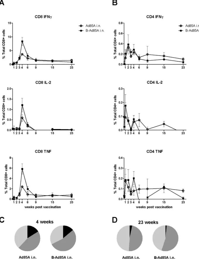
Because protection after i.n. administration of Ad85A correlates with the presence of antigen-specific cells in the lung at 4 weeks [14], we investigated whether these cells are present in the lung later after immunization. We performed intracellular staining for IFNc, IL-2 and TNFaon lung cells stimulated with a pool of peptides covering the whole protein sequence of antigen 85A (Fig. 3A). The peak of the response is 4 weeks after immunization. At this time point mice primed with BCG and boosted with Ad85A i.n. show a greater response than mice immunized with Ad85A i.n. only, implying that immunization with BCG primes for a subsequent booster response to antigen 85A. Previously we observed no priming effect of BCG on the 85A response but here we used BCG SSI as opposed to Pasteur and a different interval between priming and boosting, 8 weeks as opposed to 10 weeks in our previous study [14]. In a separate repeat experiment with a 10 week interval between the prime and boost, no priming effect of BCG on the 85A response was observed, suggesting that the priming effect is weak and inconsistent.
In BCG primed, Ad85A i.n. boosted animals at the peak of the response,,8% of the lung CD8 T cells produce IFNcin response to the pool of 85A peptides. As previously shown many fewer CD8 cells produce IL-2, while TNFaproduction is similar to IFNc[14]. The CD8 response declines from 8% to,1% of lung CD8 cells at 23 weeks. CD4 responses are relatively low and 0.4% and 0.1% of the CD4 cells are antigen-specific at 4 and 23 weeks respectively (Fig. 3B). A low response (0.5% of CD8 cells) is detected in the spleen and peripheral blood following i.n. vaccination.
Because triple cytokine-producing cells have been suggested to be important for protection against some chronic infections, including Mtb [26], we analysed the proportions of single (1+),
No difference in the frequency of cells producing different numbers of cytokines is detected between the prime and prime-boost regimes. Nor was there any change over time of the median fluorescence intensity of staining for intracellular IFNc, IL-2 and TNFain animals immunized with Ad85A i.n. only.
Parenteral i.d. immunization induces a long-lived splenic immune response which lasts for at least 23 weeks. The magnitude and duration of the antigen-specific CD8 response in the spleen of i.d. Ad85A immunized animals is very similar to the lung response in Ad85A i.n. immunized animals, with 3% of splenic antigen-Figure 1. Early control of mycobacterial growth by intranasal administration of Ad85A.BALB/c mice were immunized with BCG or primed with BCG and 10 weeks later boosted with Ad85A i.n. (A) or immunized only with Ad85A i.n. (B) Naı¨ve mice were used as controls. Mice were challenged withMtbby aerosol 4 weeks after the last immunization and sacrificed at days 1, 3, 8, 14, 21 and 28. Lung and spleen CFU were enumerated. Results are expressed as the mean counts of 5–7 mice per group. * p,0.05 Ad85A or B-Ad85A immunized mice versus Naı¨ve or BCG, **p,0.05 for B-Ad85A versus Naı¨ve, BCG versus B-Ad85A, BCG versus Naı¨ve. Similar results were obtained in a repeat experiment. Standard deviations are small, so that the error bars are within the symbols when not visible. (C) Inhibition ofMtbgrowth in macrophages by lung cellsin vitro. Murine peritoneal macrophages were infected withMtband co-cultured with lung cells from naı¨ve mice or mice immunized with Ad85A i.n. 3 weeks previously. Inhibition of mycobacterial growth was determined after 3 days. Results are expressed as the mean % inhibition+/2SEM of four experiments with four mice per group.
specific cells at the peak of the response and 1% at 23 weeks, for both the prime and prime-boost regimes. Despite this, Ad85A i.d. immunization does not reduce mycobacterial load on its own, nor does it further reduce the load when administered as a boost, confirming that the induction of a strong systemic immune response does not correlate with protection in this mouse model [14].
Having shown that i.n. immunization with Ad85A establishes a stable, long-lived protective immune response against infectious challenge, we further characterised the antigen-specific T cells. Antigen-specific IFNc+CD8 cells were identified as effector (CD62L2CD1272), effector memory (CD62L2CD127+) and
central memory cells (CD62L+
CD127+
) [27] (Fig. 4A). In the lung, antigen-specific cells are predominantly effectors, 70% and 52% respectively at 4 and 23 weeks post-immunisation and the
remaining cells are effector memory phenotype (30% and 48% respectively). In contrast, although there were very few antigen-specific cells in extra-pulmonary sites of these i.n. immunised animals, those in the spleen and blood are predominantly effector memory phenotype at 4 and 23 weeks (Fig. 4A).
When mice are immunized i.d. with Ad85A, the splenic and blood antigen-specific cells have the same phenotype as the much smaller number of cells present in these organs after i.n. immunization (Fig. 4A). Therefore irrespective of the route of immunization, blood and splenic antigen-specific cells are less activated than those in the lung, confirming previous observations that cells that home to non-lymphoid tissues display an activated phenotype [28,29].
Because down-regulation of CD27 has been associated with activation and persistent antigenic stimulation, we analysed CD27 Figure 2. Intranasal administration of Ad85A increases protection at 4 and 24 weeks.(A) Time line of the experiment. BALB/c mice were immunized with BCG, or primed with BCG and 10 weeks later boosted intranasally with Ad85A (B-Ad85A i.n.), or immunized only with Ad85A i.n. Naı¨ve mice were used as challenge controls. Mice were challenged withMtbby aerosol 4 (B) or 24 (C) weeks after the boost. Deposition in the lungs was measured 24 h after challenge and was,200 CFU/lung. Mice were sacrificed 6 weeks later and lung and spleen CFU enumerated. Symbols show
CFU counts of individual mice and the horizontal line indicates the mean. * p,0.05 versus Naı¨ve, **p,0.05 versus BCG. Similar results were obtained in two other experiments where the lungs and spleens were harvested at 4 weeks postMtbchallenge.
expression in lung cells [30].Fig. 4Bshows that antigen-specific cells in the lung lack expression of CD27 compared to total CD8 cells, suggesting that the lung might contain antigen 85A, even at 23 weeks post-immunisation.
In summary, persistent antigen-specific responses are detected in the lungs following i.n. administration of Ad85A. Long-term memory in the lungs of i.n. immunised animals is biased towards effector type CD8 T cells producing mainly IFNcand TNFa.
Presence of Antigen 85A in the Lungs
As we detect antigen-specific cells in the lungs at 23 weeks post-immunisation and because they lack CD27 surface expression, we next examined whether antigen is still present in the lungs. We used lung cells isolated 23 weeks post-Ad85A i.n. immunisation as APCs to stimulate T cells isolated from the spleens of animals immunized i.d. with Ad85A (Fig. 4C). The responding T cells were labelled with CFSE and co-cultured with unlabelled lung APCs. As more than 90% of the CD8 antigen-specific cells recognise the dominant H-2Ld antigen 85A peptide 70–78aa (MPVGGQSSF), this tetramer was used to detect antigen-specific cells responding to the lung APCs. After 5 days CFSE intensity of tetramer positive cells was analysed and compared to the same cells cultured with lung APCs from naı¨ve animals. After co-culture with APCs from Ad85A i.n. animals, tetramer binding cells undergo several cell divisions, whereas cells co-cultured with lungs cells from naı¨ve animals do not, indicating that 85A antigen, capable of inducing T cell proliferation, is present in the lungs 23 weeks post-immunization (Fig. 4C). Although we did not include an empty adenovirus control, since only the tetramer positive splenic T cells proliferated and not other activated CD8 cells in the splenic T cell population, it appears that proliferation is antigen specific, rather than a response to inflammatory cytokines.
Discussion
PulmonaryMtbinfection is unlike other respiratory infections
because innate and adaptive immune responses to Mtb are
delayed. T cell activation in the mediastinal nodes only occurs 9–10 days post-infection, while antigen-specific cells are not detected in the lungs until day 14 [18,19]. Although not fully understood, this delay may be due to low antigen dose (50–200 mycobacteria), naturally low inflammatory responses in the lung or active inhibition of antigen presentation byMtb[18]. Whatever
the reasons, a consequence is that mechanisms for promoting entry of systemic antigen-specific cells, such as up-regulation of VCAM-1, are not activated until two weeks post-infection [31]. It is therefore understandable that parenteral immunization against
Mtbprovides no protection against initial growth of mycobacteria
following pulmonary challenge [20–23]. Here we demonstrate for the first time that a protective Ad85A vaccine administered i.n. exerts its effect by suppression of mycobacterial growth during the first week afterMtbinfection, in contrast to the delayed inhibitory
effect of parenteral vaccines.
Ad85A and recombinant adenoviruses containing other anti-gens ofMtbadministered i.n. are highly protective in mice and
induce large populations of lung-resident CD8 T cells [4,9,12,14]. These remain largely confined to the lungs because of endothor-acic recirculation [29]. Even six months after immunization with Ad85A i.n, there is a significant population of antigen-specific cells in the lungs and their presence correlates with protection against
Mtb. More than 50% of lung 85A-specific CD8 cells are of effector
phenotype, even at 23 weeks post-immunization and the majority have down-regulated CD27, a phenotype that is thought to indicate continual exposure to antigen and correlates with expression of granzyme B and perforin [30]. The proliferation of 85A-specific T cells in response to lung cells of mice immunized six months previously with Ad85A, provides direct evidence for persistence of antigen, confirming other data showing persistence of adenoviral sequences up to 2 years post-intramuscular immunization [32].
Although cells from the lungs of mice immunized with Ad85A i.n. reducedMtbCFU of infected macrophagesin vitrowe do not
know the exact mechanism by which early inhibition of growthin vivois achieved. Indeed, there is little clarity regarding the cellular
and molecular mechanisms of the immune responses that limitMtb
growth after infection of naı¨ve animals, or mediate vaccine-induced protection. BCG induces mainly CD4 T cells producing TNFaand IFNcas well as multifunctional cells [24,26] and an extensive literature indicates the importance of CD4 T cells in immunity toMtb[18]. We do not know whether lung cells from
BCG-immunized mice inhibitMtbgrowth in macrophages but it
will be of interest to compare them with lung cells from Ad85A-immunized mice in future experiments. However, it is clear that CD8 T cells can also play a role in protection [15,17,18]. Administration of Ad85A i.n. induces a strong and durable antigen 85A-specific lung CD8 response of effector and effector memory phenotype and these CD8 cells may function by producing IFNc and by targeting infected cells for cytolysis, using several cytolytic pathways [17]. It is also possible that immunization with Ad85A i.n. recruits or activates other leucocytes such as neutrophils and macrophages that could contribute to inhibition of early growth of
Mtb. Evidence that IL-17 is associated with protective immunity in
cattle and mice may suggest that this is the case [6,19,22]. Further work will elucidate the exact mechanisms and genes involved in early control ofMtbgrowth in the lungs.
Recombinant adenoviruses administered i.n. are a highly effective means of inducing mucosal immunity, CD8 T cells and protection against Mtb, but there is some controversy as to the
efficacy of parenteral immunization with similar recombinant vectors. We have previously shown that i.d. boosting with Ad85A does not significantly increase protection over that afforded by Table 1.Comparison of protection after different methods of
pulmonary infection withMtb.
Lung I.N. I.T. aerosol
Naive 6.45 60.33 6.58 60.41 6.36 60.22
B 5.53 60.39* 5.36 60.60 5.75 60.32*
BA i.d. 5.41 60.36* 5.07 60.23* 5.35 60.63*
BA i.n. 4.62 60.14** 3.20 60.57** 4.95 60.48**
Spleen I.N. I.T. aerosol
Naive 4.71 60.25 4.40 60.38* 5.62 60.19
B 4.25 60.34* 4.45 60.25* 3.95 60.16*
BA i.d. 4.15 60.43* 4.10 60.43* 3.22 60.83*
BA i.n. 3.21 60.36** 2.45 60.72** 3.38 60.62*
BALB/c mice were immunized with BCG, or primed with BCG and 8 weeks later boosted with Ad85A given intradermally (BA i.d.) or intranasally (BA i.n.). Naı¨ve mice were used as controls. Mice were challenged withMtb4 weeks after the last immunization intranasally (I.N.), intratracheally (I.T.) or by aerosol. Deposition in the lungs was measured 24 h after challenge and was
,1000 CFU per lung after I.N., 620 CFU after I.T. and 880 CFU after aerosolMtb challenge. Mice were sacrificed 4 weeks later and lungs and spleen CFU enumerated. Data represent the mean+/2SD from 6–8 mice per group. *p,0.05 versus naı¨ve.
Figure 3. Cytokine responses of lung T cells to antigen 85A.Mice were immunized with Ad85A i.n. (Ad85A i.n.) or primed with BCG and boosted 8 weeks later with Ad85A intranasally (B-Ad85A i.n.). Lung cells were isolated at the indicated times after the last immunization and stimulated with pooled 85A peptides for 6 hours. The frequencies of IFNc, IL-2 and TNFaproducing cells were determined by flow cytometry on CD8 (A) and CD4 (B) gated cells. Pie charts indicate the proportions of single (light grey), dual (dark grey) and triple (black) CD8 producers of IFNc, TNFa and IL-2 in the lungs of mice 4 (C) and 23 (D) weeks after immunization. Results are expressed as the mean+/2SEM of three/four mice per group, representative of two independent experiments.
BCG alone in mice challenged by aerosol [14], but because others have used different methods we compared aerosol, intratracheal and intranasal pulmonary challenge. In all cases, an i.d. Ad85A boost after BCG priming did not significantly increase protection, although there was a trend toward lower CFU in the boosted mice. On the other hand, intramuscular (i.m.) boosting with Ad85A in mice and guinea pigs clearly provides at least transient protection [11]. In mice, it has been shown that Ad85A i.m. immunization elicits transient lung immune responses [13] but the responding cells are not localised in the airway lumen, in contrast to the situation following i.n. immunization [12]. The transient nature of pulmonary protection in these experiments is therefore most likely because cells that enter the lungs after parenteral immunization do not remain there in the absence of antigen. This explanation is supported by experiments in which recombinant 85A protein was introduced into the trachea of mice immunized parenterally with recombinant Ad85A, leading to more sustained protection against pulmonary challenge [12].
Several recent studies have demonstrated that it is possible to generate protective immunity equivalent to that of BCG by parenteral immunization. The most successful regimes employ recombinant proteins given repeatedly in adjuvants [3,7]. However, there is little evidence that such regimes provide consistently increased protection over BCG when used as boosters. We suggest that the principal reason why immunization with Ad85A i.n. consistently provides additive protection when given as a booster after BCG is that it generates a depot of antigen in the lungs that both maintains an antigen-specific population and ensures that the antigen–specific cells remain in an activated state, able to mediate early protective immune responses to pulmonaryMtb challenge.
Nevertheless it remains possible that some parenteral immunization methods may also induce cell populations that might provide protection in the lungs early after pulmonary challenge. A recent study in rhesus macaques provides some encouragement, as repeated immunization with different recombinant adenoviruses expressing SIV Gag resulted in potent, durable and functional CD8 responses at multiple mucosal surfaces [33,34].
In conclusion, our data confirm and extend previous results showing that i.n. administration of Ad85A provides robust protection against pulmonary tuberculosis in BALB/c mice. We show that protection is sustained for at least 7 months and correlates with the presence of an antigen-specific CD8 T cell response in the lungs. Intranasal immunization with Ad85A prevents an increase inMtbCFU in the lungs during the first week
after pulmonary challenge. In contrast, following parenteral immunization with BCG or an ESAT6 peptide subunit vaccine, inhibition of mycobacterial growth is only seen later after Mtb
infection (.15 days) [22,23]. Therefore we propose that an optimal immunisation strategy should target both phases of mycobacterial infection. Intranasal immunization targets the early phase because it establishes a lung antigen depot and an activated lung-resident lymphocyte population. Parenteral immunization with a protective antigen (BCG or ESAT6) establishes systemic immunity, but systemic Mtb-specific cells only enter the lungs
when the challenge Mtb has caused sufficient inflammation to
recruit circulating leucocytes [20–23].
Current prime-boost strategies assume that BCG primes forMtb
antigen(s) present in the booster vaccine and therefore generate a more powerful protective response, but even the parenteral prime-boost regimes currently entering clinical trials do not provide statistically significantly better protection than BCG alone [5–7]. In contrast we suggest that parenteral Ad85A i.n. and BCG have an additive effect because they target early and late phases ofMtb
growth, not because BCG primes for an 85A response. Formal Figure 4. Phenotype of antigen-specific cells following
intrana-sal immunization with Ad85A.Mice were immunized as described in the legend to Fig. 2 and lung cells stained for CD8, IFNc, CD62L and CD127 (A) or CD27 (B). All results are from Ad85A i.n. mice at 4 and 23 weeks. Similar data were obtained from B-Ad85A i.n. mice. Results are expressed as the mean+/2SEM from three or four mice except for blood where the PBMCs from three/four mice were pooled. Similar results were obtained from two independent experiments. (C) Persistence of antigen 85A in the lungs. Cells from the lungs of mice immunized 23 weeks previously with Ad85A i.n. (open histogram) or naı¨ve control mice (shaded histogram) were used as APCs and co-cultured with CFSE-labelled 85A-specific splenic T cells. Cells were recovered at day 5 and stained with H-2Ld 85A peptide
70–78aa (MPVGGQSSF) tetramer. The
histogram shows the CFSE profile of 85A tetramer positive CD8 cells. The result of one representative experiment of three is shown.
proof of this will come from experiments using Mtb subunit
vaccines containing different antigens to generate separate lung and systemic immune responses. Meanwhile we propose that mucosal and parenteral immunization, targeting the early and late phases of infection, most likely via different antigens, is a rational strategy to improve protective immunity toMtb.
Materials and Methods
Ethics Statement
All animal experiments were approved by the animal use ethical committee of Oxford University and fully complied with UK Home Office guidelines.
Construction of a Recombinant Adenovirus Expressing Antigen 85A
The human tissue plasminogen activator leader sequence was fused to the coding region ofMtb85A (nt 99797 to nt 98910, lacking
the leader sequence) (Geneart), subcloned into pDONR221 vector, then recombined into E1/E3 deleted AdHu5 (pAd/CMV/V5-Dest) (Invitrogen) following the manufacturer’s instructions. A replication-deficient AdHu5-85A recombinant virus clone was amplified inE.coli, digested with PacI and transfected into 293A
cells to produce an adenoviral stock. Bulk virus was produced by infecting 293A cells with the stock, then purified (PureSyn), and quantitated by spectrophotometry. This construct demonstrated similar immunogenicity and protection against aerosol Mtb
challenge as another recombinant Adenovirus-85A construct [14].
Animals and Immunizations
All experiments were performed on 6–8 week old female BALB/c mice (Harlan Orlac). BCG (SSI) was administered subcutaneously in the left hind footpad (26105colony forming units (CFU)/30ml). For
i.d. boosting, mice were anaesthetized and immunized with 25ml in each ear (26109virus particles (vp) of Ad85A per mouse), and for i.n. boosting allowed to slowly inhale 50ml of 26109vp of Ad85A. Mice were also immunized with Ad85A without prior BCG priming.
PulmonaryM. tuberculosisChallenge
Four to 30 weeks after Ad85A immunization, aerosol or intratracheal challenge with Mtb (Erdman strain, CBER/FDA)
was performed as described [14,35]. For i.n. infection, anaesthe-tized mice were allowed to inhale 50ml ofMtb. Deposition in the
lungs was measured 24 h later. Mice were sacrificed at the indicated times; theMtbload in spleens and lungs was determined
by plating 10-fold serial dilutions of tissue homogenates on Middlebrook 7H11 agar plates (E & O Laboratories Ltd).
Cell Isolation and Flow Cytometry
Lungs were perfused with PBS, minced and digested with 0.7 mg/ ml collagenase type 1 (Sigma) and 30mg/ml DNase 1 (Sigma) for 45 mins at 37uC, then passed through a cell strainer, washed and monuclear cells selected by density centrifugation on Lympholyte (Cederlane). Spleens were passed through a cell strainer and erythrocytes were lysed with RBC lysis solution (Qiagen). Peripheral blood was collected in heparinized tubes from 3–4 mice per group, diluted with DMEM and purified over Lymphoprep (Axis-Shield). Isolated lymphocytes were cultured in DMEM+10% FCS, L-glutamine, 2-mercaptoethanol, penicillin and streptomycin and stimulated with either a pool of 66 15-mer peptides overlapping by 10 amino acids encompassing the entire 85A protein, or with the dominant CD4 (Ag85A99–118aaTFLTSELPGWLQANRHVKPT) and CD8 (Ag85A70–78aaMPVGGQSSF and Ag85A145–152aa
YA-GAMSGL) peptide epitopes [36,37] at 2mg/ml (Peptide Protein Research) for 6 hours at 37uC, with the addition of GolgiPlug (BD Biosciences) 1 hour post-stimulation.
For intracellular cytokine staining, the Cytofix/Cytoperm kit (BD Biosciences) protocol was employed. Cells were washed, treated with Fc block (eBiosciences), then stained with combina-tions of CD4 (clone RM4–5), CD27 (LG.7F9), CD127 (A7R34), CD62L (MEL-14), IFNc (XMG1.2), IL-2 (JES6-5H4), TNFa (MP6-XT22) (eBioscience) or CD8 (53-6.7) depending on the experiment. The H-2Ld 85A peptide 70–78aa (MPVGGQSSF) tetramer (NIH Tetramer Facility) for the dominant 85A epitope in H-2d mice was used in some experiments to enumerate antigen-specific CD8 cells. Cells were fixed with PBS+1% paraformalde-hyde, run on an LSRII (BD Biosciences) and analysed on Flow Jo (Tree Star Inc). 3–4 individual mice were analysed per group. Cytokine producing cell frequencies and numbers presented are after background subtraction of an identically gated population of cells from the same sample, incubated without peptide.
Detection of Antigen 85A in Lung Cells
Lung cells were prepared from mice immunized with Ad85A i.n. 23 weeks previously. CD8 T cells were removed using CD8 Microbeads and the remaining antigen presenting cells (APCs) were plated in 48-well plates (86105cells per well in 1 ml of RPMI+10% FCS, L-glutamine). To generate CFSE-labeled responder cells, CD4 and CD8 T cells were positively isolated (Miltenyi) from single cell suspensions of splenocytes of mice immunized with Ad85A i.d. 20 weeks earlier, then labeled with CFSE (Invitrogen), washed and added to the APCs at a concentration of 26105cells per well. APCs and responder T cells were incubated for 5 days at 37uC. Recovered cells were labeled with H-2Ld85A peptide70–78aa(MPVGGQSSF) tetramer, CD19 (1D3), CD8 and 7AAD to exclude dead cells. Proliferation of the tetramer specific population of CFSE-labeled CD8 T cells was measured on an LSRII.
Mycobacterial Growth Inhibition Assay
The assay was performed as described [14]. Peritoneal macro-phages were plated at 15,000 cells per well in U-bottomed 96 well microtiter plates and incubated at 37uC overnight. Non-adherent cells were removed and adherent cells were infected with Mtb
overnight at a MOI of 5:1. Lung cells isolated from Ad85A i.n.-immunized or naı¨ve animals were added at an effector to target ratio of 12:1. After 72 hours co-culture, 0.2% saponin was added for 1 hour to releaseMtb. Viable organisms were quantitated by plating
on 7H11 agar. Results are expressed as % inhibition of mycobacterial growth calculated by the formula, C-E/C x100, where C is the number of CFU in cultures of infected macrophages in medium alone and E is the number of CFU in co-cultures containing lung cells from either naı¨ve or Ad85A i.n.-immunized animals.
Statistical Analysis
All results are representative of at least two independent experiments with similar results. Data were analysed using the non-parametric Mann-Whitney test.
Acknowledgments
We thank Drs Kris Huygen and Hannelie Korf for helpful advice on intratracheal infection.
Author Contributions
References
1. Skeiky YA, Alderson MR, Ovendale PJ, Guderian JA, Brandt L, et al. (2004) Differential immune responses and protective efficacy induced by components of a tuberculosis polyprotein vaccine, Mtb72F, delivered as naked DNA or recombinant protein. J Immunol 172: 7618–28.
2. Olsen AW, Williams A, Okkels LM, Hatch G, Andersen P (2004) Protective effect of a tuberculosis subunit vaccine based on a fusion of antigen 85B and ESAT-6 in the aerosol guinea pig model. Infect Immun 72: 6148–50. 3. Langermans JA, Doherty TM, Vervenne RA, van der Laan T, Lyashchenko K,
et al. (2005) Protection of macaques against Mycobacterium tuberculosis infection by a subunit vaccine based on a fusion protein of antigen 85B and ESAT-6. Vaccine 23: 2740–50.
4. Radosevic K, Wieland CW, Rodriguez A, Weverling GJ, Mintardjo R, et al. (2007) Protective immune responses to a recombinant adenovirus type 35 tuberculosis vaccine in two mouse strains: CD4 and CD8 T-cell epitope mapping and role of gamma interferon. Infect Immun 75: 4105–15. 5. Verreck FA, Vervenne RA, Kondova I, van Kralingen KW, Remarque EJ, et al.
(2009) MVA.85A boosting of BCG and an attenuated, phoP deficient M. tuberculosis vaccine both show protective efficacy against tuberculosis in rhesus macaques. PLoS One 4: e5264.
6. Vordermeier HM, Villarreal-Ramos B, Cockle PJ, Macaulay M, Rhodes SG, et al. (2009) Viral booster vaccines improve BCG-induced protection Against bovine Tuberculosis. Infect Immun 77: 3364–73.
7. Reed SG, Coler RN, Dalemans W, Tan EV, DeLa Cruz EC, et al. (2009) Defined tuberculosis vaccine, Mtb72F/AS02A, evidence of protection in cynomolgus monkeys. Proc Natl Acad Sci U S A 106: 2301–6.
8. Williams A, Hatch GJ, Clark SO, Gooch KE, Hatch KA, et al. (2005) Evaluation of vaccines in the EU TB Vaccine Cluster using a guinea pig aerosol infection model of tuberculosis. Tuberculosis (Edinb) 85: 29–38.
9. Wang J, Thorson L, Stokes RW, Santosuosso M, Huygen K, et al. (2004) Single mucosal, but not parenteral, immunization with recombinant adenoviral-based vaccine provides potent protection from pulmonary tuberculosis. J Immunol 173: 6357–65.
10. Santosuosso M, McCormick S, Zhang X, Zganiacz A, Xing Z (2006) Intranasal boosting with an adenovirus-vectored vaccine markedly enhances protection by parenteral Mycobacterium bovis BCG immunization against pulmonary tuberculosis. Infect Immun 74: 4634–43.
11. Xing Z, McFarland CT, Sallenave JM, Izzo A, Wang J, et al. (2009) Intranasal mucosal boosting with an adenovirus-vectored vaccine markedly enhances the protection of BCG-primed guinea pigs against pulmonary tuberculosis. PLoS One 4: e5856.
12. Santosuosso M, McCormick S, Roediger E, Zhang X, Zganiacz A, et al. (2007) Mucosal luminal manipulation of T cell geography switches on protective efficacy by otherwise ineffective parenteral genetic immunization. J Immunol 178: 2387–95.
13. Santosuosso M, Zhang X, McCormick S, Wang J, Hitt M, et al. (2005) Mechanisms of mucosal and parenteral tuberculosis vaccinations: adenoviral-based mucosal immunization preferentially elicits sustained accumulation of immune protective CD4 and CD8 T cells within the airway lumen. J Immunol 174: 7986–94.
14. Forbes EK, Sander C, Ronan EO, McShane H, Hill AV, et al. (2008) Multifunctional, high-level cytokine-producing Th1 cells in the lung, but not spleen, correlate with protection against Mycobacterium tuberculosis aerosol challenge in mice. J Immunol 181: 4955–64.
15. Mittrucker HW, Steinhoff U, Kohler A, Krause M, Lazar D, et al. (2007) Poor correlation between BCG vaccination-induced T cell responses and protection against tuberculosis. Proc Natl Acad Sci U S A 104: 12434–9.
16. Chen CY, Huang D, Wang RC, Shen L, Zeng G, et al. (2009) A critical role for CD8 T cells in a nonhuman primate model of tuberculosis. PLoS Pathog 5: e1000392.
17. Woodworth JS, Wu Y, Behar SM (2008) Mycobacterium tuberculosis-specific CD8+T cells require perforin to kill target cells and provide protection in vivo. J Immunol 181: 8595–603.
18. Winslow GM, Cooper A, Reiley W, Chatterjee M, Woodland DL (2008) Early T-cell responses in tuberculosis immunity. Immunol Rev 225: 284–99. 19. Cooper AM (2009) Cell-mediated immune responses in tuberculosis. Annu Rev
Immunol 27: 393–422.
20. Jung YJ, Ryan L, LaCourse R, North RJ (2005) Properties and protective value of the secondary versus primary T helper type 1 response to airborne Mycobacterium tuberculosis infection in mice. J Exp Med 201: 1915–24. 21. Serbina NV, Flynn JL (2001) CD8(+) T cells participate in the memory immune
response to Mycobacterium tuberculosis. Infect Immun 69: 4320–8. 22. Khader SA, Bell GK, Pearl JE, Fountain JJ, Rangel-Moreno J, et al. (2007)
IL-23 and IL-17 in the establishment of protective pulmonary CD4+ T cell responses after vaccination and during Mycobacterium tuberculosis challenge. Nat Immunol 8: 369–77.
23. Mollenkopf HJ, Kursar M, Kaufmann SH (2004) Immune response to postprimary tuberculosis in mice: Mycobacterium tuberculosis and Miycobac-terium bovis bacille Calmette-Guerin induce equal protection. J Infect Dis 190: 588–97.
24. Lindenstrom T, Agger EM, Korsholm KS, Darrah PA, Aagaard C, et al. (2009) Tuberculosis subunit vaccination provides long-term protective immunity characterized by multifunctional CD4 memory T cells. J Immunol 182: 8047–55.
25. Xing Z (2009) Importance of T-cell location rekindled: implication for tuberculosis vaccination strategies. Expert Rev Vaccines 8: 1465–8. 26. Darrah PA, Patel DT, De Luca PM, Lindsay RW, Davey DF, et al. (2007)
Multifunctional TH1 cells define a correlate of vaccine-mediated protection against Leishmania major. Nat Med 13: 843–50.
27. Bachmann MF, Wolint P, Schwarz K, Jager P, Oxenius A (2005) Functional properties and lineage relationship of CD8+ T cell subsets identified by expression of IL-7 receptor alpha and CD62L. J Immunol 175: 4686–96. 28. Masopust D, Vezys V, Marzo AL, Lefrancois L (2001) Preferential localization
of effector memory cells in nonlymphoid tissue. Science 291: 2413–7. 29. Beverley PC, Tchilian EZ (2008) Lessons for tuberculosis vaccines from
respiratory virus infection. Expert Rev Vaccines 7: 1165–72.
30. Baars PA, Sierro S, Arens R, Tesselaar K, Hooibrink B, et al. (2005) Properties of murine (CD8+)CD27- T cells. Eur J Immunol 35: 3131–41.
31. Feng CG, Britton WJ, Palendira U, Groat NL, Briscoe H, et al. (2000) Up-regulation of VCAM-1 and differential expansion of beta integrin-expressing T lymphocytes are associated with immunity to pulmonary Mycobacterium tuberculosis infection. J Immunol 164: 4853–60.
32. Tatsis N, Fitzgerald JC, Reyes-Sandoval A, Harris-McCoy KC, Hensley SE, et al. (2007) Adenoviral vectors persist in vivo and maintain activated CD8+T cells: implications for their use as vaccines. Blood 110: 1916–23.
33. Kaufman DR, Liu J, Carville A, Mansfield KG, Havenga MJ, et al. (2008) Trafficking of antigen-specific CD8+ T lymphocytes to mucosal surfaces following intramuscular vaccination. J Immunol 181: 4188–98.
34. Tatsis N, Lin SW, Harris-McCoy K, Garber DA, Feinberg MB, et al. (2007) Multiple immunizations with adenovirus and MVA vectors improve CD8(+) T cell functionality and mucosal homing. Virology 22: 22.
35. Yu H, Buff SM, Baatz JE, Virella-Lowell I (2008) Oral instillation with surfactant phospholipid: a reliable alternative to intratracheal injection in mouse studies. Lab Anim 42: 294–304.
36. D’Souza S, Rosseels V, Romano M, Tanghe A, Denis O, et al. (2003) Mapping of murine Th1 helper T-Cell epitopes of mycolyl transferases Ag85A, Ag85B, and Ag85C from Mycobacterium tuberculosis. Infect Immun 71: 483–93. 37. Huygen K, Lozes E, Gilles B, Drowart A, Palfliet K, et al. (1994) Mapping of