ASSOCIATION AMONG STOMATOGNATHIC FUNCTIONS,
DENTAL OCCLUSION AND TEMPOROMANDIBULAR
DISORDER SIGNS IN ASYMPTOMATIC WOMEN
Associação entre funções estomatognáticas, oclusão dentária e sinais
de disfunção temporomandibular em mulheres assintomáticas
Lais Chiodelli (1), Andrielle de Bitencourt Pacheco (1), Taiane Secretti Missau (1),
Ana Maria Toniolo da Silva (2), Eliane Castilhos Rodrigues Corrêa (3)
(1) Universidade Federal de Santa Maria, UFSM, Santa Maria,
RS, Brasil.
(2) Departamento de Fonoaudiologia e do Programa de
Pós--Graduação em Distúrbios da Comunicação Humana da Universidade Federal de Santa Maria, UFSM, Santa Maria, RS, Brasil.
(3) Departamento de Fisioterapia e do Programa de
Pós-Gra-duação em Distúrbios da Comunicação Humana da Univer-sidade Federal de Santa Maria, UFSM, Santa Maria, RS, Brasil.
Source: Coordination of Higher Education Personnel Improve-ment (CAPES)
Conlict of interest: non-existent
INTRODUCTION
The stomatognathic system is a functional unit consisted of static structures that correspond to the hyoid bone, maxillary and mandibular dental arches, maxilla, mandible and cranial bones – linked by the temporomandibular joint (TMJ) and by dynamic structures, i.e., a neuromuscular unit that provides the mobilization of the static parts. This system is responsible for sucking, chewing, swallowing,
breathing and speech functions1-4.
ABSTRACT
Purpose: to investigate the association between stomatognathic functions of mastication and
deglutition, dental occlusion and signs of temporomandibular disorders in asymptomatic women.
Methods: the stomatognathic functions were assessed by the miofuncional orofacial exam; the dental
occlusion exam included: Angle classiication; measures of overjet and overbite; presence of openbite and crossbite; and the temporomandibular joint was examined by Axis I of the Research Diagnostic
Criteria for Temporomandibular Disorders. Results: 43 women with a mean age of 23.7 years were
assessed. The miofuncional orofacial exam demonstrated changes in the mastication pattern (30.2%), atypical contractions during mastication (18.6%) and deglutition (58.1%). Regarding dental occlusion, Class I malocclusion (74.4%) was predominant, but no volunteer had an ideal occlusion. The evaluation of the temporomandibular joint showed range of motion within normal, presence of opening deviation (60.5%) and temporomandibular disorders diagnosis (16.3%). A signiicant association was found between presence of joint noises and diagnosis of temporomandibular disorders, joint noises and atypical deglutition contractions, mouth opening pattern and atypical contractions during mastication, and there was no association between occlusal Class Angle, mastication pattern and temporomandibular
disorders. Conclusion: asymptomatic volunteers showed changes in stomatognathic functions as
atypical contractions during deglutition and mastication, which were associated with the presence of joint noises and mouth opening pattern. These indings can be attributed to imbalances and lack of coordination of the muscles involved in these functions. No volunteer had ideal occlusion and no associations were found with this condition.
invited to participate voluntarily, to stomatognathic functions, dental occlusion and TMJ evaluations.
The participant’s selection included an interview, taking in account the research inclusion and exclusion criteria. Women aged between 18 and 35 years old, who signed the Consent Instrument Form were included.
It was excluded those having tooth loss (more than two teeth – excepting third molar); dentures use; psychomotor impairment signs; malformations, tumors, surgery or head and neck traumas; speech therapy and physical therapy in the orofacial motion prior or current; painful symptoms in the TMJ and mouth breathers .
The stomatognathic system was evaluated by a speech therapist with expertise in the orofacial
motion area, through myofunctional orofacial exami
-nation, according to MBGR protocol11.
For the mastication evaluation, a loaf of bread was offered. The volunteers were instructed to chew and swallow usually, and the test was repeated three times. After, it was carried out the liquid swallowing, supplied in a 200-mL disposable cup.
Mastication and swallowing functions were recorded on video, and the recordings were analyzed by three speech therapists, considering the following aspects regarding mastication: incision, crushing, chewing pattern, lip closure and atypical muscle contractions; as for the swallowing: lip closure, lip posture, food contention, atypical muscle contrac-tions and coordination.
Occlusion was evaluated by UFSM Dentistry academics and included: Angle classiication; horizontal and vertical overjet and overbite measures; openbite and crossbite presence,
maloc-clusion pattern and occlusal interferences12-15. In this
evaluation the performance of previous orthodontic treatments was also investigated.
The volunteers were irstly assessed with occluded teeth, and as for the molar key, they were classiied as: ideal occlusion, malocclusion Class I, II or III. Horizontal and vertical overjet and overbite measures were veriied with occluded teeth. The normal vertical overjet was deined when the upper incisors and canines tip covered at most one third of the jaw incisors crown length. If this distance was surpassed it was considered an overbite. The horizontal overjet was horizontally measured from the lower incisor to its antagonist, and the value considered normal was between 1-2 millimeters. Higher values were considered overbite. The cross-evaluation, i.e., the crossbite was classiied as absent or present.
In order to evaluate the malocclusion pattern and occlusal interferences, the volunteers should The TMJ along with the jaw, maxilla, teeth and
chewing muscles form a complex known as dental occlusion. A normal occlusion is characterized by a complex harmony and a normal position of dental inclined planes, which in accordance with their bone bases and muscle forces, exhibit contact points and
correct axial inclinations5.
The occlusion has a close relationship with stomatognathic functions and, despite occlusal deviations is inherent to the characteristics of the human being, it does not mean that the occlusion
is comfortable and promotes an eficient chewing6.
Furthermore, occlusion determines the movement pattern and the mandible position, and the occlusal instability may be a reason for the chewing system
overload and may also bring damages to TMJ7.
Nociceptive stimuli from the occlusion and/or TMJ may generate compensatory behaviors such as myofacial orofacial disorders, including
temporo-mandibular joint dysfunction (TMD)8.
A study7 that investigated the occlusal stability
on TMD patients found a correlation between the activity of chewing muscles and the jaw movement, i.e., the asymmetry in the chewing muscles activity can result in an abnormal jaw movement, inducing to TMD occurrence.
Also, the occlusal interference and instability may increase the functional load on TMJ altering the muscle coordination between right and left
sides9 and causing morphological changes in the
TMJ internal structure related to the coniguration,
location and function of the articular disc10.
Considering the exposed above, the aim of this research was to study the association between chewing and swallowing stomatognathic functions, the dental occlusion and TMD signs in asymp-tomatic women.
METHODS
This research consisted of an observational, cross-sectional study with a quantitative approach. The research was conducted at the Orofacial Motricity Laboratory at Speech Therapy Service (SAF-UFSM) (Santa Maria, RS), and it was part of the Project Cranio-cervico-mandibular System: diagnosis and multifactorial therapeutic approach, with the approval of Santa Maria Federal University Research Ethics Committee (UFSM) protocol number 23081.019091/2008-65, as per resolution 196/1996.
RESULTS
Forty three women, aged between 23.7 ± 4.8 years old, voluntarily participated in this study.
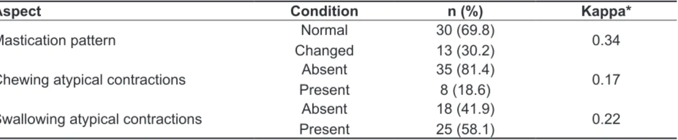
When analyzing chewing and swallowing through myofunctional orofacial examination, it was observed that all participants had incision aspects, grinding, lip closure, lip posture, food containment and coordination considered normal.
Among the volunteers assessed, mastication pattern changes were observed, as well as the presence of atypical contractions in chewing and
swallowing, the latter being found in most volun
-teers. Results also showed that there was a poor level of agreement between judges on this point (Table 1).
Regarding the occlusion, half of the women studied (51.1%) underwent previous orthodontic treatments and there was no association between this and the other variables.
Table 2 shows the assessment of dental occlusion characteristics of the volunteers in the study, where normality characteristics were predominant. No volunteer showed a completely normal occlusion. perform protrusion and lateralization movements
and the contact points were registered.
The TMJ evaluation was carried out by a trained physiotherapist through the Diagnostic Criteria for Research temporomandibular disorders (RDC/
TMD) Instrument 16. From this instrument , jaw
movement amplitudes, the presence of joint noises, mouth opening pattern and TMD diagnosis can be evaluated.
The results of the chewing and swallowing evaluations were compared to verify the agreement
degree among observers, through the Kappa coefi
-cient. These results were interpreted as follows: poor agreement (K <0), light agreement (K = 0-0.20), weak agreement (K = 0.21-0.40), moderate agreement (K= 0.41 to 0.60), substantial agreement
(K = 0.60-0.80) and excellent agreement (K>0.80)17.
The remaining analyzes were performed using the Statistic software, version 9.0 for Windows, and Chi-square and Fisher tests were used to assess the variables association: chewing pattern, chewing and swallowing atypical contractions, Angle Class, overjet, overbite, crossbite, orthodontic treatment, TMD diagnosis, articular noises and opening pattern. A level of 5% was determined signiicant.
Table 1 – Frequency of conditions found in orofacial myofunctional examination and concordance coeficient between judges
Aspect Condition n (%) Kappa*
Mastication pattern Normal
Changed
30 (69.8)
13 (30.2) 0.34
Chewing atypical contractions Absent
Present
35 (81.4)
8 (18.6) 0.17
Swallowing atypical contractions Absent
Present
18 (41.9)
25 (58.1) 0.22
Table 2 – Frequency of conditions found in dental occlusion assessment (n=43)
Aspect Condition n (%)
Angle classiication
Class I Class II Class III
32 (74.4) 5 (11.6) 6 (14.0)
Overjet Normal
Overjet
26 (60.5) 17 (39.5)
Overbite Normal
Overbite
35 (81.4) 8 (18.6)
Cross alteration Absent
Bite Crossed
40 (93.0) 3 (7.0)
Lateral malocclusion pattern Canines
Other
29 (67.4) 14 (32.6)
Protrusive malocclusion pattern Incisors
Other
35 (81.4) 8 (18.6)
Occlusion interferences Absent
Present
26 (60.5) 17 (39.5)
The TMJ evaluation trough RDC/TMD instrument observed amplitude values within the normal range of movement. The mean and standard deviation of the active opening was 42.5 ± 6.6 mm, passive
opening 51.6 ± 5.1 mm, right and left lateral devia
-tions, respectively, 9.8 ± 2.2 mm and 9.0 ± 2.3 mm, and 5.8 ± 1.9 mm in the protrusion.
Although asymptomatic, the TMD diagnosis was found in 16.3% of the volunteers (disc displacement and osteoarthritis), 60.5% had mouth opening deviation and 41.9% had noise during joint movements (Table 3).
Table 3 – Conditions found in temporomandibular joint assessment
Aspect Condition n (%)
TMD
Without diagnoses Disc displacement
Osteoarthritis
36 (83.7) 4 (9.3) 3 (7.0)
Opening
Straight 17 (39.5)
Diverted 26 (60.5)
Absent 25 (58.1)
Noises Click 14 (32.6)
Crepitus 4 (9.3)
contractions; opening pattern and chewing atypical contractions (Table 4). There was no association between variables related to the Angle Occlusal Class, chewing pattern and temporomandibular dysfunction (Table 5).
The association between chewing pattern, chewing and swallowing atypical contractions, Angle Class, overjet, overbite, TMD diagnosis, opening pattern and noise was analyzed. Results showed that there was signiicant association between TMD and joint noises; joint noises and atypical swallowing
Table 4 – Association between noises, mouth opening pattern, temporomandibular disorders, swallowing and chewing atypical contractions.
Without noises n (%)
With noises n (%)
Total
n (%) p
Without TMD With TMD Without SAC
With SAC
24 (55.8) 0 (0.0) 14 (32.6) 11 (25.5)
12 (27.9) 7 (16.3)
4 (9.3) 14 (32.6)
43 (100)
43 (100)
0.01*
0.02**
Opening straight n (%)
Opening Diverted n (%)
Total
n (%) p
Without CAC With CAC
11 (25.6) 6 (14.0)
24 (55.8)
2 (4.6) 43 (100) 0.02**
TMD: temporomandibular disorder, SAC: swallowing atypical contractions, CAC: chewing atypical contractions. * Fisher Test. **Chi
--square test.
Table 5 – Association between angle classes, mastication pattern and temporomandibular disorder
Class I n (%)
Class II n (%)
Class III
n (%) p*
Normal mastication 24 (55.8) 3 (7.0) 3 (7.0) 0.20
Altered mastication 8 (18.6) 2 (4.6) 3 (7.0)
Without TMD With TMD
27 (62.8) 5 (11.6)
4 (9.4) 1 (2.3)
5 (11.6)
1 (2.3) 0.84
TMD: temporomandibular.disorder *Chi-square test.
DISCUSSION
This research aimed to investigate the
associ-ation between chewing and swallowing stomato
-gnathic functions, dental occlusion and TMD signs in asymptomatic women.
Aspects related to mastication were normal, except for the pattern and the presence of atypical contractions. Regarding the mastication pattern, one third of the volunteers were abnormal, i.e., using predominantly a speciic side during the chewing cycle.
Although some studies have shown that healthy individuals may present a unilateral predominance, the chewing normal physiology is characterized by
unilateral cycles with periodic alternation of food on
the two sides of the dental arch18.
The alternating bilateral pattern is essential in preventing myofunctional disorders, periodontal problems and TMD, contributing as well to the maxillary bones development, arches maintenance, occlusion stability, TMJ stability, and muscular and
functional balance of the stomatognathic system19-21.
A research9 that aimed to evaluate the chewing
right and left sides, and in the jaw stability during
mastication9.
On the other hand, interferences in protrusive movements may cause morphologic alterations in
the TMJ internal structure in relation to the conigu
-ration, position and function of the articular disc10.
Although, in this study, no signiicant association was found between TMD signs and dental occlusion, the presence of mouth opening deviation may indicate the inluence of occlusal interferences in this condition.
One study25 evaluated the occlusal variables to
differentiate patients with disc displacement and osteoarthritis (n=381) on normal asymptomatic adults (n=98). By multiple logistic regression analysis, patients with disc displacement had more crossbite, and patients with osteoarthritis showed bigger overjet and smaller overbite.
TMD diagnosis was found in a low percentage of volunteers due to painful symptoms, which is typical of myogenic TMD diagnosis, has been exclusion criteria in this study. Therefore, only disc displacement and osteoarthritis diagnostics were detected through the RDC/TMD instrument, from the presence of noise during TMJ movements. This fact justiies the signiicant association between TMD diagnosis and the presence of noises. It should be emphasized that the disc displacement diagnosis can only be conirmed by magnetic
resonance image26 and, a recent study regarding
RDC instrument sensitivity and speciicity, showed low levels on these conditions, recommending
imaging tests for a more accurate diagnosis27.
Another TMD sign observed in most of the volunteers was the mouth opening deviation, which may occur due to pathological changes, joint inlammation, lack of occlusal guides, as well
as imbalance of chewing muscles26,28. It is possible
that this imbalance may also be responsible for the association between the opening pattern and atypical contractions during chewing.
As for the evaluation of TMJ range of movement , the mean values were within the normal range found
in literature29, except for the protrusion movement,
whose mean value observed was slightly below the reference levels (≥ 7mm).
The limitations of this study may be related to the myofunctional assessment protocol, which had poor agreement between judges, and the fact that a previous orthodontic treatment was not an exclusion criterion.
The relevance of this study is based on the need for an interdisciplinary approach of speech therapists, dentists and physiotherapists in the assessment and therapeutic intervention of the cranio-cervico-mandibular system. Subsequent Orofacial myofunctional assessment
demon-strated in this study the presence of chewing and swallowing atypical contractions. A recent study
on women with TMD22 justiied the changes in the
chewing muscle recruitment as a compensatory mechanism for pain symptoms relief. Besides pain, the authors also suggested that the excessive oral muscles involvement in individuals with TMD serves to counteract the tongue thrust force, which aims to prevent the food to escape from the oral cavity during swallowing. In the present research, pain does not justify atypical contractions, once the volunteers were asymptomatic.
The exaggerated participation of the orbicular and chin muscles during swallowing may be inluenced by discrepancies of bone bases, when these do not allow a normal labial occlusion. Such actions take place to ensure the lip sealing during swallowing, being observed mainly as a Class II malocclusion with overjet, due to the great anterior
posterior distance between maxilla and mandible20.
In the present study, only ive of the 43 women had occlusal Class II, and 17 had overjet, which may explain the lack of association between this variable and the myofunctional assessment, once that occlusal interference was small in magnitude.
For the swallowing to process in a normal manner, it is necessary the balance between perioral and masticatory muscles and tongue. Any disruption of this balance may lead to atypical swallowing, which may act as etiologic factors of malocclusion, once the bone is a highly plastic tissue, able of shaping to the muscle pressures. Therefore, atypical swallowing
may determine any type of malocclusion23.
Even with the a high percentage of atypical swallowing contractions, volunteers of this study showed characteristics of normal occlusion, but the ideal occlusion was not found in any of them.
Results of epidemiological studies in Brazil point out a high rate of malocclusion, ranging from 13 to 90.09%. However, these data should be viewed with caution, once, in Latin America, malocclusions researches are scarce, of regional characteristics,
and do not follow a uniform methodology14.
The relationship between occlusion and TMD
has also been discussed7,9,10,13,24, since dental
occlusion determines the movement pattern and the jaw position. Additionally, the occlusal instability may be a reason for the chewing system overload
and also leading to damages to TMJ7.
Occlusal interferences were observed in volun
mainly, atypical contractions during swallowing and chewing, which were associated with the presence of joint noises and mouth opening pattern. These indings can be attributed to imbalances and lack of coordination of the muscles involved in these functions. Regarding dental occlusion, no volunteer had an ideal occlusion and no associations were found with this assessment.
studies with larger samples and providing a deeper understanding of this system for all professionals involved, may contribute to a more comprehensive clinical practice and with more deinitive results.
CONCLUSION
The asymptomatic volunteers of this study showed some changes of stomatognathic functions,
RESUMO
Objetivo: veriicar a associação entre funções estomatognáticas de mastigação e deglutição, oclusão
dentária e sinais de disfunção temporomandibular em mulheres assintomáticas. Métodos: as funções
estomatognáticas foram avaliadas pelo exame miofuncional orofacial; o exame da oclusão dentária
compreendeu: classiicação de Angle; medidas de sobrepasse horizontal e vertical; presença de mor
-dida aberta e cruzada; e a avaliação da articulação temporomandibular foi realizada pelo instrumento
Critérios de Diagnóstico para Pesquisa de Desordens Temporomandibulares. Resultados: foram
avaliadas 43 mulheres com idade média de 23,7 anos. O exame miofuncional orofacial demonstrou
alterações no padrão de mastigação (30,2%) e contrações atípicas na mastigação (18,6%) e deglu
-tição (58,1%). Quanto à oclusão dentária, houve predomínio de classe I de Angle (74,4%), porém nenhuma voluntária apresentou uma oclusão ideal. A avaliação da articulação temporomandibular apresentou amplitude de movimento dentro da normalidade, presença de desvio na abertura da boca (60,5%) e diagnóstico de disfunção temporomandibular (16,3%). Houve associação signiicante entre presença de ruídos articulares e diagnóstico de disfunção temporomandibular e contrações atípicas na deglutição; padrão de abertura e contrações atípicas na mastigação; e não houve associação
entre a Classe Oclusal de Angle, padrão de mastigação e disfunção temporomandibular. Conclusão:
voluntárias assintomáticas apresentaram alterações das funções estomatognáticas, como contrações atípicas durante a deglutição e mastigação, as quais foram associadas com a presença de ruídos
articulares e padrão de abertura da boca. Tais achados podem ser atribuídos a desequilíbrios e inco
-ordenação dos músculos envolvidos nessas funções. Nenhuma voluntária apresentou oclusão ideal e não foram encontradas associações com esta condição.
DESCRITORES: Sistema Estomatognático; Oclusão Dentária; Transtornos da Articulação
Temporomandibular; Articulação Temporomandibular
REFERENCES
1. Marchesan IQ. Fundamentos em Fonoaudiologia: Aspectos clínicos da motricidade oral. Rio de Janeiro: Guanabara Koogan, 2005.
2. Maciel CTV, Barbosa MH, Toldo CA, Faza FCB, Chiappetta ALML. Disfunções Orofaciais nos Pacientes em Tratamento Ortodôntico. Rev CEFAC. 2006;8(4)456-66.
3. Felício CM, Ferreira CLP, Medeiros APM, Silva MAR, Tartaglia GM, Sforza C. Electromyographic indices, orofacial myofunctional status and temporomandibular disorders severity: A correlation
study. Journal of Electromyography and Kinesiology. 2012;22:266-72.
4. Franco AL, Andrade MF, Segalla JCM, Gonçalves DAG, Camparis CM. New approaches to dental occlusion: a literature update. Cranio. 2012;30(2):136-43.
5. Yamaguto OT, Vasconcelos MHF. Determinação das medidas dentárias mésio-distais em indivíduos brasileiros leucodermas com oclusão normal. R Dental Press OrtodonOrtop Facial. 2005;10(5):99-107.
brasileiros com oclusão normal natural. R Dental Press OrtodonOrtop Facial. 2006:11(1):99-106. 7. Wang C, Yin X. Occlusal risk factors associated with temporomandibular disorders in young adults with normal occlusions. Oral Surg Oral Med Oral Pathol Oral Radiol. 2012;114(4):419-23.
8. Felício CM, Oliveira MM, Silva MAMR. Effects of orofacial myofunctional therapy on temporomandibular disorders. Cranio. 2010;28(4):249-59.
9. Felício CM, Melchior MO, Silva MAMR, Celeghini RMS. Desempenho mastigatório em adultos relacionado com a desordem temporomandibular e com a oclusão. Pró-Fono R Atual Cient. 2007;19(2):151-8.
10. Gremillion HA. The relationship between occlusion and TMD: an evidence-based discussion ,JEvid Base Dent Pract. 2006;6:43-7.
11. Genaro KF, Berretin-Felix G, Rehder MIBC, Marchesan IQ. Avaliação Miofuncional Orofacial – Protocolo MBGR. Rev CEFAC. 2009;11(2):237-55. 12. Moreno I, Sanchez T, Ardizone I, Aneiros F, Celemin A. Electromyographic comparisons between clenching, swallowing and chewing in jaw muscles with varying occlusal parameters. Med Oral Patol Oral Cir Bucal. 2008;13(3):E207-13.
13. Cruz FLG, Marinho CC, Leite FPP. Relationship between abnormal horizontal or vertical dental overlap and temporomandibular disorders. Rev. odontociênc. 2009;24(3):254-7.
14. Marcomini L, Santamaria Júnior M, Lucato AS, Santos JCB, Tubel CAM. Prevalência de maloclusão e sua relação com alterações funcionais na respiração e na deglutição. Braz Dent Sci. 2010;13(8):52-8.
15. Okeson J. Tratamento das desordens temporomandibulares e oclusão. 4.ed. São Paulo: Artes Médicas; 2000.
16. Dworkin SF, Leresche L. Research diagnostic criteria for temporomandibulardisoders: Review, criteria, examinations and speciications, critique. J Craniomandibular Disorders. 1992;6:301-55.
Received on: January 22, 2014 Accepted on: July 01, 2014
Mailing address: Lais Chiodelli
R. Tuiuti, 2502/102, Centro Santa Maria – RS – Brasil CEP: 97050-420
E-mail: [email protected]
17. Viera AJ, Garret JM. Understanding Interobserver Agreement: The Kappa Statistic. Research Series. 2005;37(5):360-3.
18. Deda MRC, Picinato-Pirola MNC, de Mello-Filho FV, Trawitzki LVV. Inclinação de Cabeça Durante a Mastigação Habitual nas Deformidades Dentofaciais Classe II e III. Rev CEFAC. 2011;13(2):253-8. 19. Slavicek G, Schimmer C. Analysis of human mastigation behavior: a new approach using planar calculations of fragmented chewing sequences. J. Stomat. Occ. Med. 2010;3(1):61-7.
20. Mezzomo CL, Machado PG, Pacheco AB, Gonçalves BFT, Hoffmann CF. As implicações da classe II de Angle e da desproporção esquelética tipo classe II no aspecto miofuncional. Rev CEFAC. 2011;13(4)728-34.
21. Navarro PR, Assis GB, Souza LL, Filho EM, Azenha CR, Tessitore A. Alterações de funções orais na presença de aparelhos ortodônticos ixos com recursos intraorais. Rev CEFAC. 2013;15(5):1281-91.
22. Weber P, Corrêa ECR, Bolzan GP, Ferreira FDS, Soares JC, Silva AMT. Mastigação e deglutição em mulheres jovens com desordem temporomandibular. CoDAS. 2013;25(4):375-80.
23. Fernandes LFT, Kochenborger R, Woitchunas FE, Woitchunas DR. A inluência da deglutição atípica no padrão craniofacial e na morfologia mandibular. RFO. 2010;15(1):52-7.
24. Manfredini D, Piccotti F, Ferronato G, Guarda-Nardini L. Age peaks of different RDC/ TMD diagnoses in a patient population. J Dent. 2010;38:392-9.
25. Pullinger AG, Seligman DA. Quantiication and validation of predictive values of occlusal variables in temporomandibular disorders using a multifactorial analysis. J Prosthet Dent 2000;83:66-75.
26. Chiodelli L, Weber P, Pasinato F, Souza JA, Corrêa ECR. Manifestações clínicas de desordem temporomandibular e inclinação lateral da cabeça. Ter Man. 2012;10(50):383-8.
27. Truelove E, Pan W, Look JO, Mancl LA, Ohrbach RK, Velly AM et al. The research diagnostic criteria for temporomandibular disorders. III: validity of axis I diagnoses. J Orofac Pain. 2010;24:35-47.
28. Figueiredo VMG, Cavalcanti AL, Farias ABL, Nascimento SR. Prevalência de sinais, sintomas e fatores associados em portadores de disfunção temporomandibular. Acta. Sci. Health. Sci. 2009;31(2):159-63.