Wilson’s disease in children and adolescents:
diagnosis and treatment
Doença de Wilson em crianças e adolescentes: diagnóstico e tratamento
Stephania de Andrade Sócio1, Alexandre Rodrigues Ferreira2, Eleonora Druve T. Fagundes3, Mariza Leitão V. Roquete4,
Júlio Rocha Pimenta5, Lilian de Faria Campos6, Francisco José Penna7
Instituição: Hospital das Clínicas da Universidade Federal de Minas Gerais (UFMG), Belo Horizonte, MG, Brasil
1Mestre; Pediatra Especialista em Gastroenterologia Pediátrica pela UFMG,
Belo Horizonte, MG, Brasil
2Doutor; Professor Adjunto do Departamento de Pediatria da Faculdade de
Medicina da UFMG, Belo Horizonte, MG, Brasil
3Doutora; Professora Adjunta do Departamento de Pediatria da Faculdade
de Medicina da UFMG, Belo Horizonte, MG, Brasil
4Doutora; Professora Adjunta do Departamento de Pediatria da Faculdade
de Medicina da UFMG, Belo Horizonte, MG, Brasil
5Médico Residente de Pediatria do Hospital das Clínicas da UFMG, Belo
Horizonte, MG, Brasil
6Médica Formada pela Faculdade de Ciências Médicas de Minas Gerais,
Belo Horizonte, MG, Brasil
7Professor Titular do Departamento de Pediatria da Faculdade de Medicina
da UFMG, Belo Horizonte, MG, Brasil
Endereço para correspondência: Eleonora Druve T. Fagundes
Rua Tenente Anastácio de Moura, 740/801 – Santa Efigênia CEP 30240-390 – Belo Horizonte/MG
E-mail: eleonoradruve@uol.com.br
Conflito de interesses: nada a declarar
Recebido em: 27/5/09 Aprovado em: 31/8/09 ABSTRACT
Objective: To describe clinical symptoms, laboratory indings at diagnosis and treatment of children and adoles-cents with Wilson’s disease.
Methods: This is a descriptive and retrospective study of a series of 17 children and adolescents with Wilson’s disease, assited at the Pediatric Hepatology Ambulatory of the Hospital das Clínicas of Universidade Federal de Minas Gerais, Brazil, from 1985 to 2008. Data were collected by revision of medical charts and during clinical follow-up.
Results: Patients were 2.8 to 15.1 years old, with a mean age of 8.8±0.9 years. The disease main presentation was he-patic (53%), followed by the asymptomatic form, diagnosed by family screening. The Kayser-Fleischer ring was observed in 41% of the patients. The ceruloplasmin was altered in 15 out of 17 patients, and the urinary copper varied from 24 to 1000mcg/24h (median: 184mcg/24h). The treatment was stablished with D-penicillamine in all cases. Slight side ef-fects were observed in ive children, with no need to interrupt or change medication. Clinical and laboratory responses to treatment, with normalization of aminotransferases levels, were shown in 14 patients after a median of 10.7 months. Although treated, three patients died (one due to fulminant hepatitis and two due to severe hepatic failure).
Conclusions: Wilson’s disease is rare in the pediatric group. In children, the main presentation is the liver disease. The diagnosis can be established by reduced ceruloplasmin
levels and elevated copper excretion in the 24-hour urine, but it demands high suspicion level. There are good toler-ance and response to medical treatment.
Key-words: Wilson’s disease; hepatolenticular degenera-tion; child; hepatic insuficiency.
RESUMO
Objetivo: Descrever as formas de apresentação, as altera-ções laboratoriais ao diagnóstico e o tratamento de crianças e adolescentes com doença de Wilson.
Métodos: Estudo descritivo e retrospectivo de 17 crianças e adolescentes com doença de Wilson atendidos no Ambula-tório de Hepatologia Pediátrica do Hospital das Clínicas da Universidade Federal de Minas Gerais no período de 1985 a 2008. Os dados foram coletados dos prontuários e durante as consultas ambulatoriais.
laboratorial, com níveis normais de aminotransferases, foram evidenciadas em 14 pacientes após mediana de 10,7 meses de tratamento. Três crianças morreram (uma por hepatite fulminante e duas com complicações da insuiciência hepática grave), apesar do tratamento.
Conclusões: A doença de Wilson é rara na faixa etária pediátrica. A forma de apresentação predominante é a hepática. Seu diagnóstico se baseia principalmente em dosagem de ceruloplasmina baixa, cobre livre e cobre em urina de 24 horas elevados, mas exige alto grau de sus-peição. Apresenta boa resposta e tolerância ao tratamento medicamentoso.
Palavras-chave: doença de Wilson; degeneração hepato-lenticular; criança; insuiciência hepática.
Introduction
Wilson’s disease is an autosomal recessive condition with an estimated prevalence of one in every 40,000 people. It is caused by a mutation of the ATP 7B gene which is located on chromosome 13. The mutation reduces both the quantity of copper excreted via the biliary system and the quantity of copper bound in ceruloplasmin, which is a glycoprotein that transports the metal around the body(1). As a result, copper accumulates in several different tissues, such as the liver, central nervous system, cornea and kidneys and causes hepa-tocellular cirrhosis, dementia and neuropsychiatric disorders and affects heart and kidney function. The classic presenta-tion comprises the trio of liver disease plus neurological and ophthalmological involvement. Hepatic manifestations predominate in the pediatric age group. From 10 to 25% of cases are neurological(1), and are generally detected in adults. Copper deposited in the cornea can lead to Kayser-Fleischer (KF) rings, which is the most common ophthalmological sign, although it may be absent in children and appears to have a relationship with neuropsychiatric cases(1-3). Other, rarer symptoms are also described, such as renal (proteinuria, hematuria, lithiasis), osteoarticular (osteopenia, arthralgia, arthritis), hematological (hemolysis), cardiac (arrhythmia, ventricular hypertrophy, sudden death) and neoplastic (ad-enocarcinoma, hepatoblastoma)(1).
Wilson’s disease is a rare liver disease, but diagnosis has a great impact because a speciic treatment of proven eficacy exists and because without this treatment the disease is in-variably fatal. Early treatment averts severe complications. Diagnosis may be dificult because there is no single test
with adequate sensitivity and manifestations are not always typical, especially among children, and so it is dependent on a high index of clinical suspicion when presented with a patient with liver and/or neuropsychiatric disease(4). Diagno-sis is based on laboratory results such as: low ceruloplasmin and elevated 24-hour urine copper, free copper and copper in hepatic tissue. Observation of KF rings in an ophthalmologi-cal examination further supports the diagnosis(1).
Treatment is with copper chelating drugs. The irst-choice drug is D-penicillamine, despite the risk of neurological deterioration in up to 50% of patients and the many side ef-fects associated with it(1,4). Trientine and tetrathiomolybdate are alternative choices, and the second of these is used with patients with neurological symptoms(1). Zinc is indicated for asymptomatic cases and maintenance treatment(5).
There are few publications reporting on exclusively pedi-atric samples(4,6-10). The objective of this study was to describe the different forms of clinical presentation, laboratory ind-ings and response to treatment in children and adolescents with Wilson’s disease.
Methods
This is a retrospective descriptive study of a series of cases of children and adolescents with diagnoses of Wilson’s disease who were treated at the Pediatric Hepatology clinic at the UFMG Hospital das Clínicas between 1985 and 2008.
We included 17 patients aged less than 18 years. Data were collected by reviewing archived medical records from the UFMG Hospital das Clínicas iles and during outpatient medical consultations.
The variables studied were age at diagnosis, sex, forms of clinical presentation, laboratory tests results at diagno-sis, Kayser-Fleischer (KF) ring present/absent, abdominal ultrasonography, upper digestive endoscopy, liver biopsies, time taken for aminotransferases to drop to normal levels after starting treatment, the treatment prescribed and its side effects.
Clinical manifestations were deined as follows:
• Asymptomatic form: characterized by an absence of signs and symptoms of liver disease, or of neurological or ophthalmological involvement, but with laboratory indings compatible with Wilson’s disease.
• Acute, chronic and fulminant hepatic forms:
b) chronic hepatitis: signs of portal hypertension, he-patomegaly, splenomegaly, elevated hepatic enzyme levels, with or without jaundice;
c) fulminant hepatic failure: clinical manifestations of acute hepatitis and encephalopathy up to 8 weeks after appearance of the clinical manifestations of liver disease;
• Neurological form: characterized by neuropsychiatric symptoms such as altered behavior, psychoses, speech disorders and others.
Wilson’s disease was diagnosed on the basis of the presence of at least two of the following criteria: 1. Family history of Wilson’s disease; 2. KF rings; 3. low ceruloplasmin levels (<20mg/dL); 4. Free copper >25µg/dL [calculated as follows: free copper = serum copper (in mcg/dL) – (3 x ceruloplasmin in mg/dL)]; 5. 24-hour urine copper >100µg/24h. Our de-partment does not assay copper in dry liver tissue.
Patients were also diagnosed with the disease once other chronic hepatopathies had been ruled out, if they had chronic liver disease, had had at least one abnormal copper metabolism test result and responded well to treatment with chelating agents. Autoimmune hepatitis, chronic hepatitis B and C and α1-antitrypsin deiciency were ruled out for all patients.
All patients underwent ophthalmological examination with a slit-lamp in order to detect KF rings and sunlower
cataracts. Imaging exams such as abdominal ultrasonography and upper digestive endoscopy (UDE) were also performed where clinically indicated, which in the case of UDE was if there were signs of portal hypertension. Laboratory tests re-quested at diagnosis were: serum aspartate aminotransferase (AST) and alanine aminotransferase (ALT) assays, total and fractioned bilirubin and albumin. Aminotransferase results were considered abnormal if over the maximum reference value (MRV).
All patients were treated with D-penicillamine, starting with an initial dosage of 10mg/kg/day (or 250mg/day), increasing to 20mg/kg/day after 30 days’ treatment. The maximum recommended dosage is 1000 to 1500mg/day taken in three doses. The objective is to maintain 24-hour urine copper between 200 and 500mcg/24h and free copper below 10mcg/dL. Pyridoxine was also given simultaneously at a dosage of 25mg/day.
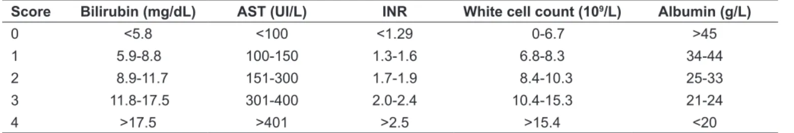
Outpatients follow-up was monthly for the irst 6 months, two-monthly from six to 12 months and three-monthly thereafter. Treatment was monitored using the following laboratory tests: 24-hour urine copper, free copper, liver func-tion assessment, hemagram, platelet count and qualitative urine analysis in order to control adverse effects. All parents and siblings of patients were screened for Wilson’s disease. The Nazer et al(11) and Dhawan et al(8) scores were calcu-lated in order to assess the severity and predict the progress of the patients with Wilson’s disease, as shown in Tables 1 and 2.
Data were analyzed using Epi-Info 6.04. Variables were expressed as means, standard deviations, medians and inter-quartile 25-75 ranges (IQ25-75%). The study was approved by the Research Ethics Committee at the UFMG.
Results
The sample comprised 17 children and adolescents, ten (59%) were female and age at diagnosis varied from 2.8 to 15.1 years, with a mean of 8.8 ± 0.9 years.
Table 1 – The Nazer et al(11) prognostic classiication based on
liver function
Score Bilirubin (mg/dL) AST (UI/L) INR
0 <5.8 <100 <1.3
1 5.9-8.8 100-150 1.3-1.6
2 8.9-11.7 151-200 1.6-1.9
3 11.8-17.5 201-300 1.9-2.4
4 >17.5 >300 >2.4
Score >7 suggests a risk of death if liver transplantation not per
-formed. INR: International Normalized Ratio.
Table 2 – The Dhawan et al mortality prediction index for Wilson’s(8)
Score Bilirubin (mg/dL) AST (UI/L) INR White cell count (109/L) Albumin (g/L)
0 <5.8 <100 <1.29 0-6.7 >45
1 5.9-8.8 100-150 1.3-1.6 6.8-8.3 34-44
2 8.9-11.7 151-300 1.7-1.9 8.4-10.3 25-33
3 11.8-17.5 301-400 2.0-2.4 10.4-15.3 21-24
4 >17.5 >401 >2.5 >15.4 <20
The majority of cases, 11/17 patients (65%), presented the hepatic form, with six cases of acute hepatitis (one fulminant) and ive of chronic hepatitis. Two patients (12%) also had glomerulonephritis. Six patients (35%) were identiied by family screening. All were asymptomatic, but with abnormal aminotransferases.
The most signiicant laboratory indings at diagnosis were: elevated AST (4.6±1.2 times MRV), ALT (3.9±0.9 times MRV) and total bilirubin (5.3±3.4mg/dL). Albumin results varied from 2.3 to 5.0, with a mean of 3.8±0.7g/dL. Urinary copper varied from 24 to 1,000mcg/24h, with a median of 184mcg/24h (p25%=106 and p75%=497) and free copper varied from 1.8 to 119µg/dL, with a median of 27µg/dL (p25%=20 and p75%=4.1). Ceruloplasmin varied from 1 to 47mg/dL, with a median of 4mg/dL (p25%=3 and p 75%=8).
All patients underwent ophthalmological examination and KF rings were detected in seven cases (41%), all with severe or chronic liver disease and with ages varying from seven to 12 years (mean: 10.6 years). The clinical and labora-tory characteristics of the patients are shown in Table 3.
During the follow-up period, 14 patients underwent abdominal ultrasonography and 64% had complications such as hepatosplenomegaly and signs of cirrhosis. Seven
underwent upper digestive endoscopy because they showed signs of portal hypertension; 43% had esophageal varices.
Just four patients had liver biopsies. One 5-year-old child had discrete and nonspeciic symptoms, a nine-year-old had erosive necrosis and cirrhosis and the other two biopsies were both on ten-year-olds, one with hepatic steatosis and discrete ibrosis and the other with chronic inlammatory hepatitis with lymphocytic iniltration and moderate ibrosis.
Drug-based treatment with D-penicillamine was given to 16 of the 17 patients (one died from fulminant hepatitis), at dosages varying from 250 to 750mg a day. Five of these 16 patients, (31%) suffered side effects such as headaches (1/16), thrombocytopenia (1/16), proteinuria (1/16), nausea and vomiting (2/16) and limb pain (1/16). However, these effects were transitory and it was not necessary to withdraw or change the medication. The age at start of treatment varied from 2.8 to 15 years with a mean of 9.9±0.9 years. The time taken for the aminotransferases of 14 of the 16 patients who started treatment to drop to normal levels varied from 1 to 24 months, with a median of 11 months (p25% 3 and p75% 12). The levels of two patients never reached normal levels and they both died from complica-tions of chronic severe liver disease. The overall mortality rate was 18% (3/17).
Table 3 - Clinical and laboratory characteristics of 17 patients with Wilson’s disease
Patient Age (Years)
Sex
(M/F)
Ceruloplasmin
(mg/dL)
Urinary copper (µg/24h)
Free copper (µg/dL)
KF
rings
Clinical presentation
1 8 M 2.0 772.5 118.7 N AH + ADGN
2 2 F 4.0 15.5 1.8 N Asymptomatic+
FH
3* 10 F 3.0 164.0 12.25 S Asymptomatic
4 14 F 2.0 240.0 26.7 N Asymptomatic
5 7 F 1.0 188.0 24.85 N Asymptomatic
6 13 M 3.0 884.4 78.55 N CH
7* 9 M 4.8 98.3 22.8 N CH+ good
response
8 10 F 5.6 183.6 30.76 S AH + ADGN
9* 10 F 7.0 1000.0 19.95 N CH
10 12 F 10.0 1000.0 80.5 S Fulminant AH
11 11 M 8.0 645.4 23.7 S AH
12* 5 F 4.4 24.39 41.14 N Asymptomatic+
FH
13 7 M 7.3 453.0 27.8 S AH
14 11 F 47.0 55.0 32.05 S CH
15 14 M 38.0 64.7 44.7 N AH+ good
response
16 11 M 3.0 497.4 20.35 S CH
17 3 F 8.89 106.0 10.0 N Asymptomatic+
FH
Table 4 shows the scores for the Nazer and Dhawan scales for the patient sample. Only one of the three pa-tients who died had a Nazer score over 7; two had Dhawan scores of 10. One of the patients with a Dhawan score of 10 improved clinically after 6 months’ treatment with D-penicillamine.
Discussion
Wilson’s disease is one of the rarer causes of liver disease in children. There are few studies that have described exclu-sively pediatric samples and those that have been published generally have small patient samples, such as the studies by Sanchez-Albisua et al(4) with 26 children, and Yuce et al(7) with 33 children. The largest sample is described by Dha-wan et al(8) from King’s College Hospital, in London, with 74 children over 37 years.
The majority of patients are diagnosed in their second decade of life and it is rare that patients less than 5 years or more than 40 years old are diagnosed(12-14). However, screen-ing for the disease among the family members of patients may reduce this age by identifying asymptomatic patients earlier in life. The earliest diagnosis made in this sample was of a two-year-old child who was asymptomatic but had abnormal aminotransferase results. The mean age at diag-nosis in this sample was 8.8 years, which is similar to other
studies with pediatric patients(4,7). Age at diagnosis in the sample described by Sanchez-Albisua et al(4) was 9.8 ±3.4 years and in Yuce et al(7) it was 10.1 ± 2.5 years.
In terms of the forms of presentation, the hepatic form is the most prevalent in this age group (65%), as observed in our study. Yuce et al(7) observed six cases of fulminant hepatitis in a sample of 33 children with Wilson’s disease. They stress the importance of investigating this disease in the light of these fulminant cases, which appear to be more common during the second decade of life(4,7). There was one case of fulminant hepatitis in our study, in a 12-year-old girl who died during the immediate postoperative period after transplantation.
We observed KF rings in 41% of the patients, with a mean age of 10.4 years. The mean age of patients who did not have rings was lower at 8.5 years. Kayser-Fleischer rings are observed less often in the pediatric age group, because they are primarily dependent on the time taken for the metal to accumulate, and incidence is 5.6% to 63% in pediatric samples(4,7). Their absence does not therefore rule out a diagnosis of Wilson’s disease. Their presence has been related to the neuropsychiatric presentation and to more severe liver disease(3). All of the patients in our sample who died had had KF rings.
The neurological form manifests with trembling, dysar-thria, ataxia, rigidity, psychiatric symptoms and others(1),
Table 4 - Nazer(11)and Dhawan(8) scores, clinical presentation and course for 17 patients
Patient Nazer score Dhawan score Presentation Course
1 2 4 Acute hepatitis + ADGN Good response
2 0 0 Asymptomatic Good response
3 0 0 Asymptomatic Good response
4 1 1 Asymptomatic Good response
5 1 4 Asymptomatic Good response
6 7 10 Chronic hepatitis Nazer 0 and Dhawan 2 after 6
months’ treatment
7 4 4 Chronic hepatitis Good response
8 1 4 Acute hepatitis + ADGN Good response
9 2 3 Chronic hepatitis Good response
10 7 10 Acute fulminant hepatitis Died
11 5 10 Acute hepatitis Died
12 0 0 Asymptomatic Good response
13 6 8 Acute hepatitis Lost to follow-up
14 8 9 Chronic hepatitis Died
15 6 7 Acute hepatitis Good response
16 2 4 Chronic hepatitis Good response
17 5 5 Asymptomatic Good response
and is observed at rates of 25%(15) to 71%(16) in adults. This presentation is less common in children and is reported at rates of 4 to 12%(4,7). None of the patients in our sample had neurological involvement.
Ceruloplasmin is the laboratory test that most often produces abnormal results (88%), followed by 24-hour urine copper (71%). In pediatric case series, ceruloplasmin has a sensitivity of 82 to 88%, while 24-hour urine copper has a sensitivity of 81 to 100%(4,7). These results emphasize the importance of testing ceruloplasmin, serum free copper and urinary copper in order to increase the sensitivity of diagnosis of suspected cases. Using just one of these tests can lead to false negatives, delaying diagnosis and impact-ing on prognosis.
Liver biopsy is not very speciic, and as such is not essential for diagnosis, with the exception of copper in tissue, which is signiicant when elevated. Notwithstanding, the copper in tissue assay can produce false-negatives in children, since it is dependent on sample size, the length of time during which the metal has been accumulating and the fact that it may be irregularly distributed(4,7,16).
The correct treatment should be initiated as early as possible in order to avoid or minimize the harmful ef-fects of copper accumulation in tissues. A diet restricting foods containing large concentrations of copper can help with treatment. Pharmacological treatment is with cop-per chelating drugs and the most widely used and studied is D-penicillamine, although it produces a series of side effects such as hypersensitivity, medullary depression, development of autoimmune diseases, neurological dete-rioration, nephrotoxicity, polyneuropathy, optic neuritis and polymyositis(2,9). Sixteen of the 17 patients in our series were started on D-penicillamine and it was well-tolerated and caused no severe side-effects that would necessitate withdrawing the drug. Dhawan et al(8) found that 3/57 patients had medullary depression after taking D-penicillamine, and needed to change to trientine. None of the patients studied here needed to change medication and all remain on D-penicillamine.
Liver transplantation is indicated where presentation is fulminant, for patients with severe liver failure who do not respond to treatment and for those with complications of portal hypertension(3).
A good response to the drug treatment is deined as when liver function test results return to normal(4,17). In our study, the median time taken to produce a clinical response
was 10.7 months after starting D-penicillamine, which is similar to what can be found in the literature(4,9,17). These data emphasize the importance of waiting suficient time to evaluate response to treatment, as long as the patient’s functions are stable.
There are severity scales designed to predict the progress of patients with Wilson’s disease and of these the prog-nostic scale proposed in 1986 by Nazer et al(11) is the most often cited. The criteria are based on research undertaken both with children and adolescents and with adults and the objective is to identify patients who will probably not respond satisfactorily to chelating treatment and are at greater risk of dying without a liver transplantation. In 2005, Dhawan et al(8) revised the Nazer prognostic criteria on the basis of a pediatric sample of 74 children (mean age of 11.9 years) and proposed adding leukocyte counts and serum albumin and changing the cutoff point (from >7 to ≥10) in order to increase the test’s speciicity. The Nazar et al(11) and Dhawan et al(8) scales can be of help with decision-making, since some studies have shown that the Child-Pugh score is not appropriate for indicating need for liver transplantation in Wilson’s disease patients(17). In our study, the Dhawan et al(8) score was more sensitive for identifying high-risk patients (two of the three patients who died had scores of 10 or more). However, one patient with a score of 10 exhibited clinical improvement after drug-based treatment. The small sample does not allow for further extrapolation, but a global assessment of the patient, together with these scales, is necessary to correctly manage these cases.
1. Roberts EA, Schilsky ML; Division of Gastroenterology and Nutrition, Hospital for Sick Children, Toronto, Ontario, Canada. A practice guideline on Wilson disease. Hepatology 2003;37:1475-92.
2. Hassan A, Masood F. Wilson’s disease: a review. J Pak Med Assoc 2004;54:479-84.
3. Schilscky ML. Diagnosis and treatment of Wilson’s disease. Pediatr Transpl 2002;6:15-9.
4. Sánchez-Albisua I, Garde T, Hierro L, Camarena C, Frauca E, de la Vega A et al. A high index of suspicion: the key to an early diagnosis of Wilson’s disease
in childhood. J Pediatr Gastroenterol Nutr 1999;28:186-90.
5. Walshe JM, Munro NA. Zinc-induced deterioration in Wilson’s disease aborted by treatment with penicillamine, dimercaprol, and a novel zero copper diet. Arch Neurol 1995;52:10-1.
6. Tissières P, Chevret L, Debray D, Devictor D. Fulminant Wilson’s disease in children: appraisal of a critical diagnosis. Pediatr Crit Care Med 2003;4:338-43. 7. Yüce A, Koçak N, Demir H, Gürakan F, Ozen H, Saltik IN et al. Evaluation of
diagnostic parameters of Wilson’s disease in childhood. Indian J Gastroenterol 2003;22:4-6.
8. Dhawan A, Taylor RM, Cheeseman P, de Silva P, Katsiyiannakis L, Mieli-Vergani G. Wilson´s disease in children: 37-year experience and revised King´s score for liver transplantation. Liver Transpl 2005;11:441-8.
9. Arnon R, Calderon JF, Schilsky M, Emre S, Shneider BL. Wilson disease in children: serum aminotransferases and urinary copper on trienthylene
tetramine dihydrochloride (trientine) treatment. J Pediatr Gastroenterol Nutr 2007;44:596-602.
10. Marcellini M, Di Ciommo V, Callea F, Devito R, Comparcola D, Sartorelli MR
et al. Treatment of Wilson’s disease with zinc from the time of diagnosis in
pediatric patients: a single-hospital, 10-year follow-up study. J Lab Clin Med 2005;145:139-43.
11. Nazer H, Ede RJ, Mowat AP, Williams R. Wilson’s disease: clinical presentation and use of prognostic index. Gut 1986;27:1377-81.
12. Hui J, Fung EL, Tang NL, Chan MH, To KF, Fok TF. Diagnosing Wilson’s disease in a 5-year-old child. J Paediatr Child Health 2002;38:
412-3.
13. Wilson DC, Phillips MJ, Cox DW, Roberts EA. Severe hepatic Wilson’s disease in preschool-aged children. J Pediatr 2000;137:719-22.
14. Ala A, Borjigin J, Rochwarger A, Schilsky M. Wilson disease in septuagenarian siblings: Raising the bar for diagnosis. Hepatology 2005;41:668-70. 15. Kumagi T, Horiike N, Michitaka K, Hasebe A, Kawai K, Tokumoto Y et al. Recent
clinical features of Wilson’s disease with hepatic presentation. J Gastroenterol 2004;39:1165-9.
16. Ferenci P, Caca K, Loudianos G, Mieli-Vergani G, Tanner S, Sternlieb I
et al. Diagnosis and phenotypic classiication of Wilson disease. Liver Int
2003;23:139-42.
17. Brewer GJ. Tetrathiomolybdate anticopper therapy for Wilson’s disease inhibits angiogenesis, ibrosis and inlammation. J Cell Mol Med 2003;7:11-20.