www.reumatologia.com.br
REVISTA BRASILEIRA DE
REUMATOLOGIA
Final elaboration 12 April 2012
Description of the evidence collection method
A review of the scientiic literature was performed with the Me-dline database. The search for evidence was based on actual cli-nical scenarios and used the following Medical Subject Headings (MeSH) terms: Arthritis, Rheumatoid, Diagnosis (Delayed Diag-nosis OR Delay OR Early Rheumatoid Arthritis OR VERA), Prog-nosis, Criteria (American College of Rheumatology/European League Against Rheumatism OR ACR/EULAR OR classiication), Comparative Study, Smoking (OR tobacco use disorder), Rheu-matoid Factor, Anti-cyclic Citrullinated Peptide (or anti-CCP), HLA-DRB1 OR PTPN22 OR EPITOPE, extra-articular OR extraar-ticular OR systemic OR ExRA, Disease Progression, Radiography OR X RAY, ULTRASONOGRAPHY, and MAGNETIC RESONANCE
Grades of recommendation and strength of evidence
A: Most consistent experimental and observational studies.
B: Less consistent experimental and observational studies.
C: Case reports (uncontrolled studies).
D: Opinion that is not substantiated by critical evaluation, based on consensus, physiological studies or animal models.
Objective
To formulate guidelines for the management of rheumatoid arthritis (RA) in Brazil, with a focus on diagnosis. The aim of the present document is to summarise the current position of the Brazilian Society of Rheumatology on this topic to orient Brazilian doctors, particularly rheumatologists, to RA diagno-sis in our country.
Introduction
Rheumatoid arthritis (RA) is a chronic, progressive, and sys-temic inlammatory disease that preferentially affects the synovial membranes of joints and eventually leads to bone and cartilage destruction1(D). RA affects 0.5%–1% of the adult
population worldwide; the disease targets patients from every ethnic background2(D) and predominately affects
fe-males (2- or 3-fold more often than fe-males). Although RA can occur at any age, it is more frequent among individuals in the fourth to sixth decades of life3(D).
A Brazilian multicentre study conducted with samples from the various macro-regions found a prevalence of up to 1% in Brazil’s adult population4(B), which corresponds to
1,300,000 people.
Guidelines
Guidelines for the diagnosis of rheumatoid arthritis
Diretrizes para o diagnóstico da artrite reumatoide
Licia Maria Henrique da Mota
a,*, Bóris Afonso Cruz
a, Claiton Viegas Brenol
a,
Ivânio Alves Pereira
a, Lucila Stange Rezende-Fronza
a, Manoel Barros Bertolo
a,
Max Vitor Carioca Freitas
a, Nilzio Antônio da Silva
a, Paulo Louzada-Junior
a,
Rina Dalva Neubarth Giorgi
a, Rodrigo Aires Corrêa Lima
a, Ronaldo Adib Kairalla
b,
Alexandre de Melo Kawassaki
b, Wanderley Marques Bernardo
c,
Geraldo da Rocha Castelar Pinheiro
aa Sociedade Brasileira de Reumatologia, São Paulo, SP, Brazil
b Sociedade Brasileira de Pneumologia e Tisiologia, Brasília, DF, Brazil c Associação Médica Brasileira, São Paulo, SP, Brazil
* Corresponding author.
E-mail: [email protected] (L.M.H Mota)
As a chronic disease that causes irreversible joint damage, RA exacts high costs from both the patients and society at large 5(B) 6,7(D).
In recent years, signiicant advances have been achieved in understanding the physiopathogenesis, diagnostic meth-ods, and therapeutic management of RA. Among these ad-vances, the recently attributed signiicance of the early disease stages or so-called early RA (irst 12 months with RA symp-toms) stands out as an acknowledged “window of therapeutic opportunity”8(B)9,10(D). However, despite all advances, the
cur-rently available (clinical, laboratory, and radiological) diagnos-tic and prognosdiagnos-tic indicators are of limited value to early diag-noses and individual progdiag-noses11(B).
The demographic and clinical features of RA vary as a func-tion of the affected populafunc-tion12(B). Most available data
corre-spond to populations in Europe and the United States13,14(D). Few
studies have been conducted on the Brazilian population15,16(B).
RA affects mostly individuals within the economically pro-ductive age range, and the disease eventually imposes signii-cant limitations on their functional ability that result in the loss of work abilities. For these reasons, the indirect costs as-sociated with RA must be included in pharmacoeconomic studies17(B).
In Brazil and industrialised countries, the costs associated with RA are high18(B). The impact of the expenses associated
with RA is more remarkable in developing countries in which the inancial resources allocated to healthcare are less robust. This situation points to the relevance of studies adapted to Brazilian conditions that assess the costs and allocation of re-sources for the diagnosis and treatment of RA19(B).
RA diagnosis is based on clinical indings and complemen-tary diagnostic tests. No single laboratory, imaging, or histo-pathological test alone can conirm a diagnosis.
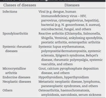
Several illnesses that present with arthritis must be con-sidered in the differential diagnosis of RA20-22(D), as described
in Table 1.
Diagnosis is easier when RA presents with the well-known pattern and the full range of typical symptoms. Diagnosis might be dificult in the early stages of disease because the characteristic serologic and radiological alterations are often absent23(D).
The clinical manifestations of RA can be classiied as ar-ticular and extra-arar-ticular. As RA is a systemic disease, general symptoms such as fever, asthenia, fatigue, myalgia, and weight loss can appear before or concomitantly with the onset of the articular manifestations24(D).
Articular manifestations
Although the articular manifestations of RA might be revers-ible in the early stages, persistent and uncontrolled synovitis leads to bone and cartilage destruction and irreversible tendon and ligament injuries.
The basic factor behind RA articular manifestations is syno-vial inlammation (synovitis), which can affect any diarthrodial joint in the body.
The clinical complaints include pain, swelling, and motion limitations of the affected joints. A physical examination will disclose the presence of pain, increased joint volume,
intra-articular effusion, heat, and eventual redness. Those ind-ings might not be evident in deep joints such as the hips and shoulders24(D).
The characteristic features of arthritis in RA are as follows24(D):
a) Polyarticular affection: usually involving more than four joints. Nevertheless, RA might begin and eventually remain as mono- or oligoarthritis.
b) Hand and wrist arthritis: affections of the wrist, metacarpo-phalangeal (MCP), and proximal intermetacarpo-phalangeal (PIP) joints are frequent from the very early disease stages. The distal interphalangeal (DIP) joints are seldom affected, a feature that distinguishes RA from other conditions such as osteo-arthritis and psoriatic osteo-arthritis.
c) Symmetric arthritis: symmetric affection of joints is a com-mon inding, although not mandatorily absolute in cases of the PIP, MCP, and metatarsophalangeal (MTP) joints. d) Cumulative or additive arthritis: arthritis usually exhibits a
cumulative pattern (progressive affection of new joints con-comitant to inlammation of the previously affected ones). e) Morning stiffness: prolonged stiffness that appears in the
morning, which is accompanied by a sensation of swelling, is near-universal feature of synovial inlammation. Unlike the short-lasting (5–10 minutes, as a rule) variety observed in osteoarthritis, in inlammatory diseases, stiffness tends to last for more than 1 hour. This phenomenon is associated with immobility that occurs concomitantly to the state of sleep or rest, rather than to a particular time of the day. The duration of stiffness tends to correlate with the degree of inlammation and is an important parameter in follow-up evaluations25(B)26(C).
Extra-articular manifestations
Although the articular manifestations are the most charac-teristic, other organs and systems can also be affected by RA.
Table 1 – Differential diagnoses of arthritis.
Classes of diseases Diseases
Infections Viral (e.g. dengue, human
immunodeiciency virus – HIV, parvovirus, cytomegalovirus, hepatitis), bacterial (e.g. N. gonorrhoeae, S. aureus), microbacterial, fungal, and others
Spondyloarthritis Reactive arthritis (Chlamydia, Salmonella,
Shigella, Yersinia), ankylosing spondylitis, psoriatic arthritis, enteropathic arthritis Systemic rheumatic
diseases
Systemic lupus erythematosus,
polymyositis/dermatomyositis, systemic sclerosis, Sjögren’s syndrome, Behçet’s disease, rheumatic polymyalgia, systemic vasculitis, and others
Microcrystalline arthritis
Gout, calcium pyrophosphate deposition disease, and others
Endocrine diseases Hypothyroidism, hyperthyroidism
Neoplastic diseases Metastatic neoplastic disease, lymphoma,
paraneoplastic syndromes, and others
Others Osteoarthritis, haemochromatosis,
The most common extra-articular manifestations of RA in-clude skin, eye, pleuropulmonary, heart, blood, neurological, and osteo-metabolic conditions. These occur more often in patients with severe and polyarticular disease, positive serol-ogy for the rheumatoid factor (RF) or cyclic citrullinated pep-tide antibodies (anti-CCP), and rheumatoid nodules27(B)28(D).
Brazilian studies conirmed that the initial manifestations of RA include polyarticular affection with persistent synovitis in the hands, long-lasting morning stiffness, a large number of painful and swollen joints, and fatigue15,16(B).
1. Is diagnosis of RA within the irst 12 months
of symptoms (early RA) associated with better
radiological and functional prognosis, compared
to later diagnosis?
The modern differentiation of RA from other joint diseases dates from 1907. As no pathognomonic traits allow a distinc-tion among the various types of arthritis in their early stages, the exact moment at which RA begins to progress as a sepa-rate entity from other articular illnesses is unknown12(B).
The deinition of early RA is important from both the theo-retical and practical perspectives, although the terms “early” and “RA” might be addressed independently, particularly be-cause the criteria applied to these classiications are based on established RA13(D).
Although controversial, early RA might be deined as the initial stage of disease or a “window of therapeutic opportu-nity” in which adequate therapy might modify the disease progression; the prognosis in this stage is better than that of later stages14(D).
The required symptom duration for the deinition of early RA varies widely in the specialised literature. Historically, any RA of a duration less than ive years has been characterised as “early”15(B). However, together with the notion of a “window
of opportunity”, the original length of early RA needed to be restricted. Starting in the early 90’s, early RA was consistently deined as the presence of symptoms for less than 24 months, with the main emphasis on the irst 12 months of clinical manifestations16(B).
The current indications are to assess patients with ar-ticular symptoms as soon as possible and to limit the early stage of RA to the irst weeks or months of symptoms (as a rule, less than 12 months). In particular, the irst 12 weeks are a critical period known as “very early” RA (VERA), while pa-tients with more than 12 weeks but fewer than 12 months of articular symptoms are classiied as so-called “late early RA” (LERA)17(B).
The proportion of rheumatologists with opportunities to assess patients within the irst six weeks of symptoms in-creased from 9% in 1997 to 17% in 2003; however, not every case is liable to such early assessment18(B).
Even while admitting imprecisions in the deinition of ear-ly RA, several authors have suggested that a substantial pro-portion of the cases with short-lasting (less than eight weeks) inlammatory arthritis exhibit spontaneous resolution, while only the few patients with persistent clinical manifestations progress into proper RA19(B)20-22(D). Thus, the establishment
of clinical, serologic, or genetic markers that can identify patients who will progress to RA at the earliest stages and consequently will need appropriate treatments to reduce the odds of developing persistent disease and articular damage is of paramount importance.
The average time for the irst visit of RA patients with a rheumatologist is 17 months, and 19 months usually elapse before the irst administration of disease-modifying antirheu-matic drugs (DMARDs). Factors such as education, the num-ber of swollen joints, age, and occupation are associated with such delays29(B).
Arthritis is characterised by articular swelling that is as-sociated with pain or stiffness. Cases that involve more than one articulation should be referred to a rheumatolo-gist, ideally within the irst six weeks following the onset of symptoms30(D).
For cases in which articular swelling was present only dur-ing the irst year of disease, the risk of articular erosion was reduced by ive years (NNT: 4), compared to those patients with joint swelling throughout the follow-up period31(B).
RA diagnosis within the irst three months of VERA was predictive of clinical (American College of Rheumatology – ACR) and radiological (Sharp score) remission32(B).
The early identiication of some factors allows clinicians to predict whether the RA lesions will exhibit radiological progression in the following 12 months. These factors include the Sharp score and modiied Total Sharp Score (mTSS), the presence of autoantibodies such as RF and anti-CCP, and in-creased acute-phase reactants such as an erythrocyte sedi-mentation rate (ESR) greater than 28 mm and an average C-reactive protein (CRP) level of 10 mg/L33(B).
The higher the erosion score at the onset of treatment, the worse the 10-year radiological prognosis (Sharp score)34(B).
Early (within the irst year) calculations of the Sharp, ero-sion, and reduced joint space scores permitted predictions of the radiological progression of RA patients who were fol-lowed-up for three years35(B).
In spite of the early (three to six months from the begin-ning of symptoms) administration of DMARD treatment, 63.6% of the patients exhibited erosion three years later due to constitutional factors such as the presence of autoantibod-ies (e.g. RF or anti-CCP) and the length of disease activity (CRP, joint swelling, and response to treatment)36(B).
The duration of RA interferes with the functional progno-sis, which is measured by means of the Health Assessment Questionnaire (HAQ) and is independent of the baseline values37(B).
When DMARD treatment was initiated within the irst year of disease (average symptom duration, six months), the radio-logical progression (Ratingen score) was reduced at the 5-year follow-up38(B).
In patients with symptom durations less than 12 weeks who were treated for RA, the radiological progression (Sharp-van der Heijde score, SHS) was reduced after six years of fol-low-up. Sustained DMARD-induced remission was 8% higher (NNT: 13) in patients with symptom durations less than 12 weeks39(B).
follow-up, compared to those who were treated one to ive years after disease onset40(B).
Recommendation
Diagnosis of RA with a symptom duration of less than 12 months (early RA) is of paramount importance because early diagnosis exerts beneicial effects on radiological and func-tional prognoses compared to later diagnosis.
2. Are the new 2010 ACR/European league against
rheumatism (EULAR) classiication criteria for RA
superior to the 1987 classiication criteria for the
early disease stage?
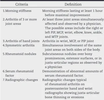
RA classiications were essentially based on the criteria for-mulated by the ACR in 198741(B), which are described in Table
2. However, those criteria did not perform well in early RA cases42(B). The ACR classiication criteria for RA were based on
individuals with long-standing disease and, until then, were considered to be the standard for the selection of patients for clinical studies. These criteria exhibit 91%–94% sensitivity and 89% speciicity for established RA. However, some of the items, such as radiological changes (erosions) and rheumatoid nodules, do not occur often in early RA. Thus, such criteria are suboptimal for the identiication of individuals with early RA (40%–90% sensitivity, and 50%–90% speciicity)43(B).
As a result, new RA classiication criteria were needed, with a special focus on the early disease stages14(D).
The new ACR/EULAR classiication criteria can be applied to any patient, provided that two basic requirements are met as follows:
1) Evidence of active clinical synovitis in at least one joint at the time of examination.
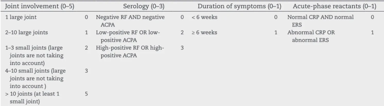
2) Synovitis cannot be better explained by another disease. The new criteria (Table 3) are based on a score system that is calculated by direct addition. The clinical manifestations are grouped into the following four domains: joint involvement, serology, duration of symptoms, and acute-phase reactants. In questionable cases, the number of involved joints can be cal-culated by the use of imaging methods such as ultrasound (US) and magnetic resonance (MRI). A score > 6 is needed to classify a patient as having deinite RA44(B). These criteria can be
ap-plied both prospectively and retrospectively, provided that the data were properly recorded.
It is worth observing that whenever a patient exhibits typi-cal erosions upon radiologitypi-cal examination and a clinitypi-cal his-tory compatible with RA (albeit non-documented), RA diagno-sis can be directly established in a manner independent of the applicability of the classiication criteria14(D).
The new 2010 criteria were not developed for the purpose of diagnosis but rather of classiication. The criteria basically serve to select homogeneous populations for studies.
Clinical RA diagnoses are extremely complex and includes multiple features that are hard to reconcile with a single scor-ing system14(D). Eventually, the formal criteria might serve to
guide clinical diagnoses.
Several features of the new criteria must be subjected to careful analysis before they can be universally accepted. In par-ticular, the criteria must be validated in different populations, including Brazilian early RA cohorts.
In patients who use methotrexate and those with persistent RA, the discriminatory powers of the 2010 ACR/EULAR criteria were 76% and 87%, respectively, when the score was at least 6, and 63% and 46%, respectively, when it was < 645(B).
Assuming the need for methotrexate, a diagnostic gold standard, during the irst year of follow-up, the 2010 ACR/EU-LAR criteria were able to diagnose 86% of the cases for which the score was at least 6 and 49% when it was < 645, compared
to 87% and 41%, respectively, when the 1987 ACR criteria were used46(B).
A comparison of the 2010 ACR/EULAR (score of at least 6) and 1987 ACR (score > 4) criteria relative to the diagnosis of pa-tients with a disease duration of less than 12 months showed positive predictive values of 70.7% and 65.3%, respectively, and negative predictive values of 76.1% and 79.1%, respectively47(B).
The discriminatory powers of the 2010 ACR/EULAR and 1987 ACR criteria during an 18-month follow-up period were com-pared and are shown in Table 4.
The application of the 2010 ACR/EULAR criteria at disease onset detected more patients who required DMARD treatment than did the 1987 ACR criteria; the values were 62% and 38%, respectively, and more particularly with regard to the use of methotrexate during the 18-month follow-up, the values were 68% versus 42%, respectively. However, the 2010 ACR/EULAR criteria were associated with a higher rate of false-positive cases (8% versus 2% for the 1987 ACR criteria)48(B).
Table 2 – 1987 American College of Rheumatology classiication criteria for rheumatoid arthritis.
Criteria Deinition
1. Morning stiffness Morning stiffness lasting at least 1 hour
before maximal improvement 2. Arthritis of 3 or more
joint areas
At least three joint areas simultaneously affected and observed by a physician. The possible areas include the right or left PIP, MCP, wrist, elbow, knee, ankle, and MTP joints.
3. Arthritis of hand joints Arthritis in wrist, MCP, or PIP joint
4. Symmetric arthritis Simultaneous involvement of the same
joint areas on both sides of the body.
5. Rheumatoid nodules Subcutaneous nodules over bony
prominences, extensor surfaces, or in juxta-articular regions as observed by a physician
6. Serum rheumatoid factor
Demonstration of abnormal amounts of serum rheumatoid factor.
7. Radiographic changes Radiographic changes typical of rheumatoid arthritis on posteroanterior hand and wrist radiographs showing juxta-articular bone thinning or erosions
In cases for which the 1987 ACR criteria had been used to deine RA (without radiological aid), the 2010 ACR/EULAR cri-teria were diagnostic of disease in 59% of the cases (positive predictive value), and ruled out the diagnosis in 93% (negative predictive value). The rate of false-positive results for the 2010 ACR/EULAR criteria was 17%. If RA was considered a chronic disease (ive years of follow-up), the discriminatory power of the 2010 ACR/EULAR criteria fell to 68% when the score was at least 6 and to 61% when the score was < 6. Nevertheless, the 1987 ACR criteria identiied 11.3% fewer cases as RA than did the 2010 ACR/EULAR criteria49(B).
Recommendation
The 2010 CR/EULAR criteria identify more patients with early RA than do the 1987 ACR criteria. However, the rate of
false-positive cases is higher with the newer criteria. When follow-up criteria such as use of DMARDs or disease persis-tence are used, the discriminatory powers of the 2010 CR/ EULAR and the 1987 ACR criteria are similar.
3. Is smoking associated with a poorer prognosis
for articular disease in RA patients?
Smoking was found to increase the risk of non-response (ACR50) by 18.3% [number needed to harm (NNH): 6] in pa-tients with early RA (24 weeks of symptom duration)50(B).
According to the EULAR criteria, RA patients who were smokers were less likely to achieve a good response at three months of treatment, compared to the non-smokers (NNH: 11). The patients who continued to smoke exhibited lower odds
Table 3 – 2010 ACR/EULAR classiication criteria for rheumatoid arthritis.
Target population (Who should be tested?) Patients who meet the following criteria:
1) Have at least 1 joint with deinite clinical synovitis (swelling)* 2) Present with synovitis that is not better explained by another disease
*Differential diagnoses might include conditions such as systemic lupus erythematosus, psoriatic arthritis, and gout. If the relevant differential diagnoses to consider are unclear, an expert rheumatologist should be consulted.
Joint involvement (0–5) Serology (0–3) Duration of symptoms (0–1) Acute-phase reactants (0–1)
1 large joint 0 Negative RF AND negative
ACPA
0 < 6 weeks 0 Normal CRP AND normal
ERS
0
2–10 large joints 1 Low-positive RF OR
low-positive ACPA
2 ≥ 6 weeks 1 Abnormal CRP OR
abnormal ERS
1
1–3 small joints (large joints are not taking into account)
2 High-positive RF OR
high-positive ACPA
3
4–10 small joints (large joints are not taking into account )
3
> 10 joints (at least 1 small joint)
5
A score ≥ 6 is needed for the classiication of a patient with deinite RA. “Joint involvement” refers to any swollen or tender joint on examination, which might be conirmed by imaging evidence of synovitis. Distal interphalangeal joints, irst carpometacarpal joints, and irst metatarsophalangeal joints are excluded from assessment. “Small joints” refers to the metacarpophalangeal joints, proximal interphalangeal joints, second through ifth metatarsophalangeal joints, thumb interphalangeal joints, and wrists. “Large joints” refers to the shoulders, elbows, knees, and ankles. In the category “> 10 joints” at least one of the involved joints must be a small joint; the other joints can include any combination of large and additional small joints, as well as joints that are not speciically listed elsewhere (e.g., temporomandibular, acromioclavicular, sternoclavicular).
In “Serology”, negative refers to IU values of the rheumatoid factor or anti-citrullinated protein antibody that are less than equal to the upper limit of normal (ULN) for the laboratory and assay, low-positive refers to IU values that are higher than the ULN, but ≤ 3 times the ULN for the laboratory and assay, and high-positive refers to IU values that are ≥ 3 times the ULN for the laboratory and assay.
“Duration of symptoms” refers to patient self-reports of the duration of signs or symptoms of joint synovitis that are clinically involved at the time of assessment.
“Acute-phase reactants” (erythrocyte sedimentation rate, and C-reactive protein) are considered to be normal/abnormal according to the local laboratory standards.
Modiied from: Aletaha D, Neogi T, Silman AJ, Funovits J, Felson DT, Bingham CO 3rd. 2010 rheumatoid arthritis classiication criteria: an American College of Rheumatology/European League Against Rheumatism collaborative initiative. Ann Rheum Dis. 2010; 69(9):1580-8.
Table 4 – Positive and negative predictive values from the 1987 ACR and 2010 ACR/EULAR criteria for patients with rheumatoid arthritis who are using disease-modifying anti-rheumatic drugs at the onset of disease and 18 months later.
Measurement Cohort onset 18 months later
2010 ACR/EULAR 1987 ACR 2010 ACR/EULAR 1987 ACR
+ predictive value 75% 85% 73% 81%
of good treatment responses during a 5-year follow up period. That difference in good treatment responses was somewhat higher in patients who were treated with anti-tumour necrosis factor (TNF) (14%; NNH: 7)51(B).
Smokers tend to exhibit more extra-articular manifesta-tions of RA (pleuritic, pericarditis, interstitial lung disease, neuropathy, glomerulonephritis, vasculitis), compared to non-smokers, as well as higher average Disease Activity Score (DAS-28), and Health Assessment Questionnaire (HAQ) values 52(B).
Smoking increased the use of DMARDs in RA patients and reduced their clinical responses (ACR50) by 16% (NNH: 6), par-ticularly in smokers of more than 20 packages/year53(B).
The radiological progression of RA was similar in smokers and non-smokers after three years, which did not agree with the poorer clinical responses exhibited by the former54(B).
RA patients who were smokers exhibited greater disease ac-tivity (joint pain and swelling) when compared to non-smokers after 24 months of treatment. The pain scores [(on a visual ana-logue scale – VAS) were also higher among the smokers. How-ever, radiological progression did not differ between smokers and non-smokers55(B).
Disease activity (measured as joint swelling, pain, and DAS-28) was greater in patients with an average symptom duration of seven months who were smokers when compared to non-smokers after a 5-year follow-up56(B).
Recommendation
Smoking increases the disease activity of RA and reduces clini-cal and functional responses over time. However, there is no suficient evidence regarding its inluence on radiological dis-ease progression.
4. Is measurement of the rheumatoid factor
a reliable test for diagnosis and prognostic
stratiication in RA?
RF is an antibody that targets the Fc fragment of IgG57(D). RF
is classically associated with RA, is found in the serum of ap-proximately 70% of RA patients, and is signiicantly correlated with a poorer prognosis. High RF levels are associated with aggressive disease, the presence of rheumatoid nodules, and extra-articular manifestations58(D).
The diagnostic value of RF alone is limited because 30%– 50% of patients might be seronegative during the early stage of RA57(D). In addition to its low sensitivity, its speciicity is
lim-ited. Individuals without RA might test positive for RF, and its prevalence increases with age59(B). Patients with other medical
conditions, both rheumatologic and not, might also test posi-tive for RF44,60(B). Therefore, negative RF serology does not rule
out a diagnosis of RA, whereas the interpretation of positive results must be carefully checked against the clinical indings.
Brazilian data (incident early RA cohort) indicate a RF preva-lence of approximately 50% in patients 61(B).
RF-positive patients with RA exhibited a 17% increase of mortality (NNH: 6) and cardiovascular mortality (NNH: 6) after 20 years of follow-up62(A).
The mortality of RA patients with RF-positive serology did not differ from that of seronegative patients after 14 years of
follow-up. However, when the results were analysed according to the number of expected events in the population, the mor-tality, and more speciically the cardiovascular mormor-tality, was elevated in the RF-positive patients 63(B).
A 10-year follow-up study of RA patients, in which 24% of the cases tested positive for RF of the IgM and IgA isotypes, found that radiological progression was associated with and could be predicted by the serological indings (e.g. IgM or IgA)64(B).
In a population of RA patients, 51% of whom were RF-posi-tive, the presence of RF was predictive of radiological sion in 69% of the cases, whereas its absence ruled out progres-sion in 83%.
RF was predictive of radiological progression (Larsen score) in RA patients after 5 years of follow-up65(B).
RF was predictive of radiological progression (Sharp or Lars-en scores)35,66(B) and the need for biological therapy67(B) in RA
patients after three years of follow-up.
The risk of radiological progression was 24.3% (NNH: 4) high-er among RF-positive patients vhigh-ersus RF-negative patients68(A).
With a pre-test RA probability rate of 35%, positive RF (IgM, IgA, and IgG isotypes), measured by ELISA, increased the diag-nostic probability rate to 94%, while negative serology ruled out RA with an 85% certainty rate69(B).
In a population of patients with a 35% probability rate of RA, RF (IgM, IgA, and IgG isotypes) increased the post-test prob-ability to 96%70(B).
Recommendation
RF measurement contributes estimations of prognosis for RA patients, particularly with regard to radiological progression and mortality. Positive RF serology, particularly in populations with a pre-test probability rate of 35%, increased the diag-nostic probability to 94%–96%, whereas negative RF serology ruled RA out with a post-test probability of 85%.
5. Is anti-CCP investigation superior to
rheumatoid factor investigation for RA diagnosis?
Recently, several anti-citrullinated protein antibodies (ACPA) were shown to behave as important diagnostic tools for RA; these had a similar sensitivity and superior speciicity to RF, in addition to their possible participation in disease physiopathogenesis71(B). Their possible roles as markers of RA
activity are controversial72(B).
Cyclic citrullinated peptide antibodies
Among the investigated antibodies that target ilaggrin-ci-trulline system antigens, anti-CCP exhibits the widest clini-cal applicability, with 70%–75% sensitivity and approximate-ly 95% speciicity. Anti-CCP anaapproximate-lyses are particularapproximate-ly useful for patients with early RA and negative RF serology73(B).
Other antibodies
Other antibodies are also used to investigate RA. The aim is to develop methods with sensitivities and speciicities sat-isfactory for early disease diagnosis, as well as more reli-able markers for activity and prognosis. These antibodies include anti-mutated citrullinated vimentin (anti-MCV) 80-82(B), anti-keratin (AKA), anti-perinuclear factor (APF)83(B),
anti-ilaggrin84(B), anti-citrullinated ibrinogen (ACF)85(B),
anti-protein A2 of the heterogeneous nuclear ribonucleoanti-protein complex (anti-RA33)83(B), anti-interleukin 1 (anti-IL1)86(B),
anti-1-α-enolase87(B), and anti-advanced glycation
end-prod-uct (AGE)88(B). The speciicities of these antibodies are
gen-erally satisfactory for RA diagnosis, but their sensitivities is generally lower than that of anti-CCP.
The 2010 ACR/EULAR criteria14(D) include only RF and ACPA
under the heading “autoantibodies”, and the values of these antibodies are described as negative, low, or high titres. As the values of both RF and anti-CCP are expressed as interna-tional units (IU), the results are rated negative when they are equal to or higher than the upper limit of normal (ULN) in the corresponding laboratory; low-positive when they are higher than the ULN, but equal to or lower than 3 times the ULN; and high-positive when they are higher than three times the ULN. Positive anti-CCP correlated with the MRI swelling and ero-sion score at a 4-year follow-up, whereas negative anti-CCP correlated with the synovitis score89(B).
Anti-CCP was superior to RF for predictions of the progres-sion of undifferentiated arthritis into RA (diagnostic certain-ties of 93% and 68%, respectively). The former also permitted better estimates of the severity of disease at a 7-year follow-up90(B).
The risk of positive anti-CCP serology in patients with ac-tive RA is 23% higher than that of patients in the period before disease. The anti-CCP alterations did not change after seven years of follow-up91(B).
With regard to the use of anti-CCP (second generation, anti-CCP2) and data from 15 recent RA cohorts, it was con-cluded that a single positive result permits a diagnosis of RA (likelihood ratio, LR+ 12.7), but that a single negative result does not rule out RA (LR− 0.45). Upon comparing RF and anti-CCP2, we found that their sensitivities are similar (56% and 58%, respectively), but the speciicity of anti-CCP2 is superior (96% versus 86% for RF). The sensitivity and speciicity of anti-CCP2 are higher than those of anti-CCP1. The combination of positive RF and anti-CCP2 only slightly increases the diagnos-tic certainty, compared to anti-CCP2 alone (LR+ 27 versus 22, respectively). An analysis of global evidence allows us to esti-mate the sensitivity of anti-CPP2 a 67%, and the speciicity as 96%. Assuming a prevalence of 42% in RA patients according to the 1987 ACR criteria, positive anti-CCP2 serology increases the diagnostic certainty to 90%, and negative serology rules out RA with a certainty of 75%92(B).
Recommendation
The sensitivity of anti-CCP is similar to that of RF, but the speci-icity of the former is superior, especially in the early disease stages. Anti-CCP evaluations are recommended in patients with a clinical suspicion of RA and negative RF test serology.
6. Are genetic markers
(
evaluations of HLA-DRB1
shared epitope alleles and PTPN22 genes
)
useful
for characterisations of RA patients with poorer
prognosis?
Although countless genetic markers have been described in as-sociation with RA, only the HLA-DRB1 shared epitope (SE) 10The presence of SE (HLA-DRB1) in RA patients did not correlate with radiological disease progression105,107(B). However, according to
some data, SE alleles and anti-CCP antibody levels might be as-sociated with the severity of joint damage (erosion and radio-logical damage score) in RA patients108(B). The HLA SE had no
predictive value relative to radiological RA progression109(B).
The frequency of HLA-DRB1 alleles with SE was found to be high in Latin American RA patients110(B).
The presence of SE alleles (DRB1) might be predictive of mor-tality, including cardiovascular mormor-tality, in RA patients with RA111,112(B).
An association was found between the DRB1 genotype and RF-positive RA patients with a 3.0%–3.7% (NNH: 30) risk increase113(B).
Recommendation
The PTPN22 gene polymorphism is associated with RA. Al-though it is not predictive of speciic therapeutic responses to biological therapy, it is predictive of remission when as-sociated with anti-CCP. Alone or in combination with HLA-DRB1 (SE), the PTPN22 polymorphism permits estimations of radiological progression or disease activity. The HLA-DRB1 al-lele seems to play a more important role in the prediction of poor prognosis relative to the progression, activity, severity, and mortality of RA.
7. Does the occurrence of extra-articular
manifestations denote a more aggressive disease
progression?
Although articular manifestations are the most characteristic, RA can also affect other organs and systems. The most fre-quent extra-articular manifestations include skin, eye, pleu-ropulmonary, heart, blood, neurological, and osteo-metabolic conditions. These occur more often in patients with severe and polyarticular disease, positive RF or anti-CCP serology, and rheumatoid nodules27(B)28(D).
The incidence of extra-articular manifestations in RA is 47.5%, which includes cardiovascular, blood, eye, and lung af-fections. Such manifestations are associated with a greater likelihood of the use of biological agents114(B).
Clinically signiicant lung interstitial disease occurs in 10% of RA patients115(B). Patient mortality depends on the type of
lung affection and is greater when the affection is diffuse116(B).
Pulmonary ibrosis-related mortality is approximately 6%115(B).
The average survival of patients with interstitial pneumonia is 3.2 years, and thus is generally lower compared to that of other varieties of interstitial disease (6.6 years)116(B). In RA patients
The mortality rate of RA patients with lung interstitial dis-ease is 7%, and the average survival duration after diagnosis is three years. In spite of the association between interstitial lung disease and RA activity, the latter was only denoted by increased ESR in that study118(B).
The presence of kidney dysfunction in RA patients is not as-sociated with the activity, progression, dysfunction, or severity of the disease119(B).
RA patients with extra-articular manifestations exhibit a 20% increase in the risk of cardiovascular events (including acute myocardial infarction, angina, coronary disease, and stroke) (NNH: 5)120(B).
The survival of patients with extra-articular manifestations of RA (18% of cases) is lower than that of patients with exclu-sive articular manifestations, and the relative risk of death in the former increases by 27% after seven years of follow-up. Similar to the extra-articular manifestations, comorbidities also increase mortality, particularly cardiovascular conditions because these cause 31% of patient deaths. Increased mortal-ity correlates with greater disease activmortal-ity (RF), worse function (HAQ), and increased radiological progression121(B).
In RA patients with extra-articular manifestations, the scores that assess disease activity, such as DAS28 and HAQ, and the Larsen radiological score tend to be poorer, thus de-noting a greater disease severity. Only 4.1% of such patients achieved remission122(B).
After 15 years of follow-up, mortality increased only in the patients with extra-articular manifestations (relative risk in-crease: 51%), compared to those without such conditions; peri-carditis was the most signiicant of the manifestations123(B).
The mortality rate of RA patients with extra-articular mani-festations (7.9% prevalence) was one death per 4.3 patients per year, whereas the rate of patients without articular manifesta-tions was one death per 11.4 patients per year124(B).
The risk of severe gastrointestinal diseases is elevated in RA patients with extra-articular manifestations (4.6% prevalence). In such patients, the disease intensity (ACR criteria) and the signs of radiological progression are also greater125(B).
Recommendation
RA progression is more severe in patients with extra-articular manifestations. These patients have more intense disease ac-tivity with reduced functional capacities, responses to treat-ment (less occurrence of remission), and life expectancies, compared to those with exclusive articular manifestations.
8. Is conventional radiography an appropriate test
for RA diagnosis?
Conventional radiography is the most widely used imaging method for assessments of structural joint damage in RA. In addition to its diagnostic utility, conventional radiography plays an important role in the monitoring of disease progres-sion, provided that it is performed at regular intervals126(D).
The initial radiographic signs include increased amounts of soft tissues and juxta-articular osteopenia. More charac-teristic signs of RA, such as reduced joint space and bone ero-sion, appear later in the disease course.
The presence of bone erosion during the early stages of RA represents a risk factor for the development of persistent arthritis127(B). This factor is associated with functional
limita-tion and thus with poorer prognosis128(B).
When erosions are identiied by radiography (15% preva-lence), the diagnostic probability increases to 100%. However, as negative indings do not reduce the probability (18%), they do not rule out a RA diagnosis129(B).
In patients with strong clinical suspicion of RA but nega-tive RF serology and radiography, the presence of anti-CCP antibodies and erosions on MRI are highly speciic for RA diagnosis130(B).
In RA patients, the sensitivity of MRI for the detection of erosions is greater than that of conventional radiogra-phy. Conventional radiography detected 89% of erosions in the MCP joint bones and 15.8% in the wrist bones; these were lower than the MRI detection rates of 100% and 69%, respectively131(B).
The diagnostic accuracy of conventional radiography in the detection of wrist bone erosions in RA patients was 63%, whereas the accuracy of MRI was 77%132(B).
The diagnostic sensitivity of radiography for the detection of MCP joint bone erosions in RA patients was 14%, compared to 66% with MRI133(B).
In RA patients who were followed-up for two years, radi-ography identiied damage progression in 40% of the cases (total Larsen score) and 15% of the MCP joint bones (Larsen score). The accuracy of plain radiography in the identiication of damage progression was similar to that of MRI134(B).
Detection of erosions by means of the E score in RA pa-tients was lower on radiographic assessment (13.1 ± 8.3) than on MRI (28.8 ± 10.0)135(B).
In a population of RA patients with joint erosions (95% prevalence), radiography identiied 59% of the cases, com-pared to 95% by MRI136(B).
Radiography of the hands of RA patients identiied 50% fewer erosions than MRI, although the identiication of radio-logical progression was similar with both methods137(B).
In a population of RA patients with a 43% prevalence of erosions, radiography increased the diagnostic probability to 80% for cases with positive indings, and ruled out a diagnosis in 85% for those with negative indings. After a 3-year follow-up period, the identiication of erosions on radiography de-creased to 81% and 60%, respectively138(B).
In a population of patients with arthritis, 36% of whom were diagnosed with RA, diagnostic radiography increased the probability of RA to 50%, but when the results were nega-tive, the probability decreased to 33%139(B).
Radiographic assessment of RA patients only slightly in-creased the probability of distinguishing between RF seropos-itive and seronegative cases. In a population with a 59% prev-alence of RF seropositive cases, positive radiographic indings (destruction) increased the probability to 66%, and a lack of radiographic indings decreased it to 47%140(B).
Recommendation
9. Is ultrasound superior to conventional
radiography in the diagnosis and establishment
of prognosis of RA?
The sensitivity of musculoskeletal US and MRI for the detec-tion of structural damage was superior to that of conven-tional radiography141(D).
US is useful for early detection, as well as the monitor-ing of inlammatory activity and signs of joint destruction when performed by an operator with signiicant experience in musculoskeletal diseases135(B).
Compared to MRI, the cost of US is lower, and it is not contraindicated for patients with metallic implants or claus-trophobia. Additionally, US permits dynamic assessments of the joints and bilateral comparisons, as well as evaluations of other anatomical structures134(B)141,143(D).
The use of power and colour Doppler might provide com-plementary information and thus contribute to characteri-sations of inlammatory activity144(D).
Positive and negative US indings, when used to identify joint inlammation in RA patients, permits deinite diagno-ses in 79% and 55% of the cadiagno-ses, respectively. These results are similar those of radiography (Sharp score) when it ex-hibits positive indings (74%), but superior when the radio-graphic indings are negative (38%)145(B).
Using MRI as the gold-standard (as in the present study), US was superior to radiography in the detection of bone ero-sions in patients with recent RA, whereby the LR+ values were 31 and 20, respectively. Based on a lesion prevalence of 50%, in cases with positive indings, the diagnostic probabili-ties of US and radiography increase to 99% and 97%, respec-tively. Therefore, the utility of both methods is similar135(B).
Relative to the detection of erosions in RA patients, when US exhibits positive indings, it achieves a diagnostic certain-ty of 82% and for negative indings, a certaincertain-ty rate of 61%, compared to 95% and 55%, respectively, for radiography146(B).
The sensitivity and speciicity of US in the detection of inlammatory signs and interphalangeal joint destruction in RA patients were 59% and 98%, respectively, compared to 42% and 99% for radiography, respectively. The diagnostic certainties relative to US and conventional radiography were 97% and 98%, respectively, when those results were positive and 71% and 63%, respectively, when they were negative147(B).
The sensitivity and speciicity of US in the detection of MCP joint erosions in the of RA patients were 79% and 97%, respectively, compared to 32% and 98% for conventional ra-diography, respectively. The diagnostic certainties relative to US and conventional radiography were 96% and 94%, respec-tively, when those results were positive and 82% and 46%, respectively, when they were negative148(B).
The sensitivity and speciicity of US in the detection of glenohumeral joint erosions in RA patients were 74% and 75%, respectively, compared to 67% and 100% for radiogra-phy, respectively. The diagnostic certainties relative to US and conventional radiography were 75% and 100%, respec-tively, when those results were positive and 74% and 75%, respectively, when they were negative149(B).
The diagnostic accuracy of US in the identiication of ero-sions in RA patients was 84% and was thus superior to that
of radiography (73%). However, when only the tests with positive indings were considered for analysis, the LR of US was lower (5 versus 13), which indicates less diagnostic certainty150(B).
In patients with early RA, US found erosions that were not identiied by radiography in 19.3% of the cases, but failed to diagnose 8.8% of the cases that were identiied by radiography. The combination of both methods permit-ted the diagnosis of 45.6% of the lesions in that patient population151(B).
In patients with early RA, US correlated with disease ac-tivity (DAS28) and functional capacity (HAQ) scores at 12 months of follow-up152(B).
In patients with early RA, US increased the detection of erosions in 42.0% of the cases at the time of diagnosis and after 9 months of follow-up, compared to radiography153(B).
The detection of joint lesions in RA patients was greater with US versus radiography; speciically, US detection was 5% greater at the time of diagnosis, and 23% greater after seven years of follow-up154(B). However, in another study,
radiography identiied a larger number of erosions in RA patients, compared to US (37% and 30%, respectively). After six months, the rates were 48% and 41%, respectively155(B).
After accounting for the number of humeral erosions (greater tuberosity, anteromedial, and posterolateral) in RA patients, the diagnostic certainties of US and radiography were 90% and 40%, respectively, when the indings were positive and 96% and 39%, respectively, when the indings were negative152(B).
Recommendation
US might contribute to the diagnosis of joint erosions in RA patients, as well to the monitoring of disease progression.
10. Is magnetic resonance superior to
conventional radiography and ultrasound for the
diagnosis and establishment of prognosis of RA?
MRI is the most sensitive method with which to detect the changes associated with the early stages of RA. It permits the assessment of structural alterations of the soft tissues, bone, and cartilage, in addition to erosions at an earlier stage than conventional radiography138(B).
In addition to the features identiied by conventional radiography, MRI is further able to detect bone swelling, which was shown to be a predictor of bone erosion135(B).
In Brazil, factors such as the high cost and lack of stan-dardisation limit the use of MRI in clinical practice.
The results of MRI relative to RA diagnosis vary widely as a function of the applied criteria and the investigated population. Thus, the sensitivity varies from 20% to 100%, and the speciicity from 0% to 100%136,156-158(B). Additionally,
the results of MRI relative to RA progression vary widely, with a sensitivity range from 18% to 100% and a speciic-ity range from 6% to 97%156,159-161(B). Furthermore, the use
In RA patients, MRI (Outcome Measures in Rheumatology – OMERACT - deinition) permits the diagnosis of erosions (hands or wrists) with 35%–90% sensitivity and 35%–90% speciicity, bone swelling (hands, wrists, or MCP joints) with 32.5%–65% sensitivity and 82.5%–100% speciicity, and synovi-tis (hands or wrists) with 40-80% sensitivity and 57.7%–92.5% speciicity163(B).
Compared to MRI, when the indings were positive, con-ventional radiography could diagnose MCP and PIP joint ero-sions with certainty in 98%–100% of the cases, and US in 86%– 7% of the cases. When the indings were negative, the rates were 84% and 93%, respectively 135,155(B).
The diagnostic accuracy of Doppler US for the identiica-tion of joint inlammaidentiica-tion in RA patients was 75%, compared to MRI164(B).
Using computed tomography (CT) as the gold-standard for the diagnosis of erosions in the wrists of RA patients, when the indings were positive, MRI accurately diagnosed 90% of the cases, compared to conventional radiography138,154(B).
Using high-ield MRI as the gold-standard for the diagnosis of erosions in the wrists and MCP joints of RA patients, when the indings were positive, limb MRI accurately diagnosed 88% to 93% of the cases, compared to conventional radiography (94%–98%) and US (82%)165,166(B).
A combination of MRI synovitis, swelling, and erosion scores permitted the identiication of RA patient responses to TNF-α inhibitor treatment at a 12-month follow-up167(B).
As a method for long-term functional assessments in RA patients, MRI identiied improvements only in 29% of the cases, compared to the functional status (assessed by doctors and patients)168(B).
MRI (bone swelling) and US (inlammation) exhibited simi-lar abilities to identify the progression of erosion in RA pa-tients (using the Rheumatoid Arthritis MRI Scoring System – RAMRIS – as the gold standard) over a 12-month follow-up period152(B).
The progression of erosion was identiied by MRI (OMER-ACT) in 23% of patients with RA over a 5-year period, compared with 40% by conventional radiography (Larsen score)140(B).
Recommendation
MRI is the most sensitive method with which to detect the changes associated with the early stages of RA. It permits the assessment of structural alterations of the soft tissues, bone, and cartilage, in addition to erosions at an earlier stage than conventional radiography. In Brazil, factors such as the high cost and lack of standardisation have limited the use of MRI in clinical practice.
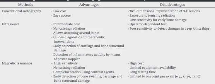
Table 5 summarises the advantages and disadvantages of the imaging methods used to assess RA patients.
Conclusion
The present guidelines were elaborated by the Commission of Rheumatoid Arthritis of the Brazilian Society of Rheuma-tology to formulate recommendations for the diagnosis and initial assessment of RA in Brazil. Due to the country’s territo-rial extension and the diversity of its macro-regions, local dif-ferences relative to differential diagnoses and access to some (laboratory or imaging) technologies might occur.
RA diagnosis is of paramount importance, especially in the earliest stages.
Lack of diagnosis means a lack of appropriate treatment and, consequently, an increased risk of the development of persistent inlammation and progressive joint damage. Rheu-matologists must be included as early as possible in assess-ments of patients with arthritis due to their wider experience and familiarity with the possible differential diagnoses and investigational approaches.
Despite the recent publication of guidelines for the diagno-sis of RA, a revision of this subject that accounts for particular Brazilian features is relevant.
Therefore, the establishment of recommendations for RA ultimately seeks to deine and provide a solid basis for Brazil-ian rheumatologists with data from controlled trials to pro-mote a homogeneous approach to diagnosis within the Bra-zilian socioeconomic context.
Table 5 – Advantages and disadvantages of the imaging methods used to assess patients with rheumatoid arthritis.
Methods Advantages Disadvantages
Conventional radiography - Low cost
- Easy access
- Two-dimensional representation of 3-D lesions - Exposure to ionising radiation
- Low sensitivity for early bone damage
Ultrasound - Intermediate cost
- No ionising radiation - Allows assessing several joints - Guides diagnostic and therapeutic
interventions
- Early detection of cartilage and bone structural damage
- Detection of inlammatory activity by means of power Doppler
- Operator-dependent test
- Poor sensitivity to detect changes in deep joints (hips)
Magnetic resonance - High sensitivity
- No ionising radiation
- Complementation using contrast agents - Early detection of bone swelling, cartilage and
bone structural damage
- High cost
- Limited equipment availability - Long testing time
Because the knowledge relative to RA increases rapidly, the corresponding recommendations should be updated on a periodic and regular basis.
Conlicts of interest
Mota LMH: Participated in clinical and/or experimental stud-ies sponsored by Roche and Mantecorp; was given personal or institutional grants by Abbott, AstraZeneca, MSD, Roche, and Pizer; and was a guest lecturer at meetings and other activities sponsored by Abbott, MSD, Novartis, Roche, and Wyeth.
Cruz BA: Participated in clinical and/or experimental stud-ies sponsored by Roche; was given personal or institutional grants by Abbott, Bristol-Myers Squibb, Mantecorp, MSD, No-vartis, Roche, Wyeth, and Pizer; and was a guest lecturer at meetings and other activities sponsored by Abbott, MSD, Man-tecorp, Novartis, Roche, and Wyeth.
Brenol CV: Participated in clinical and/or experimental studies sponsored by Bristol-Myers Squibb, Pizer, Roche, and Wyeth; was given personal or institutional grants by Abbott, Bristol-Myers Squibb, Mantecorp, MSD, Roche, and Wyeth; and was a guest lecturer at meetings and other activities sponsored by Abbott and Roche.
Pereira IA: Participated in clinical and/or experimental stud-ies sponsored by Roche; was given personal or institutional grants by Abbott, MSD, Roche, BMS, Jansen, and Pizer; was a guest lecturer at meetings and other activities sponsored by Abbott, MSD, BMS, Pizer, Roche, and Janssen; is a member of the consultant or executive boards of pharmaceutical compa-nies or the normative committees of scientiic studies spon-sored by Abbott, BMS, Janssen, Roche, Pizer, and MSD.
Rezende-Fronza LS: Participated in clinical and/or experi-mental studies sponsored by Bristol-Myers Squibb, Pizer, and Roche; and wrote scientiic papers for journals sponsored by Pizer.
Bertolo MB: Was a guest lecturer at meetings and other ac-tivities sponsored by Abbott, Pizer, and Sanoi Aventis.
Freitas MVC: Was given personal or institutional grants by Abbott, MSD, Pizer, Roche, and Wyeth; was a guest lecturer at meetings and other activities sponsored by Abbott, MSD, Pizer, Roche, and Wyeth; is a member of the consultant or executive boards of pharmaceutical companies or the normative com-mittees of scientiic studies sponsored by AstraZeneca, MSD, and Wyeth; and wrote scientiic papers for journals sponsored by Abbott, AstraZeneca, Bristol-Myers Squibb, and Wyeth.
Silva NA: Participated in clinical and/or experimental stud-ies sponsored by Bristol-Myers Squibb and Roche; was given personal or institutional grants by Abbott, MSD, Pizer, Roche, and Wyeth; and was a guest lecturer at meetings and other ac-tivities sponsored by Janssen, Mantecorp, MSD, and Roche.
Louzada-Junior P: Participated in clinical and/or experimen-tal studies sponsored by Merck and Roche; was given personal or institutional grants by Abbott; and was a guest lecturer at meetings and other activities sponsored by Bristol-Meyers-Squibb, Pizer, and Roche.
Giorgi RD: Was given personal or institutional grants by Bristol-Myers Squibb and Roche; and was a guest lecturer at meetings and other activities sponsored by Bristol-Myers Squibb and Roche.
Lima RAC: Participated in clinical and/or experimental stud-ies sponsored by Mantecorp and Roche; was given personal or institutional grants by Acteion, Lilly, and Pizer; and was a guest lecturer at meetings and other activities sponsored by Acteion, Lilly, and Pizer.
Pinheiro GRC: Was given personal or institutional grants by Janssen and Roche.
R E F E R E N C E S
1. Lee DM, Weinblatt ME. Rheumatoid arthritis. Lancet. 2001;358:903-11.
2. Alarcón GS. Epidemiology of rheumatoid arthritis. Rheum Dis Clin North Am. 1995;21:589-604.
3. Silman AJ, Pearson JE. Epidemiology and genetics of rheumatoid arthritis. Arthritis Res. 2002;4:S265-72. 4. Marques-Neto JF, Gonçalves ET, Langen LFOB, Cunha MFL,
Radominski S, Oliveira SM, et al. Multicentric study of the prevalence of adult rheumatoid arthritis in Brazilian population samples. Rev Bras Reumatol. 1993;33:169-73. 5. Chehata JC, Hassell AB, Clarke SA, Mattey DL, Jones
MA, Jones W, et al. Mortality in rheumatoid arthritis: relationship to single and composite measures of disease activity. Rheumatology (Oxford). 2001;40:447-52.
6. Emery P. The optimal management of early rheumatoid arthritis: the key to preventing disability. Br J Rheumatol. 1994;33:765-8.
7. Sokka T. Work disability in early rheumatoid arthritis. Clin Exp Rheumatol. 2003;21:S71-4.
results of a special early arthritis clinic compared to routine patient care. Br J Rheumatol. 1998;37:1084-8. 9. Majithia V, Geraci SA. Rheumatoid arthritis: diagnosis and
management. Am J Med. 2007;120:936-9.
10. Haque UJ, Bathon JM. The role of biological in early rheumatoid arthritis. Best Pract Res Clin Rheum. 2005;19:179-89.
11. Cabral D, Katz JN, Weinblatt ME, Ting G, Avorn J, Solomon DH. Development and assessment of indicators of rheumatoid arthritis severity: results of a Delphi panel. Arthritis Rheum. 2005;53:61-6.
12. Sokka T, Kautiainen H, Pincus T, Verstappen SM, Aggarwal A, Alten R, et al. Work disability remains a major
problem in rheumatoid arthritis in the 2000s: data from 32 countries in the QUEST-RA study. Arthritis Res Ther. 2010;12:R42.
13. Saag KG, Teng GG, Patkar NM, Anuntiyo J, Finney C, Curtis JR, et al. American College of Rheumatology. American College of Rheumatology 2008 recommendations for the use of nonbiologic and biologic disease-modifying antirheumatic drugs in rheumatoid arthritis. Arthritis Rheum. 2008;59(6):762-84.
14. Smolen JS, Landewé R, Breedveld FC, Dougados M, Emery P, Gaujoux-Viala C, et al. EULAR recommendations for the management of rheumatoid arthritis with synthetic and biological disease-modifying antirheumatic drugs. Ann Rheum Dis. 2010;69:964-75.
15. da Mota LM, Laurindo IM, dos Santos-Neto LL. Demographic and clinical characteristics of a cohort of patients with early rheumatoid arthritis. Rev Bras Reumatol. 2010;50:235-48.
16. Louzada-Junior P, Souza BD, Toledo RA, Ciconelli RM. Descriptive analysis of the demographical and clinical characteristics of the patients with rheumatoid arthritis in the State of São Paulo, Brazil. Rev Bras Reumatol. 2007;47:84-90.
17. Schoels M, Wong J, Scott DL, Zink A, Richards P, Landewé R, et al. Economic aspects of treatment options in rheumatoid arthritis: a systematic literature review informing the EULAR recommendations for the management of rheumatoid arthritis. Ann Rheum Dis. 2010;69:995-1003.
18. de Azevedo AB, Ferraz MB, Ciconelli RM. Indirect costs of rheumatoid arthritis in Brazil. Value Health. 2008;11:869-77. 19. Chermont GC, Kowalski SC, Ciconelli RM, Ferraz MB. Resource
utilization and the cost of rheumatoid arthritis in Brazil. Clin Exp Rheumatol. 2008;26:24-31.
20. Mease PJ. Inlammatory musculoskeletal disease:
identiication and assessment. J Rheumatol. 2011;38:557-61. 21. Scott DL, Wolfe F, Huizinga TW.Rheumatoid arthritis. Lancet.
2010;376:1094-108.
22. da Mota LM, Cruz BA, Brenol CV, Pereira IA, Fronza LS, Bertolo MB, ET al. Consenso da Sociedade Brasileira de Reumatologia 2011 para o diagnóstico e avaliação inicial da artrite reumatoide. Rev Bras Reumatol. 2011;51:199-219. 23. Dixon WG, Symmons DP. Does early rheumatoid arthritis
exist? Best Pract Res Clin Rheumatol. 2005;19:37-53.
24. Woolf AD. How to assess musculoskeletal conditions. History and physical examination. Best Pract Res Clin Rheumatol. 2003;17:381-402.
25. Yazici Y, Pincus T, Kautiainen H, Sokka T. Morning stiffness in patients with early rheumatoid arthritis is associated more strongly with functional disability than with joint swelling and erythrocyte sedimentation rate. J Rheumatol. 2004;31:1723-6.
26. Hazes JM, Hayton R, Silman AJ. A reevaluation of the symptom of morning stiffness. J Rheumatol. 1993;20: 1138-42.
27. Goeldner I, Skare TL, de Messias Reason IT, Nisihara RM, Silva MB, da Rosa Utiyama SR. Association of anticyclic
citrullinated peptide antibodies with extra-articular
manifestations, gender, and tabagism in rheumatoid arthritis patients from southern Brazil. Clin Rheumatol. 2011;30:975-80.
28. Turesson C, Eberhardt K, Jacobsson LT, Lindqvist E. Incidence and predictors of severe extra-articluar disease manifestations in an early rheumatoid arthritis inception cohort. Ann Rheum Dis. 2007;66:1543-44.
29. Hernández-García C, Vargas E, Abásolo L, Lajas C, Bellajdell B, Morado IC, et al. Lag time between onset of symptoms and access to rheumatology care and DMARD therapy in a cohort of patients with rheumatoid arthritis. J Rheumatol. 2000;27:2323-8.
30. Combe B, Landewe R, Lukas C, Bolosiu HD, Breedveld F, Dougados M, et al. EULAR recommendations for the management of early arthritis: report of a task force of the European Standing Committee for International Clinical Studies Including Therapeutics (ESCISIT). Ann Rheum Dis. 2007;66:34-45.
31. Luukkainen R, Sokka T, Kautiainen H, Hannonen P, Laasonen L, Leirisalo-Repo M, et al. Prognosis of 5-year radiographic erosions of the wrist according to early, late, and persistent wrist swelling or tenderness in patients with early rheumatoid arthritis. J Rheumatol. 2007;34:50-3. 32. Bosello S, Fedele AL, Peluso G, Gremese E, Tolusso B,
Ferraccioli G. Very early rheumatoid arthritis is the major predictor of major outcomes: clinical ACR remission and radiographic non-progression. Ann Rheum Dis. 2011;70:1292-5.
33. Mouterde G, Lukas C, Logeart I, Flipo RM, Rincheval N, Daurès JP, et al. Predictors of radiographic progression in the ESPOIR cohort: the season of irst symptoms may inluence the short-term outcome in early arthritis. Ann Rheum Dis. 2011;70:1251-6.
34. Courvoisier N, Dougados M, Cantagrel A, Goupille P, Meyer O, Sibilia J, et al. Prognostic factors of 10-year radiographic outcome in early rheumatoid arthritis: a prospective study. Arthritis Res Ther. 2008;10:R106.
35. Combe B, Dougados M, Goupille P, Cantagrel A, Eliaou JF, Sibilia J, et al. Prognostic factors for radiographic damage in early rheumatoid arthritis: a multiparameter prospective study. Arthritis Rheum. 2001;44:1736-43.
36. Machold KP, Stamm TA, Nell VP, Plugbeil S, Aletaha D, Steiner G, et al. Very recent onset rheumatoid arthritis: clinical and serological patient characteristics associated with radiographic progression over the irst years of disease. Rheumatology (Oxford). 2007;46:342-9.
37. Welsing PM, Fransen J, van Riel PL. Is the disease course of rheumatoid arthritis becoming milder? Time trends since 1985 in an inception cohort of early rheumatoid arthritis. Arthritis Rheum. 2005;52:2616-24.
38. Kyburz D, Gabay C, Michel BA, Finckh A; physicians of SCQM-RA. The longterm impact of early treatment of rheumatoid arthritis on radiographic progression: a population-based cohort study. Rheumatology (Oxford). 2011;50:1106-10. 39. van der Linden MP, le Cessie S, Raza K, van der Woude D,
Knevel R, Huizinga TW, et al. Long-term impact of delay in assessment of patients with early arthritis. Arthritis Rheum. 2010;62:3537-46.
40. Keysser M, Keysser C, Keitel W, Keysser G. Loss of functional capacity caused by a delayed onset of DMARD therapy in rheumatoid arthritis. Long-term follow-up results of the Keitel function test. Brief deinite report. Z Rheumatol. 2001;60:69-73.